

Effect of Extracellular Matrix Membrane on Bone Formation in a Rabbit Tibial Defect Model
- PMID: 27047963
- PMCID: PMC4800078
- DOI: 10.1155/2016/6715295
Effect of Extracellular Matrix Membrane on Bone Formation in a Rabbit Tibial Defect Model
Abstract
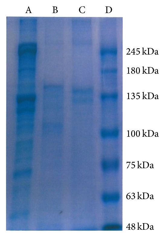
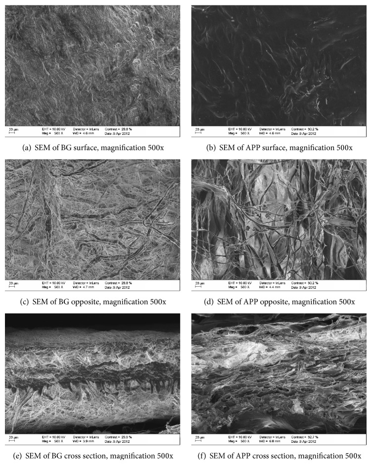
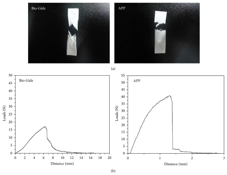
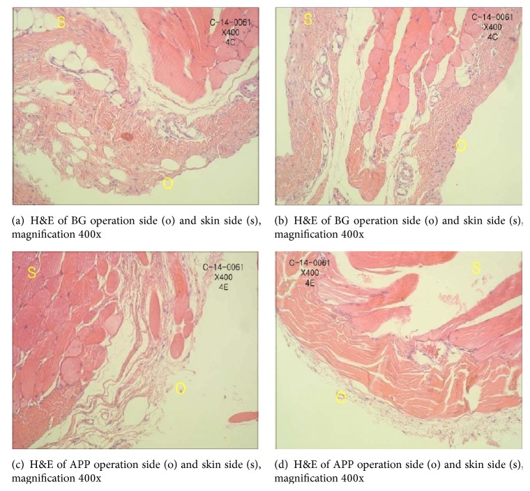
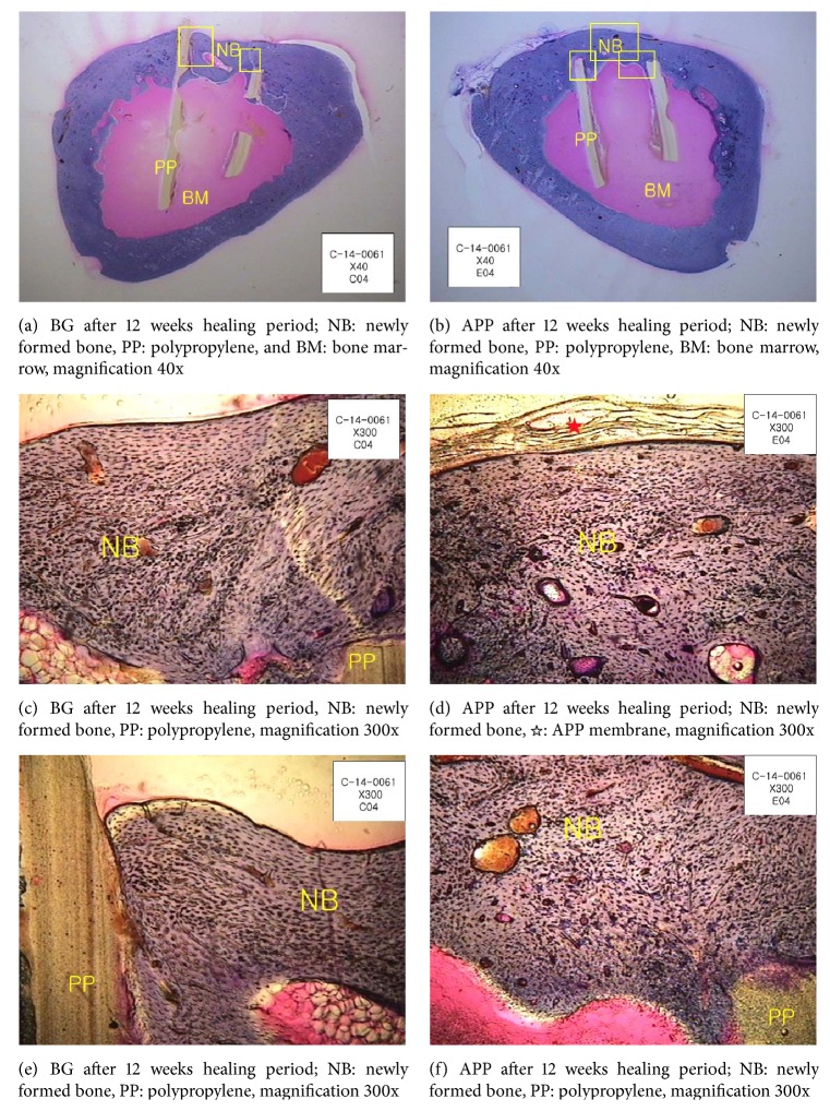
Absorbable extracellular matrix (ECM) membrane has recently been used as a barrier membrane (BM) in guided tissue regeneration (GTR) and guided bone regeneration (GBR). Absorbable BMs are mostly based on collagen, which is more biocompatible than synthetic materials. However, implanted absorbable BMs can be rapidly degraded by enzymes in vivo. In a previous study, to delay degradation time, collagen fibers were treated with cross-linking agents. These compounds prevented the enzymatic degradation of BMs. However, cross-linked BMs can exhibit delayed tissue integration. In addition, the remaining cross-linker could induce inflammation. Here, we attempted to overcome these problems using a natural ECM membrane. The membrane consisted of freshly harvested porcine pericardium that was stripped from cells and immunoreagents by a cleaning process. Acellular porcine pericardium (APP) showed a bilayer structure with a smooth upper surface and a significantly coarser bottom layer. APP is an ECM with a thin layer (0.18-0.35 mm) but with excellent mechanical properties. Tensile strength of APP was 14.15 ± 2.24 MPa. In in vivo experiments, APP was transplanted into rabbit tibia. The biocompatible material was retained for up to 3 months without the need for cross-linking. Therefore, we conclude that APP could support osteogenesis as a BM for up to 3 months.
Figures
Similar articles
-
Effect of Multilaminate Small Intestinal Submucosa as a Barrier Membrane on Bone Formation in a Rabbit Mandible Defect Model.Biomed Res Int. 2018 Jun 19;2018:3270293. doi: 10.1155/2018/3270293. eCollection 2018. Biomed Res Int. 2018. PMID: 30018978 Free PMC article.
-
Acellular pericardium: A naturally hierarchical, osteoconductive, and osteoinductive biomaterial for guided bone regeneration.J Biomed Mater Res A. 2021 Feb;109(2):132-145. doi: 10.1002/jbm.a.37011. Epub 2020 Jun 16. J Biomed Mater Res A. 2021. PMID: 32441432
-
Guided bone regeneration using acellular bovine pericardium in a rabbit mandibular model: in-vitro and in-vivo studies.J Periodontal Res. 2014 Aug;49(4):499-507. doi: 10.1111/jre.12129. Epub 2013 Sep 12. J Periodontal Res. 2014. PMID: 24024647
-
[Use of native and cross-linked collagen membranes for guided tissue and bone regeneration].Schweiz Monatsschr Zahnmed. 2006;116(11):1112-23. Schweiz Monatsschr Zahnmed. 2006. PMID: 17144624 Review. German.
-
Advances in Modification Methods Based on Biodegradable Membranes in Guided Bone/Tissue Regeneration: A Review.Polymers (Basel). 2022 Feb 23;14(5):871. doi: 10.3390/polym14050871. Polymers (Basel). 2022. PMID: 35267700 Free PMC article. Review.
Cited by
-
Effect of Multilaminate Small Intestinal Submucosa as a Barrier Membrane on Bone Formation in a Rabbit Mandible Defect Model.Biomed Res Int. 2018 Jun 19;2018:3270293. doi: 10.1155/2018/3270293. eCollection 2018. Biomed Res Int. 2018. PMID: 30018978 Free PMC article.
-
Enhancing bone formation using absorbable AZ31B magnesium alloy membranes during distraction osteogenesis: A new material study.Heliyon. 2023 Jul 11;9(8):e18032. doi: 10.1016/j.heliyon.2023.e18032. eCollection 2023 Aug. Heliyon. 2023. PMID: 37534007 Free PMC article.
-
Morphogenetically-Active Barrier Membrane for Guided Bone Regeneration, Based on Amorphous Polyphosphate.Mar Drugs. 2017 May 17;15(5):142. doi: 10.3390/md15050142. Mar Drugs. 2017. PMID: 28513544 Free PMC article.
-
Bone Augmentation of Peri-Implant Dehiscence Defects Using Multilaminated Small Intestinal Submucosa as a Barrier Membrane: An Experimental Study in Dogs.Biomed Res Int. 2019 Nov 16;2019:8962730. doi: 10.1155/2019/8962730. eCollection 2019. Biomed Res Int. 2019. PMID: 31828142 Free PMC article.
-
[Research progress on the modification of guided bone regeneration membranes].Hua Xi Kou Qiang Yi Xue Za Zhi. 2019 Jun 1;37(3):325-329. doi: 10.7518/hxkq.2019.03.019. Hua Xi Kou Qiang Yi Xue Za Zhi. 2019. PMID: 31218871 Free PMC article. Review. Chinese.
References
-
- Zhang Z., Li G., Shi B. Physicochemical properties of collagen, gelatin and collagen hydrolysate derived from bovine limed split wastes. Journal of the Society of Leather Technologies and Chemists. 2006;90(1):23–28.
Publication types
MeSH terms
Substances
LinkOut - more resources
Full Text Sources
Other Literature Sources
