

Evaluation of post-mortem lateral cerebral ventricle changes using sequential scans during post-mortem computed tomography
- PMID: 27048214
- PMCID: PMC4976059
- DOI: 10.1007/s00414-016-1327-2
Evaluation of post-mortem lateral cerebral ventricle changes using sequential scans during post-mortem computed tomography
Abstract
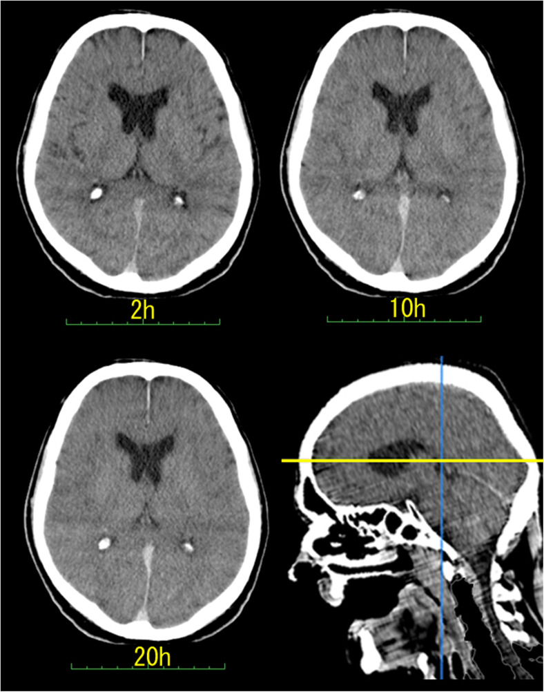
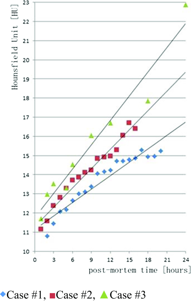
In the present study, we evaluated post-mortem lateral cerebral ventricle (LCV) changes using computed tomography (CT). Subsequent periodical CT scans termed "sequential scans" were obtained for three cadavers. The first scan was performed immediately after the body was transferred from the emergency room to the institute of legal medicine. Sequential scans were obtained and evaluated for 24 h at maximum. The time of death had been determined in the emergency room. The sequential scans enabled us to observe periodical post-mortem changes in CT images. The series of continuous LCV images obtained up to 24 h (two cases)/16 h (1 case) after death was evaluated. The average Hounsfield units (HU) within the LCVs progressively increased, and LCV volume progressively decreased over time. The HU in the cerebrospinal fluid (CSF) increased at an individual rate proportional to the post-mortem interval (PMI). Thus, an early longitudinal radiodensity change in the CSF could be potential indicator of post-mortem interval (PMI). Sequential imaging scans reveal post-mortem changes in the CSF space which may reflect post-mortem brain alterations. Further studies are needed to evaluate the proposed CSF change markers in correlation with other validated PMI indicators.
Keywords: Lateral cerebral ventricle; Post mortem interval; Post-mortem change; Post-mortem computed tomography; Sequential scan.
Figures


Similar articles
-
Estimation of the time of death by measuring the variation of lateral cerebral ventricle volume and cerebrospinal fluid radiodensity using postmortem computed tomography.Int J Legal Med. 2021 Nov;135(6):2615-2623. doi: 10.1007/s00414-021-02698-6. Epub 2021 Sep 25. Int J Legal Med. 2021. PMID: 34562107 Free PMC article.
-
The radiodensity of cerebrospinal fluid and vitreous humor as indicator of the time since death.Forensic Sci Med Pathol. 2016 Sep;12(3):248-56. doi: 10.1007/s12024-016-9778-9. Epub 2016 Apr 27. Forensic Sci Med Pathol. 2016. PMID: 27117292 Free PMC article.
-
Quantitative estimation of a ratio of intracranial cerebrospinal fluid volume to brain volume based on segmentation of CT images in patients with extra-axial hematoma.Neuroradiol J. 2017 Feb;30(1):10-14. doi: 10.1177/1971400916678227. Epub 2016 Nov 11. Neuroradiol J. 2017. PMID: 27837185 Free PMC article.
-
Post-mortem CT imaging of the lungs: pathological versus non-pathological findings.Radiol Med. 2017 Dec;122(12):902-908. doi: 10.1007/s11547-017-0802-2. Epub 2017 Aug 23. Radiol Med. 2017. PMID: 28836139 Review.
-
Post-mortem CT and MRI: appropriate post-mortem imaging appearances and changes related to cardiopulmonary resuscitation.Br J Radiol. 2016;89(1058):20150851. doi: 10.1259/bjr.20150851. Epub 2015 Nov 12. Br J Radiol. 2016. PMID: 26562099 Free PMC article. Review.
Cited by
-
Post mortem computed tomography meets radiomics: a case series on fractal analysis of post mortem changes in the brain.Int J Legal Med. 2022 May;136(3):719-727. doi: 10.1007/s00414-022-02801-5. Epub 2022 Mar 3. Int J Legal Med. 2022. PMID: 35239030 Free PMC article.
-
Essence of postmortem computed tomography for in-hospital deaths: what clinical radiologists should know.Jpn J Radiol. 2023 Oct;41(10):1039-1050. doi: 10.1007/s11604-023-01443-w. Epub 2023 May 17. Jpn J Radiol. 2023. PMID: 37193920 Free PMC article. Review.
-
Exploring radiomic features of lateral cerebral ventricles in postmortem CT for postmortem interval estimation.Int J Legal Med. 2025 Mar;139(2):667-677. doi: 10.1007/s00414-024-03396-9. Epub 2024 Dec 20. Int J Legal Med. 2025. PMID: 39702800
-
Estimation of the time of death by measuring the variation of lateral cerebral ventricle volume and cerebrospinal fluid radiodensity using postmortem computed tomography.Int J Legal Med. 2021 Nov;135(6):2615-2623. doi: 10.1007/s00414-021-02698-6. Epub 2021 Sep 25. Int J Legal Med. 2021. PMID: 34562107 Free PMC article.
-
Radiomic analysis of postmortem lung changes: a PMCT-based approach for estimating the postmortem interval.Forensic Sci Med Pathol. 2025 Aug 27. doi: 10.1007/s12024-025-01071-y. Online ahead of print. Forensic Sci Med Pathol. 2025. PMID: 40864404
References
-
- Yen K, Lövblad KO, Scheurer E, Ozdoba C, Thali MJ, Aghayev E, Jackowski C, Anon J, Frickey N, Zwygart K, Weis J, Dirnhofer R. Post-mortem forensic neuroimaging: correlation of MSCT and MRI findings with autopsy results. Forensic Sci Int. 2007;173:21–35. doi: 10.1016/j.forsciint.2007.01.027. - DOI - PubMed
MeSH terms
LinkOut - more resources
Full Text Sources
Other Literature Sources
