. 2016 Jun 15;473(12):1703-18.
doi: 10.1042/BCJ20160203. Epub 2016 Apr 5.

Septin oligomerization regulates persistent expression of ErbB2/HER2 in gastric cancer cells

Affiliations

Septin oligomerization regulates persistent expression of ErbB2/HER2 in gastric cancer cells

Elizabeth A Marcus et al. Biochem J. .

Abstract

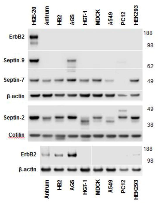
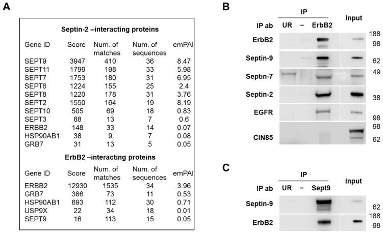
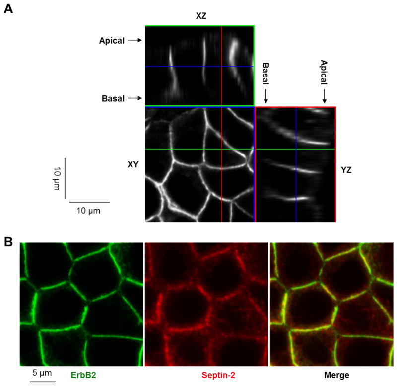
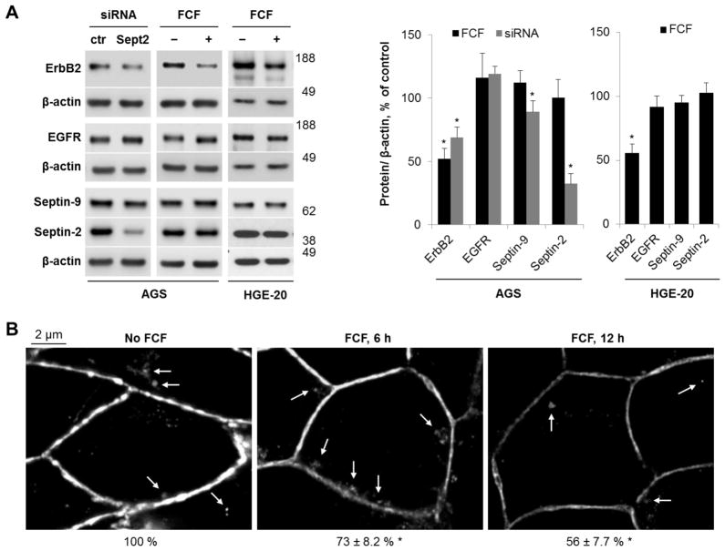
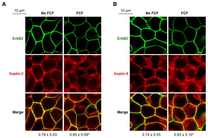
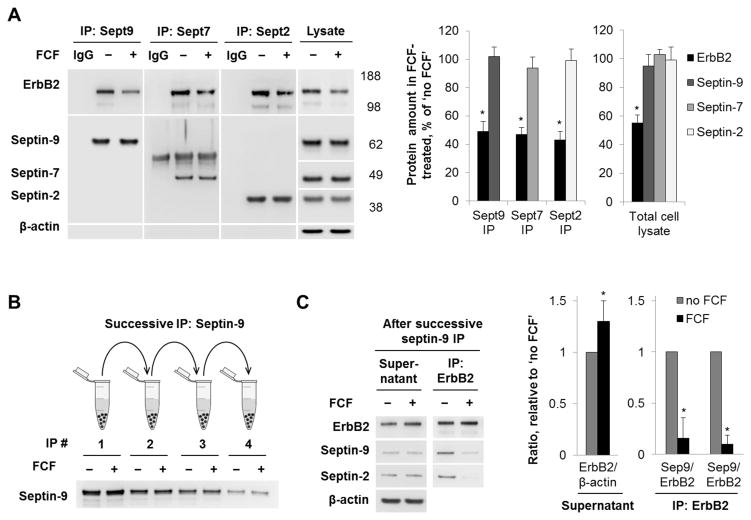
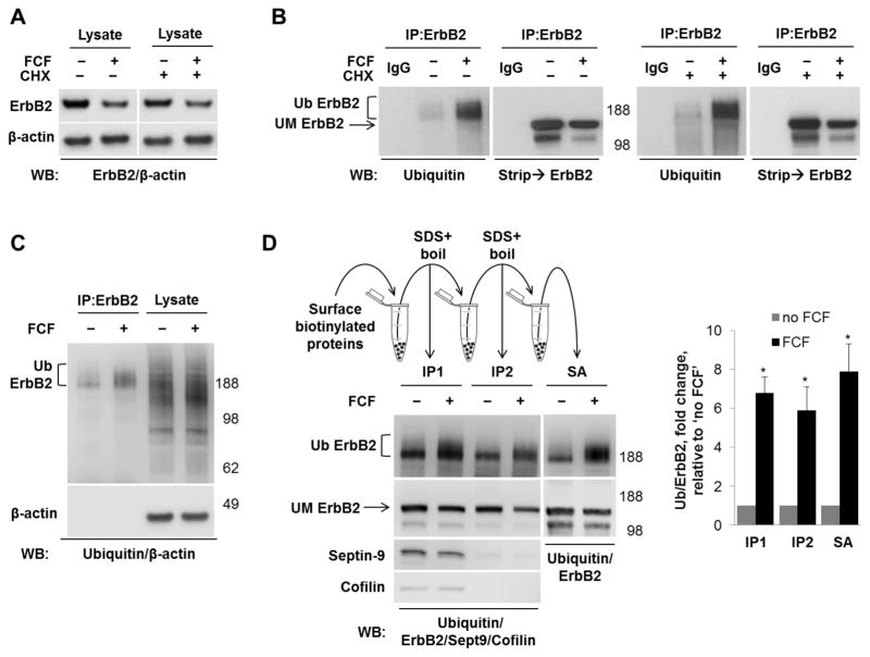
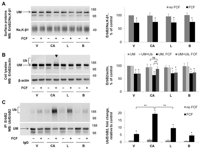
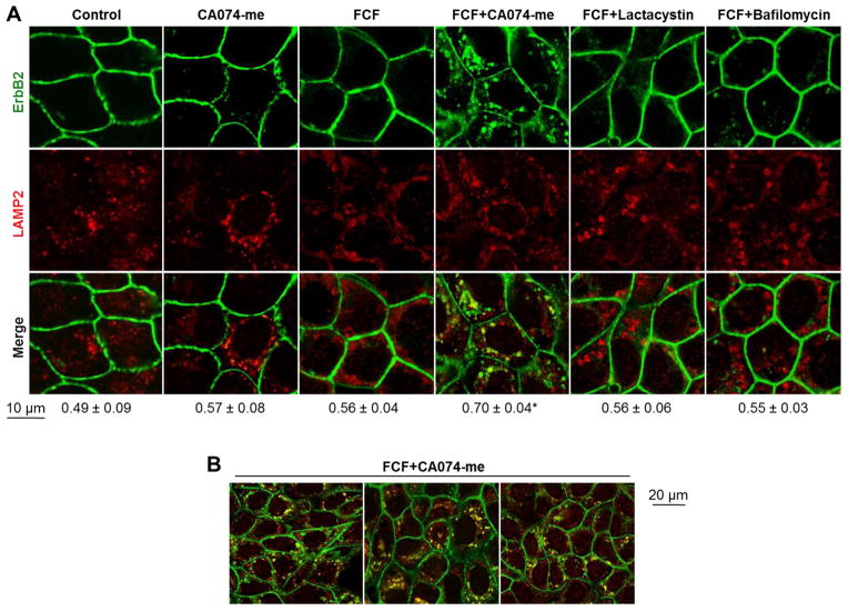
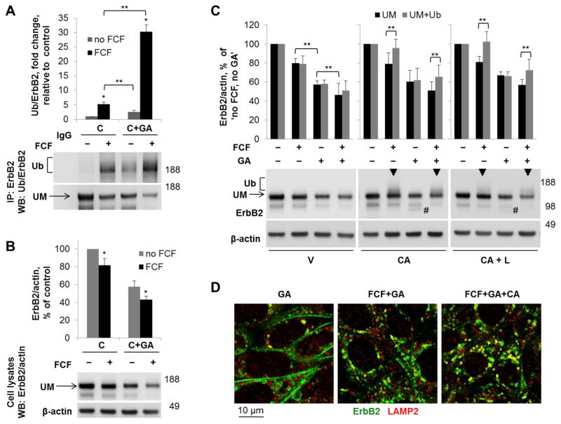
Septins are a family of cytoskeletal GTP-binding proteins that assemble into membrane-associated hetero-oligomers and organize scaffolds for recruitment of cytosolic proteins or stabilization of membrane proteins. Septins have been implicated in a diverse range of cancers, including gastric cancer, but the underlying mechanisms remain unclear. The hypothesis tested here is that septins contribute to cancer by stabilizing the receptor tyrosine kinase ErbB2, an important target for cancer treatment. Septins and ErbB2 were highly over-expressed in gastric cancer cells. Immunoprecipitation followed by MS analysis identified ErbB2 as a septin-interacting protein. Knockdown of septin-2 or cell exposure to forchlorfenuron (FCF), a well-established inhibitor of septin oligomerization, decreased surface and total levels of ErbB2. These treatments had no effect on epidermal growth factor receptor (EGFR), emphasizing the specificity and functionality of the septin-ErbB2 interaction. The level of ubiquitylated ErbB2 at the plasma membrane was elevated in cells treated with FCF, which was accompanied by a decrease in co-localization of ErbB2 with septins at the membrane. Cathepsin B inhibitor, but not bafilomycin or lactacystin, prevented FCF-induced decrease in total ErbB2 by increasing accumulation of ubiquitylated ErbB2 in lysosomes. Therefore, septins protect ErbB2 from ubiquitylation, endocytosis and lysosomal degradation. The FCF-induced degradation pathway is distinct from and additive with the degradation induced by inhibiting ErbB2 chaperone Hsp90. These results identify septins as novel regulators of ErbB2 expression that contribute to the remarkable stabilization of the receptor at the plasma membrane of cancer cells and may provide a basis for the development of new ErbB2-targeting anti-cancer therapies.

Keywords: ErbB2; endocytosis; gastric cancer; lysosomal degradation; septin; ubiquitylation.


Conflict of interest statement

Declaration of interest: The authors declare that they have no conflicts of interest with the contents of this article.

Figures

Figure 1
Figure 1. ErbB2 and septin-9 are highly expressed in gastric cancer cells
ErbB2, septin-9, septin-2, and septin-7 expression were determined by western blot analysis of total lysates of various cell lines and human gastric antrum, β-actin was used as a control. Cofilin was used as a control for septin-2 given the size similarity between septin-2 and β-actin. ErbB2 is very highly expressed in HGE-20 gastric cancer cells, to the extent that the band is not visible in other cell types with reasonable exposure of the HGE-20 cell band. Once HGE-20 cells were removed from the ErbB2 blot, expression was also seen in human gastric antrum, HB2 breast cancer cells, and AGS gastric cancer cells. Septin-9 similarly showed increased expression in gastric cancer cells compared to other cell types.
Figure 2
Figure 2. ErbB2 interacts with septins in HGE-20 gastric cancer cells
Proteins immunoprecipitated using antibodies against septin-2 or ErbB2 from HGE-20 cell lysates were analyzed by mass spectrometry. ErbB2, known ErbB2-associated proteins, were detected in septin-2 immunoprecipitate and septin-9 was detected in ErbB2 immunoprecipitate (A). Interaction of ErbB2 with septins and a known dimerization partner, EGFR, was confirmed by western blot, following immunoprecipitation of ErbB2 in HGE-20 cell lysate. Ubiquitin ligase c-cbl binding protein, CIN85, a protein that mediates the interaction between septin-9 and EGFR, was not seen in ErbB2 IP (B). Interaction between septin-9 and ErbB2 was again confirmed by western blot following IP of HGE-20 cell lysates with septin-9 antibody (C). emPAI-exponentially modified protein abundance index, UR-unrelated antibody negative control (anti-human influenza hemagglutinin for ErbB2 IP and anti-green fluorescent protein for septin-9 IP).
Figure 3
Figure 3. ErbB2 is localized to the plasma membrane and co-localizes with septins
ErbB2 protein is localized mainly to the basolateral membrane of HGE-20 cells as shown by confocal microscopy (A). ErbB2 and septin-2 co-localize at the lateral membrane in HGE-20 cells, as shown by immunofluorescence using antibodies against ErbB2 and septin-2 (B).
Figure 4
Figure 4. siRNA silencing and interruption of septin dynamics with FCF decrease ErbB2 levels in gastric cancer cells
AGS cells were transiently transfected with septin-2-specific or control (ctr) siRNA or AGS and HGE-20 cells were incubated with FCF for 12 hours. Western blot was performed on clarified lysates. ErbB2 protein levels were decreased with siRNA silencing of septin-2. Use of FCF led to a similar decrease in ErbB2 protein in both cell lines. The structurally similar EGFR was not affected, demonstrating specificity of response. β-actin was used as a loading control (A). Immunofluroescence with ErbB2 antibodies in HGE-20 cells showed accumulation of ErbB2 in intracellular compartments following 6 hour incubation with FCF, with overall decreased expression and minimal intracellular appearance of ErbB2 at 12 hours, suggestive of intracellular degradation (B). Arrows in the images point to intracellular accumulation of ErbB2. Numbers below images indicate the intensity of total fluorescence as % of control as calculated by analyzing at least 10 microscopic fields for each condition using Zen 2009 software. n=3 independent experiments, errors or error bars represent standard deviation (s.d.), *-significant difference from condition without siRNA or FCF, p<0.05, Student’s t-test.
Figure 5
Figure 5. FCF decreases co-localization and co-immunoprecipitation of ErbB2 with septins
HGE-20 cells were incubated with FCF or vehicle for 12 hours, followed by fixation and immunofluorescence. The images show an altered distribution of septin-2 (A) and septin-9 (B) and a decrease in co-localization between septins and ErbB2 (A-B) in the presence of FCF. Coefficients of co-localization as calculated using Zen 2009 software are shown below the images. Errors bars represent s.d., n=3 independent experiments, at least 12 fields were analyzed for each condition; *-significant difference from no FCF control, P <0.05, Student’s t-test.
Figure 6
Figure 6. FCF decreases the interaction between septins and ErbB2
HGE-20 cells were incubated with FCF for 12 hours, followed by immunoprecipitation with antibodies against septin proteins. ErbB2 was decreased in septin immunoprecipitates and cell lysates in the presence of FCF, while septin protein levels were unchanged. IgG refers to immunoprecipitation procedure without cell lysate (A). HGE-20 cells were incubated with or without FCF for 12 hours and lysed. The level of septin-9 was depleted in HGE-20 cell lysates by 4 consecutive immunoprecipitations with septin-9 antibodies, each using the supernatanat of the prior preparation. The level of septin-9 was clearly depleted by the 4th immunoprecipitation (B). Western blot analysis of the final supernatant from (B) detects more ErbB2 in a sample from FCF-treated cells as compared to control. Western blot analysis of ErbB2 immunoprecipitates from the final supernatant from (B) shows less septin-2 and septin-9 bound to ErbB2 in the sample from FCF-treated cells (C). n=3 independent experiments, error bars represent s.d., *-significant difference from no FCF condition, P<0.05, Student’s t-test.
Figure 7
Figure 7. FCF induces ubiquitylation of ErbB2 at the plasma membrane
There was no difference in FCF-induced decrease in ErbB2 in the presence or absence of cycloheximide, as shown by western blot of total HGE-20 cell lysate, suggesting the effect of FCF is on mature ErbB2 protein (A). Incubation with FCF or vehicle was completed for 12 hours with or without cycloheximide, followed by immunoprecipitation with ErbB2 antibodies. Western blot using ubiquitin antibodies, followed ErbB2 antibodies, showed increased ubiquitylation of ErbB2 in the presence of FCF, independent of new protein synthesis (B). Ubiquitylation induced by FCF is specific to ErbB2, as shown by western blot comparison of ErbB2 immunoprecipitate and total cell lysate using ubiquitin antibodies; β-actin was used as a loading control (C). Cells were incubated with FCF or vehicle, surface proteins were biotinylated from basolateral side, followed by immunoprecipitation with ErbB2 antibodies (IP1), then boiling of eluted proteins in SDS to remove interacting proteins, followed by dilution in non-ionic detergent and repeat immunoprecipitation with ErbB2 antibodies (IP2). Loss of septin-9 and cofilin signals on western blot in IP2 confirms dissociation of ErbB2 from interacting proteins. Increased ubiquitylation of ErbB2 is still seen in the absence of interacting proteins, demonstrating that ErbB2 itself is ubiquitylated. Proteins eluted from the second IP by boiling in SDS were diluted in non-ionic detergent and biotinylated ErbB2 was isolated by streptavidin extraction (SA). As shown by western blot with ubiquitin antibodies, the increased ubiquitylation of ErbB2 induced by FCF is seen in this fraction, demonstrating ubiquitylation occurs at the plasma membrane. The graph shows the FCF-increased ubiquitylation seen in each experiment as compared to control (D). Ub-ubiquitylated ErbB2, UM-unmodified ErbB2, IgG-immunoprecipitation control without cell lysate, SA-streptavidin.
Figure 8
Figure 8. Cathepsin B inhibitor increases the amount of FCF-induced ubiquitylated forms of ErbB2 in cell lysates but not in the plasma membrane fraction
HGE-20 cells were incubated with the indicated inhibitors for 6 hours, followed by basolateral biotinylation of surface proteins, streptavidin extraction, and western blot using anti-ErbB2 antibodies. Na+,K+-ATPase β1 subunit was used as a loading control. ErbB2 protein levels in the membrane fraction were not protected by any of the indicated inhibitors in the presence of FCF (A). In total cell lysates, ubiquitylated ErbB2, seen as an increased density running above the main ErbB2 band (arrowhead), was increased in the presence of cathepsin B inhibitor CA074-me (B). ErbB2 was immunoprecipitated, followed by western blot using anti-ubiquitin, then anti-ErbB2 antibodies, confirming a significant increase in ubiquitylated forms of ErbB2 in the presence of CA074-me and also showing a moderate increase in the presence of lactacystin (C). Quantification for each blot is shown to the right. Error bars, s.d., n=4 independent experiments, statistics done by Student’s t-test, ns-not significant, * - significant difference from the no-FCF control, P<0.05, ** - significant difference between indicated conditions, p<0.05, CA-CA074-me, L-lactacystin, B-bafilomycin, V-vehicle, Ub-ubiquitylated form of ErbB2, UM-unmodified form of ErbB2.
Figure 9
Figure 9. Incubation of HGE-20 cells with FCF and cathepsin B inhibitor leads to accumulation of ErbB2 in lysosomes
HGE-20 cells were incubated with the indicated inhibitors for 6 hours, then fixed. Immunofluorescence was performed using antibodies against ErbB2 and the lysosomal protein LAMP2. There was an increase in co-localization of ErbB2 with lysosomes in the presence of FCF and cathepsin B inhibitor CA074-me. Coefficient of overlap between ErbB2 and LAMP2 as calculated using Zen 2009 software with the confocal microscope is shown below the images (A). Low magnification images of HGE-20 cells incubated with FCF and CA074-me show that lysosomal accumulation of ErbB2 occurs in the majority of cells in a cell monolayer (B). Error bars, s.d., n=3 independent experiments, at least 12 fields were analyzed for each condition, * - significant difference from no-FCF condition, P = 0.0007, Student’s t-test.
Figure 10
Figure 10. FCF augments the effect of Hsp90 inhibitor geldanamycin on ubiquitylation and degradation of ErbB2 and works via a distinct pathway
HGE-20 cells were incubated for 6 hours with cycloheximide and with or without FCF and geldanamycin. ErbB2 was immunoprecipitated, followed by western blot with antibodies against ubiquitin, and then ErbB2. Ubiquitylation of ErbB2 in the presence of FCF and geldanamycin was greater than with either agent alone (A). Western blot of total cell lysates obtained following the same incubation conditions demonstrates an additive effect of both agents on the level of mature ErbB2. β-actin was used as a loading control (B). HGE-20 cells were incubated with FCF and/or geldanamycin and the indicated inhibitors of protein degradation for 6 hours, followed by western blot using antibodies against ErbB2, with β-actin as a loading control. FCF-induced ubiquitylated forms of ErbB2, shown as an increased density above the main band (arrowheads), are increased by CA074-me in the presence and absence of geldanamycin. CA074-me protected geldanamycin-induced ErbB2 degradation products (indicated by # on the blot) independent of FCF. Lactacystin did not augment the effects of CA074-me on the FCF or geldanamycin-induced changes, demonstrating that proteasomes are not directly involved in ErbB2 degradation (C). HGE-20 cells were incubated with the indicated inhibitors for 6 hours, fixed, and immunofluorescence was completed using antibodies against ErbB2 and LAMP2. Co-localization of ErbB2 with lysosomes was increased in the presence of both FCF and geldanamycin (D). Error bars, s.d., n=3 independent experiments, * - significant difference from no-FCF condition, Student’s t-test, P<0.05, ** - significant difference between indicated conditions, Student’s t-test, P<0.05, C-cycloheximide, GA-geldanamycin, UM-unmodified ErbB2, V-vehicle, CA-CA074-me, L-lactacystin.

Similar articles

Cited by

References

    1. Hall PA, Russell SE. Mammalian septins: dynamic heteromers with roles in cellular morphogenesis and compartmentalization. The Journal of pathology. 2012;226:287–299. - PubMed
    1. Russell SE, Hall PA. Do septins have a role in cancer? British journal of cancer. 2005;93:499–503. - PMC - PubMed
    1. Connolly D, Abdesselam I, Verdier-Pinard P, Montagna C. Septin roles in tumorigenesis. Biol Chem. 2011;392:725–738. - PubMed
    1. Kocevar N, Odreman F, Vindigni A, Grazio SF, Komel R. Proteomic analysis of gastric cancer and immunoblot validation of potential biomarkers. World J Gastroenterol. 2012;18:1216–1228. - PMC - PubMed
    1. Scott M, Hyland PL, McGregor G, Hillan KJ, Russell SE, Hall PA. Multimodality expression profiling shows SEPT9 to be overexpressed in a wide range of human tumours. Oncogene. 2005;24:4688–4700. - PubMed

Publication types

MeSH terms