

Stem cells and bFGF in tendon healing: Effects of lentiviral gene transfer and long-term follow-up in a rat Achilles tendon defect model
- PMID: 27048602
- PMCID: PMC4822291
- DOI: 10.1186/s12891-016-0999-6
Stem cells and bFGF in tendon healing: Effects of lentiviral gene transfer and long-term follow-up in a rat Achilles tendon defect model
Abstract
Background: The influence of stem cells and lentiviral expression of basic fibroblastic growth factor (bFGF) on tendon healing and remodelling was investigated in an in-vivo long-term (12 weeks) rat Achilles tendon defect model.
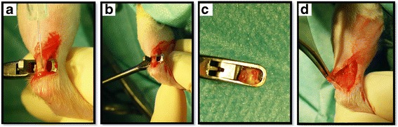
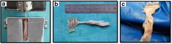
Methods: In sixty male Lewis rats, complete tendon defects (2.4 mm) were created and either left untreated (PBS) or treated by injection of stem cells lentivirally expressing the enhanced green fluorescence marker gene eGFP (MSC-LV-eGFP) or basic fibroblast growth factor bFGF (MSC-LV-bFGF). Tendons were harvested after 12 weeks and underwent biomechanical and (immuno)-histological analysis.
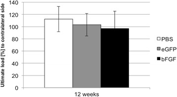
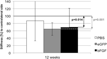
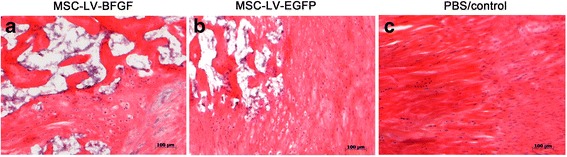
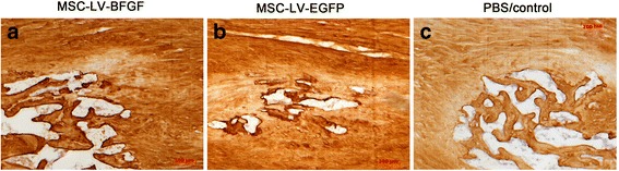
Results: After 12 weeks the mean ultimate load to failure ratio (treated side to contralateral side) in biomechanical testing reached 97 % in the bFGF-group, 103 % in the eGFP-group and 112 % in the PBS-group. Also in the stiffness testing both MSC groups did not reach the results of the PBS group. Histologically, the MSC groups did not show better results than the control group. There were clusters of ossifications found in all groups. In immunohistology, only the staining collagen-type-I was strongly increased in both MSC groups in comparison to PBS control group. However, there were no significant differences in the (immuno)-histological results between both stem cell groups.
Conclusion: The biomechanical and (immuno)-histological results did not show positive effects of the MSC groups on tendon remodelling in a long-term follow-up. Interestingly, in later stages stem cells had hardly any effects on biomechanical results. This study inspires a critical and reflected use of stem cells in tendon healing.
Keywords: Growth factor; Lentiviral vector; Mesenchymal stem cells (MSCs); Tendon healing; bFGF.
Figures
References
-
- Muller SA, Todorov A, Heisterbach PE, Martin I, Majewski M. Tendon healing: an overview of physiology, biology, and pathology of tendon healing and systematic review of state of the art in tendon bioengineering. Knee Surg Sports Traumatol Arthrosc. 2015;23(7):2097–105. doi: 10.1007/s00167-013-2680-z. - DOI - PubMed
Publication types
MeSH terms
Substances
LinkOut - more resources
Full Text Sources
Other Literature Sources
Medical
