. 2016 Apr;30(4):285-303.
doi: 10.1007/s10822-016-9899-y. Epub 2016 Apr 5.

Computational analysis of EBNA1 "druggability" suggests novel insights for Epstein-Barr virus inhibitor design

Affiliations

Computational analysis of EBNA1 "druggability" suggests novel insights for Epstein-Barr virus inhibitor design

Eleonora Gianti et al. J Comput Aided Mol Des. 2016 Apr.

Abstract

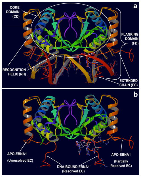
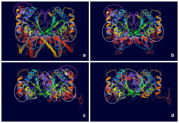
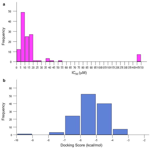
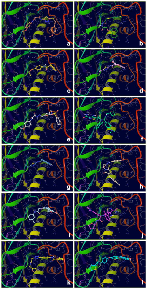
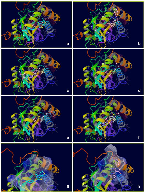
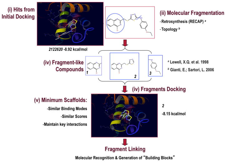
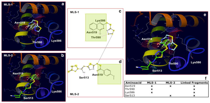
The Epstein-Barr Nuclear Antigen 1 (EBNA1) is a critical protein encoded by the Epstein-Barr Virus (EBV). During latent infection, EBNA1 is essential for DNA replication and transcription initiation of viral and cellular genes and is necessary to immortalize primary B-lymphocytes. Nonetheless, the concept of EBNA1 as drug target is novel. Two EBNA1 crystal structures are publicly available and the first small-molecule EBNA1 inhibitors were recently discovered. However, no systematic studies have been reported on the structural details of EBNA1 "druggable" binding sites. We conducted computational identification and structural characterization of EBNA1 binding pockets, likely to accommodate ligand molecules (i.e. "druggable" binding sites). Then, we validated our predictions by docking against a set of compounds previously tested in vitro for EBNA1 inhibition (PubChem AID-2381). Finally, we supported assessments of pocket druggability by performing induced fit docking and molecular dynamics simulations paired with binding affinity predictions by Molecular Mechanics Generalized Born Surface Area calculations for a number of hits belonging to druggable binding sites. Our results establish EBNA1 as a target for drug discovery, and provide the computational evidence that active AID-2381 hits disrupt EBNA1:DNA binding upon interacting at individual sites. Lastly, structural properties of top scoring hits are proposed to support the rational design of the next generation of EBNA1 inhibitors.

Keywords: Computational approaches to pocket finding; EBNA1 “druggability” assessment; Molecular docking; Structure-based drug design.


Figures

Fig. 1
Fig. 1
Structural Organization of the EBNA1 Protein. a Functional EBNA1 dimer bound to DNA: the tri-nucleotide (tri-nt) bp sequence (3′-TGC-5′)113 located in the center of the DNA Binding Site is shown in stick rendering. The Core Domain (CD, residues 504–607), comprising the Recognition Helix (RH), as well as the Flanking Domain (FD, residues 461–503), constituted by the helix perpendicular to DNA forming the Extended Chain (EC) loop that embraces the DNA molecule, are shown. b Superimposition of apo (PDB ID 1VHI) and DNA-bound (PDB ID 1B3T) crystal structures of EBNA1. In the apo conformation, the EC loop is unstructured and (partially) unsolved. In the bound-conformation, the EC loop adopts a conformation embracing the DNA helix (the DNA molecule was removed from the visualization). The tri-nucleotide (tri-nt) bp sequence (3′-TGC-5′)113 located in the center of the DNA Binding Site is shown
Fig. 2
Fig. 2
Four Putative Binding Sites identified on EBNA1. Maps were generated using SiteMap (www.schrodinger.com). Blue ovals indicate the Primary Binding Site. White ovals indicate the Secondary Binding Site. Yellow ovals indicate the Tertiary Binding Site. Pink ovals indicate the Quaternary Binding Site. a, b show the crystal structure of EBNA1 co-crystallized in complex with DNA (PDB ID 1B3T). In b, the DNA molecule was removed to show the maps generated by SiteMap (blue ovals). c, d show the crystal structure of EBNA1 unbound to DNA (PDB ID 1VHI and The Wistar Institute proprietary structure, respectively)
Fig. 3
Fig. 3
Histogram Distributions of Activity and Score Values for AID 2381 Compounds. a Experimental Activities (μM IC50); b Docking Scores (kcal/mol)
Fig. 4
Fig. 4
Docking Complexes of Top Scoring Compounds against EBNA1 Primary Binding Site. Chemical structures, docking scores and IC50 values are reported in Table 1. a 15944862; b 3167530; c 5973599; d 3186499; e 5826369; f 3185712; g 1432744; h 1919699; i 7256141; j 3185717; k 1452563; l 1107365
Fig. 5
Fig. 5
Docking complexes of top scoring compounds against EBNA1 secondary binding site. Chemical structures, docking scores and IC50 values are reported in Table 2. a 2122620; b 647423; c 1335528; d 1272394; e 3186499; f 4879718; g 2122620 shown within the surface binding site; h 647423 shown within the surface binding site
Fig. 6
Fig. 6
Minimum Ligand Scaffold (MLS) Approach used to Target the Secondary Binding Site (I). Compounds from initial docking (Hits) were fragmented into smaller “fragment-like” molecules, which were successively docked against the Secondary Site. If they obeyed the Minimum Scaffold Requirements, then a new molecular framework was designed directly within cavity by properly linking them together
Fig. 7
Fig. 7
Minimal Ligand Scaffold (MLS) Approach used to Target the Secondary Binding Site (II). Two active compounds, classified by multiple binding-sites docking as putative Secondary Site binders, were fragmented to generate MLSs 1 and 2. Ideally, by joining them together upon exploring their synthetic feasibility (or eventually looking for similar synthetically accessible building blocks), the expectation is to obtain compounds demonstrating new binding interactions (Linked Fragments column). Overlapping substructures shown in green

References

    1. Young LS, Rickinson AB. Nat Rev Cancer. 2004;4:757–768. - PubMed
    1. Linder SE, Sugden B. Plasmid. 2007;58:1–12. - PMC - PubMed
    1. Kempkes B, Pich D, Zeidler R, Hammerschmidt W. Proc Natl Acad Sci USA. 1995;92:5875–5879. - PMC - PubMed
    1. Humme S, Reisbach G, Feederle R, Delecluse HJ, Bousset K, Hammerschmidt W, Schepers A. Proc Natl Acad Sci USA. 2003;100:10989–10994. - PMC - PubMed
    1. Bochkarev A, Barwell JA, Pfuetzner RA, Furey W, Jr, Edwards AM, Frappier L. Cell. 1995;83:39–46. - PubMed

Publication types

MeSH terms

LinkOut - more resources