

Rapamycin reverses the senescent phenotype and improves immunoregulation of mesenchymal stem cells from MRL/lpr mice and systemic lupus erythematosus patients through inhibition of the mTOR signaling pathway
- PMID: 27048648
- PMCID: PMC4931856
- DOI: 10.18632/aging.100925
Rapamycin reverses the senescent phenotype and improves immunoregulation of mesenchymal stem cells from MRL/lpr mice and systemic lupus erythematosus patients through inhibition of the mTOR signaling pathway
Abstract
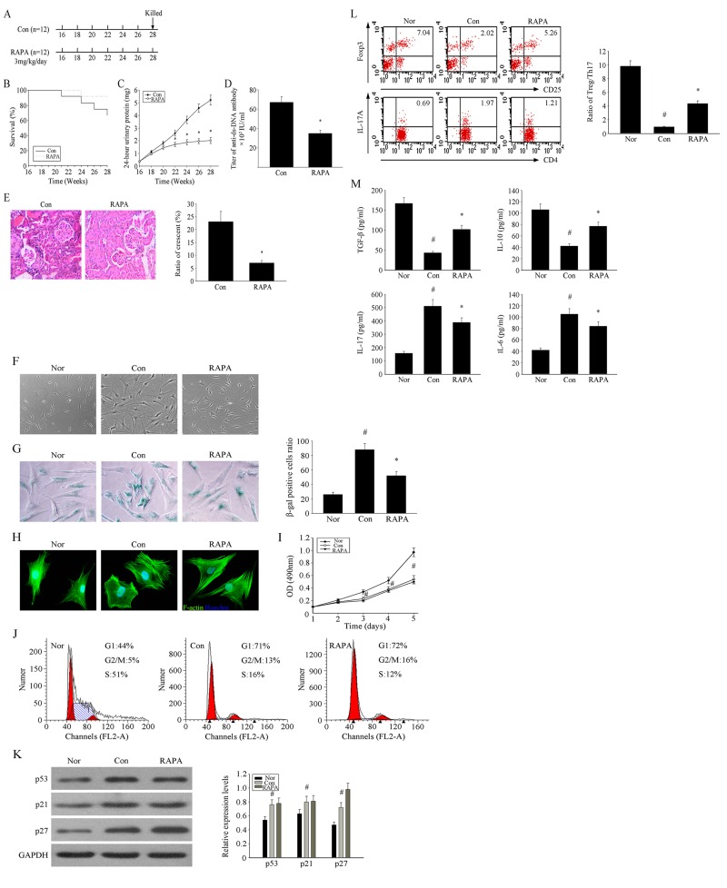
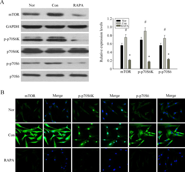
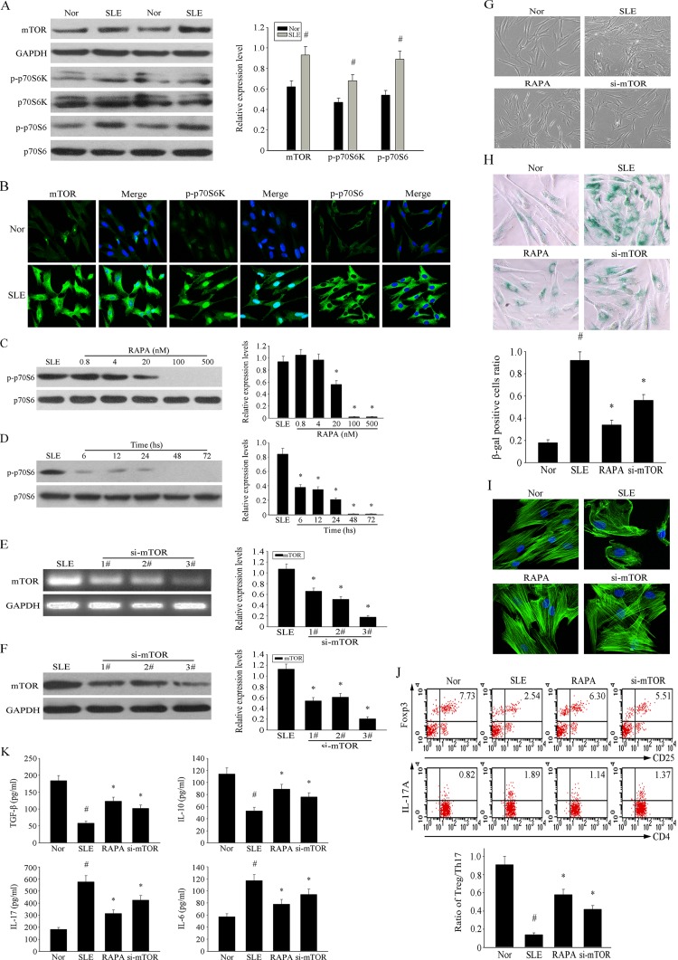
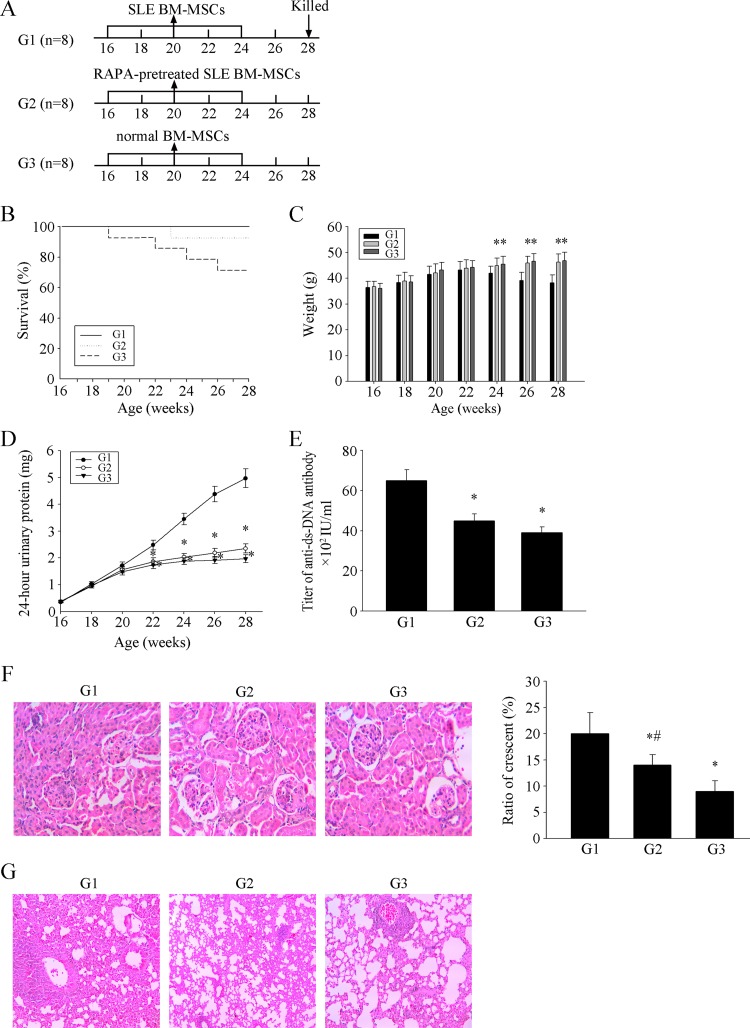
We have shown that bone marrow (BM)-derived mesenchymal stem cells (BM-MSCs) from SLE patients exhibit senescent behavior and are involved in the pathogenesis of SLE. The aim of this study was to investigate the effects of rapamycin (RAPA) on the senescences and immunoregulatory ability of MSCs of MRL/lpr mice and SLE patients and the underlying mechanisms. Cell morphology, senescence associated β-galactosidase (SA-β-gal) staining, F-actin staining were used to detect the senescence of cells. BM-MSCs and purified CD4+ T cells were co-cultured indirectly. Flow cytometry was used to inspect the proportion of regulatory T (Treg) /T helper type 17 (Th17). We used small interfering RNA (siRNA) to interfere the expression of mTOR, and detect the effects by RT-PCR, WB and immunofluorescence. Finally, 1x10⁶ of SLE BM-MSCs treated with RAPA were transplanted to cure the 8 MRL/lpr mice aged 16 weeks for 12 weeks. We demonstrated that RAPA alleviated the clinical symptoms of lupus nephritis and prolonged survival in MRL/lpr mice. RAPA reversed the senescent phenotype and improved immunoregulation of MSCs from MRL/lpr mice and SLE patients through inhibition of the mTOR signaling pathway. Marked therapeutic effects were observed in MRL/lpr mice following transplantation of BM-MSCs from SLE patients pretreated with RAPA.
Keywords: immunoregulation; mesenchymal stem cells (MSCs); rapamycin (RAPA); senescence; systemic lupus erythematosus (SLE).
Conflict of interest statement
No potential conflicts of interest were disclosed.
Figures
Similar articles
-
Targeting HMGB1 by ethyl pyruvate ameliorates systemic lupus erythematosus and reverses the senescent phenotype of bone marrow-mesenchymal stem cells.Aging (Albany NY). 2019 Jul 14;11(13):4338-4353. doi: 10.18632/aging.102052. Aging (Albany NY). 2019. PMID: 31303606 Free PMC article.
-
Human umbilical cord-derived mesenchymal stem cell therapy ameliorates lupus through increasing CD4+ T cell senescence via MiR-199a-5p/Sirt1/p53 axis.Theranostics. 2021 Jan 1;11(2):893-905. doi: 10.7150/thno.48080. eCollection 2021. Theranostics. 2021. PMID: 33391511 Free PMC article.
-
PTEN/Akt-p27(kip1) Signaling Promote the BM-MSCs Senescence and Apoptosis in SLE Patients.J Cell Biochem. 2015 Aug;116(8):1583-94. doi: 10.1002/jcb.25112. J Cell Biochem. 2015. PMID: 25649549
-
Bone marrow transplantation: a new strategy for intractable diseases.Drugs Today (Barc). 2002 Feb;38(2):103-11. doi: 10.1358/dot.2002.38.2.820106. Drugs Today (Barc). 2002. PMID: 12532188 Review.
-
mTOR signaling: A pivotal player in Treg cell dysfunction in systemic lupus erythematosus.Clin Immunol. 2022 Dec;245:109153. doi: 10.1016/j.clim.2022.109153. Epub 2022 Oct 17. Clin Immunol. 2022. PMID: 36265758 Review.
Cited by
-
Senescence in Human Mesenchymal Stem Cells: Functional Changes and Implications in Stem Cell-Based Therapy.Int J Mol Sci. 2016 Jul 19;17(7):1164. doi: 10.3390/ijms17071164. Int J Mol Sci. 2016. PMID: 27447618 Free PMC article. Review.
-
Key Signaling Pathways in Aging and Potential Interventions for Healthy Aging.Cells. 2021 Mar 16;10(3):660. doi: 10.3390/cells10030660. Cells. 2021. PMID: 33809718 Free PMC article. Review.
-
Gut microbiota in SLE: from animal models to clinical evidence and pharmacological perspectives.Lupus Sci Med. 2023 Feb;10(1):e000776. doi: 10.1136/lupus-2022-000776. Lupus Sci Med. 2023. PMID: 36813473 Free PMC article. Review.
-
mTOR Signaling in Kidney Diseases.Kidney360. 2020 Sep 3;1(11):1319-1327. doi: 10.34067/KID.0003782020. eCollection 2020 Nov 25. Kidney360. 2020. PMID: 35372878 Free PMC article. Review.
-
Mesenchymal stem cells and connective tissue diseases: From bench to bedside.J Transl Int Med. 2022 Nov 15;11(1):30-45. doi: 10.2478/jtim-2022-0028. eCollection 2023 Mar. J Transl Int Med. 2022. PMID: 37533846 Free PMC article.
References
-
- Rahman A, Isenberg DA. Systemic lupus erythematosus. The New England journal of medicine. 2008;358:929–939. - PubMed
-
- Jehl F, Quoix E, Leveque D, Pauli G, Breillout F, Krikorian A, Monteil H. Pharmacokinetic and preliminary metabolic fate of navelbine in humans as determined by high performance liquid chromatography. Cancer research. 1991;51:2073–2076. - PubMed
-
- Rosado MM, Diamanti AP, Capolunghi F, Carsetti R. B cell modulation strategies in autoimmunity: the SLE example. Current pharmaceutical design. 2011;17:3155–3165. - PubMed
-
- Smith KG, Jones RB, Burns SM, Jayne DR. Long-term comparison of rituximab treatment for refractory systemic lupus erythematosus and vasculitis: Remission, relapse, and re-treatment. Arthritis and rheumatism. 2006;54:2970–2982. - PubMed
MeSH terms
Substances
LinkOut - more resources
Full Text Sources
Other Literature Sources
Medical
Research Materials
Miscellaneous
