. 2016 May 27;291(22):11596-607.
doi: 10.1074/jbc.M115.712539. Epub 2016 Apr 5.

Cysteine S-Glutathionylation Promotes Stability and Activation of the Hippo Downstream Effector Transcriptional Co-activator with PDZ-binding Motif (TAZ)

Affiliations

Cysteine S-Glutathionylation Promotes Stability and Activation of the Hippo Downstream Effector Transcriptional Co-activator with PDZ-binding Motif (TAZ)

Rajesh Kumar Gandhirajan et al. J Biol Chem. .

Abstract

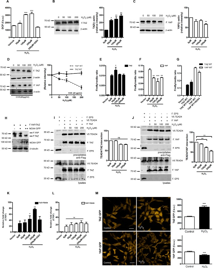
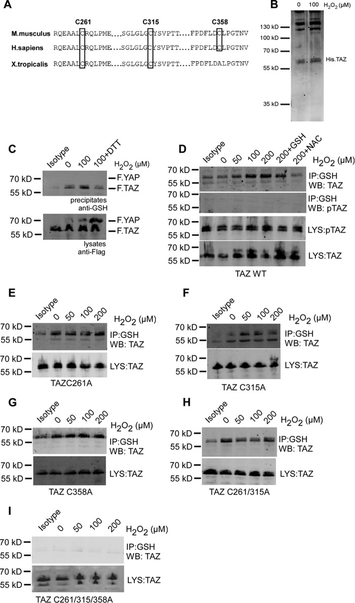
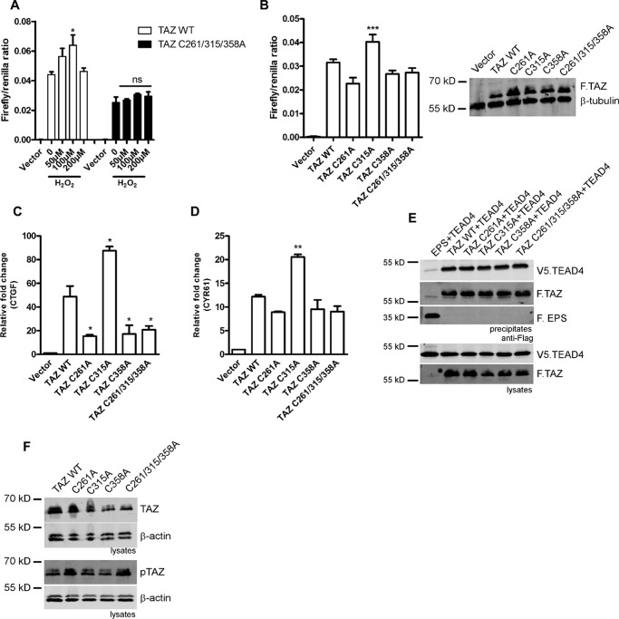
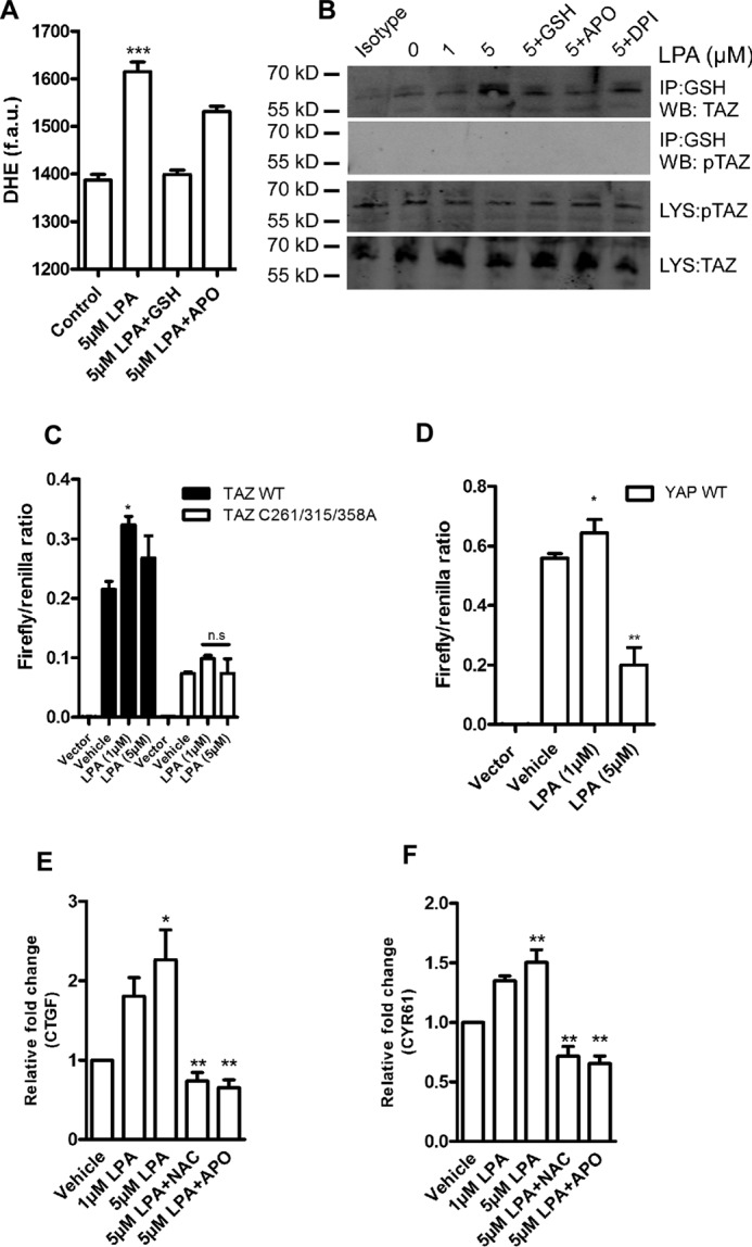
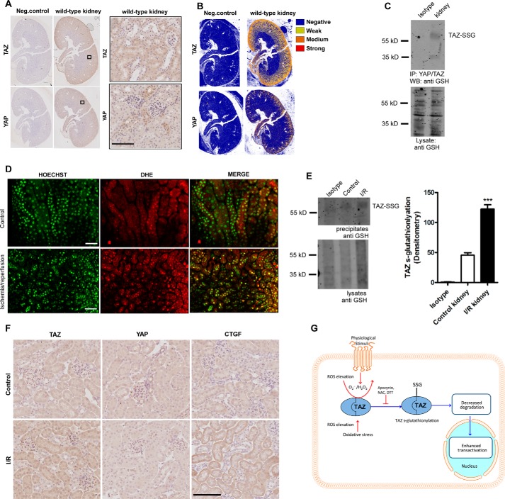
Transcriptional co-activator with PDZ-binding motif (TAZ) and Yes-associated protein (YAP) are critical transcriptional co-activators downstream of the Hippo pathway involved in the regulation of organ size, tissue regeneration, proliferation, and apoptosis. Recent studies suggested common and distinct functions of TAZ and YAP and their diverse impact under several pathological conditions. Here we report differential regulation of TAZ and YAP in response to oxidative stress. H2O2 exposure leads to increased stability and activation of TAZ but not of YAP. H2O2 induces reversible S-glutathionylation at conserved cysteine residues within TAZ. We further demonstrate that TAZ S-glutathionylation is critical for reactive oxygen species (ROS)-mediated, TAZ-dependent TEA domain transcription factor (TEAD) trans-activation. Lysophosphatidic acid, a physiological activator of YAP and TAZ, induces ROS elevation and, subsequently, TAZ S-glutathionylation, which promotes TAZ-mediated target gene expression. TAZ expression is essential for renal homeostasis in mice, and we identify basal TAZ S-glutathionylation in murine kidney lysates, which is elevated during ischemia/reperfusion injury in vivo This induced nuclear localization of TAZ and increased expression of connective tissue growth factor. These results describe a novel mechanism by which ROS sustains total cellular levels of TAZ. This preferential regulation suggests TAZ to be a redox sensor of the Hippo pathway.

Keywords: Hippo pathway; S-glutathionylation; Yes-associated protein (YAP); connective tissue growth factor (CTGF); oxidative stress; reactive oxygen species (ROS); transcriptional co-activator with PDZ-binding motif (TAZ).


Figures

FIGURE 1.
FIGURE 1.
H2O2 elevates total protein levels of TAZ and increases TAZ/TEAD activity. A, HEK 293T cells were challenged with 50, 100, and 200 μm H2O2 for 1 h. After treatment, cells were loaded with a cellular ROS indicator (H2DCFDA), and changes in fluorescence arbitrary units (f.a.u.) were quantified using a microplate reader. B and C, HEK 293T cells expressing FLAG.TAZ (F.TAZ) or FLAG.YAP (F.YAP) were challenged with 50, 100, and 200 μm H2O2 for 1 h. Western blotting and densitometry analysis reveals elevated protein levels of TAZ but not YAP. D, HEK 293T cells expressing FLAG.TAZ or FLAG.YAP were pretreated with cycloheximide (CHX, 40 μg/ml) for 1 h. Cells were then challenged with 50, 100, and 200 μm H2O2 for an additional 1 h. Western blotting and densitometry analysis reveals stable protein levels of TAZ but not YAP. E and F, HEK 293T cells were treated with 50, 100, and 200 μm H2O2 for 6 h. H2O2 enhances the activity of the transcriptional coactivator TAZ (E) but not YAP (F) in TEAD reporter assays, as described under “Experimental Procedures.” G, HEK 293T cells expressing FLAG.TAZ or FLAG.YAP were co-transfected with NOX4. NOX4 enhances the activity of the transcriptional coactivator TAZ but not YAP in TEAD reporter assays. H, immunoblotting of whole cell lysates reveals that increasing amounts of NOX4 degrades YAP in a dose-dependent manner but not TAZ. I and J, HEK 293T cells expressing FLAG.TAZ and V5.TEAD4 or FLAG.YAP and V5.TEAD4 were treated with increasing concentrations of H2O2 for 1 h. Cells expressing FLAG.EPS and V5.TEAD4 served as negative control. Co-immunoprecipitation reveals that H2O2 does not significantly alter the TAZ/TEAD4 interaction but that the YAP/TEAD4 interaction is perturbed. K and L, CTGF expression levels analyzed by qPCR reveal that TAZ induces CTGF expression following 6-h H2O2 treatment, which is reversed by antioxidant pretreatment. In contrast, YAP-induced CTGF expression is not significantly altered by H2O2. M, NIH-3T3 TAZ or YAP GFP flp-in stable cells were exposed to 250 μm H2O2 for 1 h. Shown are representative confocal images and quantification of GFP fluorescence. Scale bars = 50 μm. Data are mean ± S.E. from three independent experiments (*, p < 0.05; **, p < 0.01; ***, p < 0.001; ns, not significant).
FIGURE 2.
FIGURE 2.
H2O2 induces S-glutathionylation of TAZ at three cysteine residues. A, sequence alignment of mouse TAZ (NP_598545.2) reveals conserved cysteine residues at position Cys-261, Cys-315, and Cys-358. B, Coomassie staining of recombinant His.TAZ protein following 100 μm H2O2 exposure for 30 min in vitro. C, HEK 293T cells transiently co-transfected with FLAG.TAZ and FLAG.YAP were treated with 100 μm H2O2 for 1 h. 1 mm DTT was added to cells 30 min after H2O2 exposure. After immunoprecipitation (IP) with anti-GSH antibody, Western blotting analysis revealed that wild-type FLAG.TAZ (55 kDa) undergoes S-glutathionylation but not YAP (72 kDa). D, HEK 293T cells transiently transfected with FLAG.TAZ were treated with 50, 100, and 200 μm H2O2 for 1 h. After IP with anti-GSH antibody, Western blotting (WB) analyses of lysates (LYS) and precipitates (IP) revealed that wild-type FLAG.TAZ undergoes S-glutathionylation in a dose-dependent manner, which was inhibited by the antioxidants GSH and N-acetylcysteine. E–I, HEK 293T cells transiently transfected with FLAG.TAZ cysteine mutants as indicated were treated with 50, 100, and 200 μm H2O2 for 1 h. After IP with anti-GSH antibody, Western blotting analysis revealed that TAZ undergoes S-glutathionylation in all mutants analyzed (C261A (E), C315A (F), C358A (G), C261A/C315A (H), and C261A/C315A/C358A (I)), indicating the presence of multiple S-glutathionylation sites within TAZ. Mouse IgG (isotype) is shown as a control. Shown are representative data from three independent experiments.
FIGURE 3.
FIGURE 3.
H2O2-mediated activation of TAZ depends on S-glutathionylation at three cysteine residues. A, TEAD reporter assay showing increased activity of TAZ wild-type, whereas the TAZ triple mutant fails to elicit the H2O2-induced response. B, basal TEAD reporter activity of TAZ cysteine mutants. Immunoblotting of whole cell extracts from the luciferase assay shows basal TAZ protein levels. C and D, qPCR analysis reveals increased basal CTGF and CYR61 expression in the TAZ C315A mutant compared with the wild type in HEK 293 cells. E, HEK293T cells expressing FLAG.TAZ (F.TAZ) and V5.TEAD4 were immunoprecipitated using M2 beads; F.EPS served as negative control. Co-immunoprecipitation reveals that TAZ cysteine mutants do not alter the TAZ/TEAD4 interaction. F, basal total and phosphorylated (Ser-89) levels of TAZ cysteine mutants in HEK 293 cells. Data are presented as mean ± S.E. from three independent experiments (*, p < 0.05; **, p < 0.01; ***, p < 0.001; ns, not significant).
FIGURE 4.
FIGURE 4.
LPA induces TAZ S-glutathionylation. A, HEK 293T cells were treated with 5 μm LPA for 1 h. After treatment, cells were loaded with DHE, and superoxide elevation was quantified as fluorescence arbitrary units (f.a.u.) in a fluorescence plate reader. APO, apocynin. B, HEK 293T transiently expressing FLAG.TAZ were challenged with 1 and 5 μm LPA for 1 h. After IP with anti-GSH antibody, Western blotting (WB) analyses revealed that wild-type TAZ undergoes S-glutathionylation in a dose-dependent manner, which was inhibited with pretreatment of antioxidants and the pan NOX inhibitor apocynin or diphenyliodonium (DPI). Mouse IgG IP (isotype) is shown as a control. C, TEAD reporter activity of TAZ wild-type and the cysteine triple mutant 6 h following LPA treatment. D, TEAD reporter activity of YAP wild-type 6 h following LPA exposure. E and F, HEK 293T cells transiently expressing FLAG.TAZ were challenged with 1 or 5 μm LPA for 6 h. TAZ target gene CTGF and CYR61 levels were quantified by qPCR. LPA induces TAZ-mediated CTGF and CYR61 expression, which is attenuated with pretreatment with 2 mm N-acetylcysteine (NAC) and 10 μm apocynin. Data are mean ± S.E. from three independent experiments (*, p < 0.05; **, p < 0.01; ***, p < 0.001; ns, not significant).
FIGURE 5.
FIGURE 5.
TAZ S-glutathionylation is induced by ischemia/reperfusion in the kidney. A, immunohistochemical staining of wild-type mouse kidneys with total TAZ and YAP antibodies. Neg, negative. B, positive pixel count analysis of sections in A, showing expression levels of TAZ and YAP (n = 3). C, TAZ/YAP was immunoprecipitated using total anti-YAP/TAZ antibody from whole kidney lysates of wild-type mice. S-Glutathionylated TAZ (TAZ-SSG) was detected using anti-GSH antibody under non-reducing conditions. WB, Western blot. D, in vivo elevation of superoxide (nuclear DHE) in tubular epithelial cells upon renal I/R injury by two-photon microscopy (n = 3). E, whole kidney lysates from healthy and I/R kidney reveal increased TAZ glutathionylation following I/R injury (n = 3). Rabbit IgG IP (Isotype) is shown as a control. F, immunohistochemical staining of wild-type mouse kidneys following I/R injury showing increased cytoplasmic and nuclear TAZ and CTGF levels in I/R kidneys compared with the control (n = 3). G, proposed model in which ROS derived from physiological or oxidative stress pathways induce TAZ glutathionylation, leading to activation and stability. Data are mean ± S.E. from three independent experiments (*, p < 0.05; ***, p < 0.001). Scale bars = 200 μm.

Similar articles

Cited by

References

    1. Piccolo S., Dupont S., and Cordenonsi M. (2014) The biology of YAP/TAZ: Hippo signaling and beyond. Physiol. Rev. 94, 1287–1312 - PubMed
    1. Zhang H., Liu C. Y., Zha Z. Y., Zhao B., Yao J., Zhao S., Xiong Y., Lei Q. Y., and Guan K. L. (2009) TEAD transcription factors mediate the function of TAZ in cell growth and epithelial-mesenchymal transition. J. Biol. Chem. 284, 13355–13362 - PMC - PubMed
    1. Wu S., Liu Y., Zheng Y., Dong J., and Pan D. (2008) The TEAD/TEF family protein Scalloped mediates transcriptional output of the Hippo growth-regulatory pathway. Dev. Cell 14, 388–398 - PubMed
    1. Zhao B., Ye X., Yu J., Li L., Li W., Li S., Yu J., Lin J. D., Wang C. Y., Chinnaiyan A. M., Lai Z. C., and Guan K. L. (2008) TEAD mediates YAP-dependent gene induction and growth control. Genes Dev. 22, 1962–1971 - PMC - PubMed
    1. Yu F. X., Zhao B., Panupinthu N., Jewell J. L., Lian I., Wang L. H., Zhao J., Yuan H., Tumaneng K., Li H., Fu X. D., Mills G. B., and Guan K. L. (2012) Regulation of the Hippo-YAP pathway by G-protein-coupled receptor signaling. Cell 150, 780–791 - PMC - PubMed

Publication types

MeSH terms

LinkOut - more resources