

Three-dimensional direct measurement of cardiomyocyte volume, nuclearity, and ploidy in thick histological sections
- PMID: 27048757
- PMCID: PMC4822151
- DOI: 10.1038/srep23756
Three-dimensional direct measurement of cardiomyocyte volume, nuclearity, and ploidy in thick histological sections
Abstract
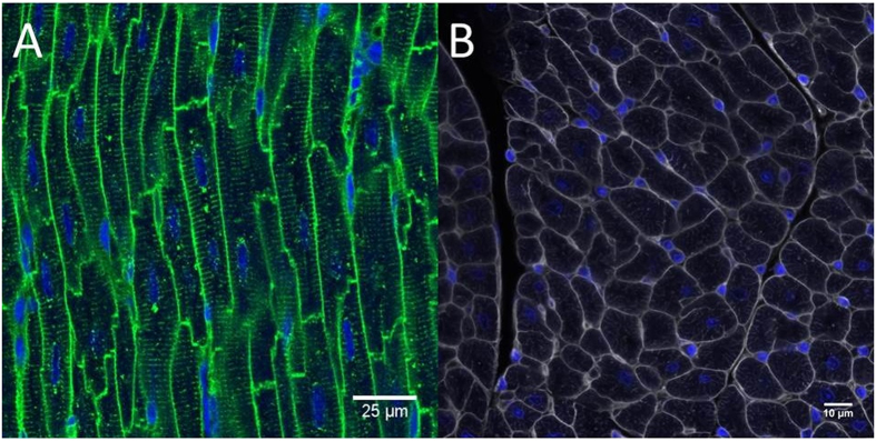
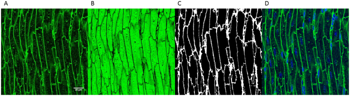
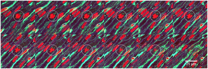
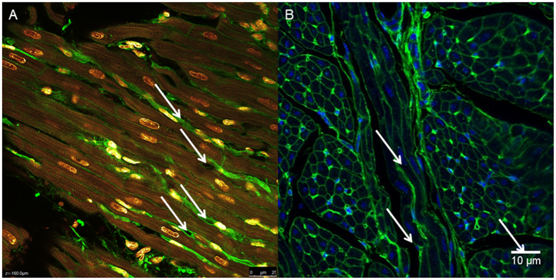
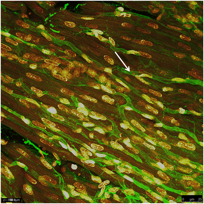
Quantitative assessment of myocardial development and disease requires accurate measurement of cardiomyocyte volume, nuclearity (nuclei per cell), and ploidy (genome copies per cell). Current methods require enzymatically isolating cells, which excludes the use of archived tissue, or serial sectioning. We describe a method of analysis that permits the direct simultaneous measurement of cardiomyocyte volume, nuclearity, and ploidy in thick histological sections. To demonstrate the utility of our technique, heart tissue was obtained from four species (rat, mouse, rabbit, sheep) at up to three life stages: prenatal, weaning and adulthood. Thick (40 μm) paraffin sections were stained with Wheat Germ Agglutinin-Alexa Fluor 488 to visualise cell membranes, and DAPI (4',6-diamidino-2-phenylindole) to visualise nuclei and measure ploidy. Previous methods have been restricted to thin sections (2-10 μm) and offer an incomplete picture of cardiomyocytes. Using confocal microscopy and three-dimensional image analysis software (Imaris Version 8.2, Bitplane AG, Switzerland), cardiomyocyte volume, nuclearity, and ploidy were measured. This method of staining and analysis of cardiomyocytes enables accurate morphometric measurements in thick histological sections, thus unlocking the potential of archived tissue. Our novel time-efficient method permits the entire cardiomyocyte to be visualised directly in 3D, eliminating the need for precise alignment of serial sections.
Figures
Similar articles
-
Three-dimensional confocal laser scanning DNA ploidy cytometry in thick histological sections.J Pathol. 1996 Oct;180(2):214-22. doi: 10.1002/(SICI)1096-9896(199610)180:2<214::AID-PATH633>3.0.CO;2-P. J Pathol. 1996. PMID: 8976883
-
Isolation of Cardiomyocytes from Fixed Hearts for Immunocytochemistry and Ploidy Analysis.J Vis Exp. 2020 Oct 7;(164):10.3791/60938. doi: 10.3791/60938. J Vis Exp. 2020. PMID: 33104059 Free PMC article.
-
Identification of cardiomyocyte nuclei and assessment of ploidy for the analysis of cell turnover.Exp Cell Res. 2011 Jan 15;317(2):188-94. doi: 10.1016/j.yexcr.2010.08.017. Epub 2010 Sep 7. Exp Cell Res. 2011. PMID: 20828558
-
Simultaneous Assessment of Cardiomyocyte DNA Synthesis and Ploidy: A Method to Assist Quantification of Cardiomyocyte Regeneration and Turnover.J Vis Exp. 2016 May 23;(111):53979. doi: 10.3791/53979. J Vis Exp. 2016. PMID: 27285379 Free PMC article.
-
Modelling and analysis of 3-D arrangements of particles by point processes with examples of application to biological data obtained by confocal scanning light microscopy.J Microsc. 1991 Mar;161(Pt 3):405-33. doi: 10.1111/j.1365-2818.1991.tb03100.x. J Microsc. 1991. PMID: 2046088 Review.
Cited by
-
Multi-omic analysis reveals VEGFR2, PI3K, and JNK mediate the small molecule induction of human iPSC-derived cardiomyocyte proliferation.iScience. 2024 Jul 11;27(8):110485. doi: 10.1016/j.isci.2024.110485. eCollection 2024 Aug 16. iScience. 2024. PMID: 39171295 Free PMC article.
-
Long-Term Transcriptomic Changes and Cardiomyocyte Hyperpolyploidy after Lactose Intolerance in Neonatal Rats.Int J Mol Sci. 2023 Apr 11;24(8):7063. doi: 10.3390/ijms24087063. Int J Mol Sci. 2023. PMID: 37108224 Free PMC article.
-
Mylk3 null C57BL/6N mice develop cardiomyopathy, whereas Nnt null C57BL/6J mice do not.Life Sci Alliance. 2020 Mar 25;3(4):e201900593. doi: 10.26508/lsa.201900593. Print 2020 Apr. Life Sci Alliance. 2020. PMID: 32213617 Free PMC article.
-
p47phox-Dependent Oxidant Signalling through ASK1, MKK3/6 and MAPKs in Angiotensin II-Induced Cardiac Hypertrophy and Apoptosis.Antioxidants (Basel). 2021 Aug 26;10(9):1363. doi: 10.3390/antiox10091363. Antioxidants (Basel). 2021. PMID: 34572995 Free PMC article.
-
3D Microwell Platforms for Control of Single Cell 3D Geometry and Intracellular Organization.Cell Mol Bioeng. 2020 Aug 20;14(1):1-14. doi: 10.1007/s12195-020-00646-9. eCollection 2021 Feb. Cell Mol Bioeng. 2020. PMID: 33643464 Free PMC article.
References
-
- Banerjee I., Fuseler J. W., Price R. L., Borg T. K. & Baudino T. A. Determination of cell types and numbers during cardiac development in the neonatal and adult rat and mouse. American journal of physiology. Heart and circulatory physiology 293, H1883–1891, doi: 10.1152/ajpheart.00514.2007 (2007). - DOI - PubMed
Publication types
MeSH terms
Substances
LinkOut - more resources
Full Text Sources
Other Literature Sources
