

Regulation of necrotic cell death: p53, PARP1 and cyclophilin D-overlapping pathways of regulated necrosis?
- PMID: 27048819
- PMCID: PMC5490387
- DOI: 10.1007/s00018-016-2202-5
Regulation of necrotic cell death: p53, PARP1 and cyclophilin D-overlapping pathways of regulated necrosis?
Abstract
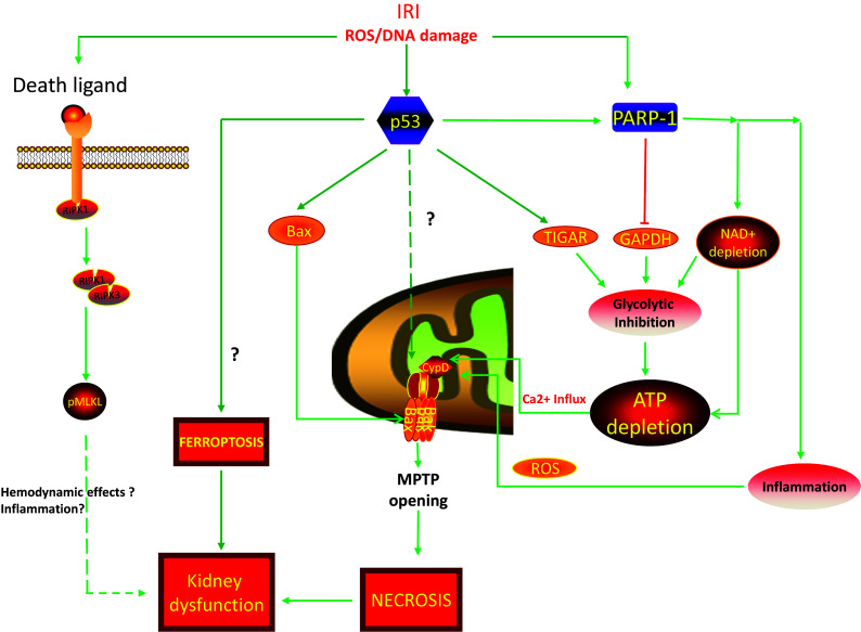
In contrast to apoptosis and autophagy, necrotic cell death was considered to be a random, passive cell death without definable mediators. However, this dogma has been challenged by recent developments suggesting that necrotic cell death can also be a regulated process. Regulated necrosis includes multiple cell death modalities such as necroptosis, parthanatos, ferroptosis, pyroptosis, and mitochondrial permeability transition pore (MPTP)-mediated necrosis. Several distinctive executive molecules, particularly residing on the mitochondrial inner and outer membrane, amalgamating to form the MPTP have been defined. The c-subunit of the F1F0ATP synthase on the inner membrane and Bax/Bak on the outer membrane are considered to be the long sought components that form the MPTP. Opening of the MPTP results in loss of mitochondrial inner membrane potential, disruption of ATP production, increased ROS production, organelle swelling, mitochondrial dysfunction and consequent necrosis. Cyclophilin D, along with adenine nucleotide translocator and the phosphate carrier are considered to be important regulators involved in the opening of MPTP. Increased production of ROS can further trigger other necrotic pathways mediated through molecules such as PARP1, leading to irreversible cell damage. This review examines the roles of PARP1 and cyclophilin D in necrotic cell death. The hierarchical role of p53 in regulation and integration of key components of signaling pathway to elicit MPTP-mediated necrosis and ferroptosis is explored. In the context of recent insights, the indistinct role of necroptosis signaling in tubular necrosis after ischemic kidney injury is scrutinized. We conclude by discussing the participation of p53, PARP1 and cyclophilin D and their overlapping pathways to elicit MPTP-mediated necrosis and ferroptosis in acute kidney injury.
Keywords: Bax; Cyclophilin D; Necroptosis; PARP1; Regulated necrosis; p53.
Figures
Similar articles
-
The mitochondrial permeability transition pore regulates nitric oxide-mediated apoptosis of neurons induced by target deprivation.J Neurosci. 2011 Jan 5;31(1):359-70. doi: 10.1523/JNEUROSCI.2225-10.2011. J Neurosci. 2011. PMID: 21209222 Free PMC article.
-
The mitochondrial ATP synthase is a negative regulator of the mitochondrial permeability transition pore.Proc Natl Acad Sci U S A. 2023 Dec 19;120(51):e2303713120. doi: 10.1073/pnas.2303713120. Epub 2023 Dec 13. Proc Natl Acad Sci U S A. 2023. PMID: 38091291 Free PMC article.
-
Oxidative stress alters mitochondrial bioenergetics and modifies pancreatic cell death independently of cyclophilin D, resulting in an apoptosis-to-necrosis shift.J Biol Chem. 2018 May 25;293(21):8032-8047. doi: 10.1074/jbc.RA118.003200. Epub 2018 Apr 6. J Biol Chem. 2018. PMID: 29626097 Free PMC article.
-
Physiologic functions of cyclophilin D and the mitochondrial permeability transition pore.Circ J. 2013;77(5):1111-22. doi: 10.1253/circj.cj-13-0321. Epub 2013 Mar 29. Circ J. 2013. PMID: 23538482 Free PMC article. Review.
-
Regulation and pharmacology of the mitochondrial permeability transition pore.Cardiovasc Res. 2009 Jul 15;83(2):213-25. doi: 10.1093/cvr/cvp151. Epub 2009 May 15. Cardiovasc Res. 2009. PMID: 19447775 Free PMC article. Review.
Cited by
-
SIRT3 a Major Player in Attenuation of Hepatic Ischemia-Reperfusion Injury by Reducing ROS via Its Downstream Mediators: SOD2, CYP-D, and HIF-1α.Oxid Med Cell Longev. 2018 Nov 13;2018:2976957. doi: 10.1155/2018/2976957. eCollection 2018. Oxid Med Cell Longev. 2018. PMID: 30538800 Free PMC article. Review.
-
Proximal tubule cyclophilin D mediates kidney fibrogenesis in obstructive nephropathy.Am J Physiol Renal Physiol. 2021 Oct 1;321(4):F431-F442. doi: 10.1152/ajprenal.00171.2021. Epub 2021 Aug 16. Am J Physiol Renal Physiol. 2021. PMID: 34396791 Free PMC article.
-
Mechanism and treatment of intracerebral hemorrhage focus on mitochondrial permeability transition pore.Front Mol Neurosci. 2024 Jul 31;17:1423132. doi: 10.3389/fnmol.2024.1423132. eCollection 2024. Front Mol Neurosci. 2024. PMID: 39156127 Free PMC article. Review.
-
Urea transport B gene induces melanoma B16 cell death via activation of p53 and mitochondrial apoptosis.Cancer Sci. 2018 Dec;109(12):3762-3773. doi: 10.1111/cas.13825. Epub 2018 Nov 11. Cancer Sci. 2018. PMID: 30290033 Free PMC article.
-
MS275 as Class I HDAC inhibitor displayed therapeutic potential on malignant ascites by iTRAQ-based quantitative proteomic analysis.BMC Gastroenterol. 2022 Jan 21;22(1):29. doi: 10.1186/s12876-022-02101-7. BMC Gastroenterol. 2022. PMID: 35062876 Free PMC article.
References
-
- Galluzzi L, Pietrocola F, Bravo-San Pedro JM, Amaravadi RK, Baehrecke EH, Cecconi F, Codogno P, Debnath J, Gewirtz DA, Karantza V, Kimmelman A, Kumar S, Levine B, Maiuri MC, Martin SJ, Penninger J, Piacentini M, Rubinsztein DC, Simon HU, Simonsen A, Thorburn AM, Velasco G, Ryan KM, Kroemer G. Autophagy in malignant transformation and cancer progression. EMBO J. 2015;34(7):856–880. doi: 10.15252/embj.201490784. - DOI - PMC - PubMed
-
- Galluzzi L, Bravo-San Pedro JM, Vitale I, Aaronson SA, Abrams JM, Adam D, Alnemri ES, Altucci L, Andrews D, Annicchiarico-Petruzzelli M, Baehrecke EH, Bazan NG, Bertrand MJ, Bianchi K, Blagosklonny MV, Blomgren K, Borner C, Bredesen DE, Brenner C, Campanella M, Candi E, Cecconi F, Chan FK, Chandel NS, Cheng EH, Chipuk JE, Cidlowski JA, Ciechanover A, Dawson TM, Dawson VL, De Laurenzi V, De Maria R, Debatin KM, Di Daniele N, Dixit VM, Dynlacht BD, El-Deiry WS, Fimia GM, Flavell RA, Fulda S, Garrido C, Gougeon ML, Green DR, Gronemeyer H, Hajnoczky G, Hardwick JM, Hengartner MO, Ichijo H, Joseph B, Jost PJ, Kaufmann T, Kepp O, Klionsky DJ, Knight RA, Kumar S, Lemasters JJ, Levine B, Linkermann A, Lipton SA, Lockshin RA, Lopez-Otin C, Lugli E, Madeo F, Malorni W, Marine JC, Martin SJ, Martinou JC, Medema JP, Meier P, Melino S, Mizushima N, Moll U, Munoz-Pinedo C, Nunez G, Oberst A, Panaretakis T, Penninger JM, Peter ME, Piacentini M, Pinton P, Prehn JH, Puthalakath H, Rabinovich GA, Ravichandran KS, Rizzuto R, Rodrigues CM, Rubinsztein DC, Rudel T, Shi Y, Simon HU, Stockwell BR, Szabadkai G, Tait SW, Tang HL, Tavernarakis N, Tsujimoto Y, Vanden Berghe T, Vandenabeele P, Villunger A, Wagner EF, Walczak H, White E, Wood WG, Yuan J, Zakeri Z, Zhivotovsky B, Melino G, Kroemer G. Essential versus accessory aspects of cell death: recommendations of the NCCD 2015. Cell Death Differ. 2015;22(1):58–73. doi: 10.1038/cdd.2014.137. - DOI - PMC - PubMed
Publication types
MeSH terms
Substances
Grants and funding
LinkOut - more resources
Full Text Sources
Other Literature Sources
Research Materials
Miscellaneous
