. 2016 Jun;15(6):1163-76.
doi: 10.1158/1535-7163.MCT-15-0973. Epub 2016 Apr 5.

A Novel Plant Sesquiterpene Lactone Derivative, DETD-35, Suppresses BRAFV600E Mutant Melanoma Growth and Overcomes Acquired Vemurafenib Resistance in Mice

Affiliations

A Novel Plant Sesquiterpene Lactone Derivative, DETD-35, Suppresses BRAFV600E Mutant Melanoma Growth and Overcomes Acquired Vemurafenib Resistance in Mice

Jia-Hua Feng et al. Mol Cancer Ther. 2016 Jun.

Abstract

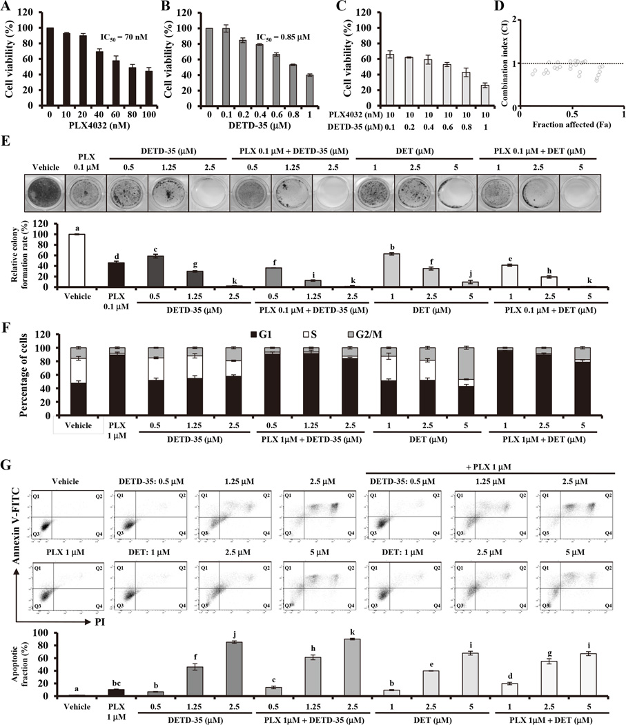
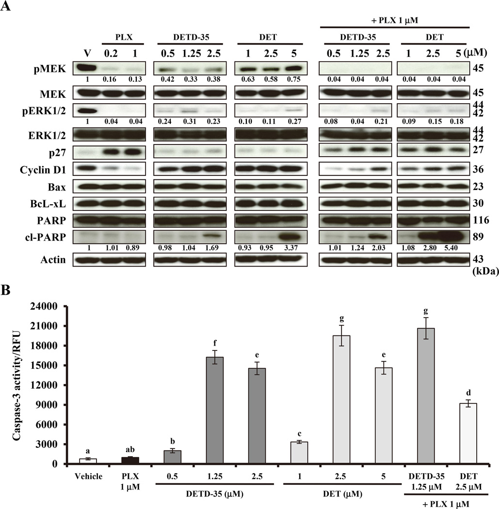
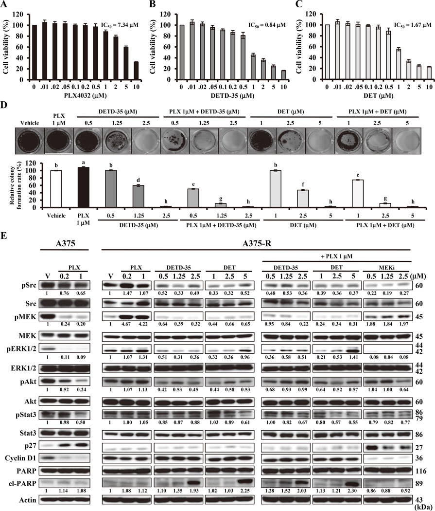
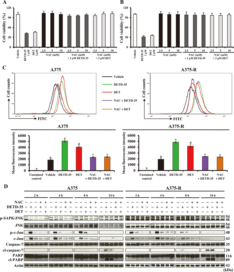
Acquired resistance to vemurafenib develops through reactivation of RAF/MEK/ERK signaling or bypass mechanisms. Recent combination therapies such as a MEK inhibitor combined with vemurafenib show improvement in major clinical end points, but the percentage of patients with adverse toxic events is higher than with vemurafenib monotherapy and most patients ultimately relapse. Therefore, there is an urgent need to develop new antimelanoma drugs and/or adjuvant agents for vemurafenib therapy. In this study, we created a novel semiorganically modified derivative, DETD-35, from deoxyelephantopin (DET), a plant sesquiterpene lactone demonstrated as an anti-inflammatory and anti-mammary tumor agent. Our results show that DETD-35 inhibited proliferation of a panel of melanoma cell lines, including acquired vemurafenib resistance A375 cells (A375-R) established in this study, with superior activities to DET and no cytotoxicity to normal melanocytes. DETD-35 suppressed tumor growth and reduced tumor mass as effectively as vemurafenib in A375 xenograft study. Furthermore, DETD-35 also reduced tumor growth in both acquired (A375-R) and intrinsic (A2058) vemurafenib resistance xenograft models, where vemurafenib showed no antitumor activity. Notably, the combination of DETD-35 and vemurafenib exhibited the most significant effects in both in vitro and in vivo xenograft studies due to synergism of the compound and the drug. Mechanistic studies suggested that DETD-35 overcame acquired vemurafenib resistance at least in part through deregulating MEK-ERK, Akt, and STAT3 signaling pathways and promoting apoptosis of cancer cells. Overall, our results suggest that DETD-35 may be useful as a therapeutic or adjuvant agent against BRAF(V600E) mutant and acquired vemurafenib resistance melanoma. Mol Cancer Ther; 15(6); 1163-76. ©2016 AACR.


Conflict of interest statement

Conflict of interests

The authors declare no conflict of interests.

Figures

Figure 1
Figure 1
DETD-35 shows synergism with vemurafenib in vitro. A, B and C, A375 melanoma cells were treated with the indicated concentrations of PLX4032, DETD-35 and PLX4032***35 for 72 h, respectively. Cell viability was determined using MTT assay. D, compound-drug combination study using CalcuSyn software. CI < 1 indicates synergism; CI = 1 indicates additive effect; CI > 1 indicates antagonism between the two compounds tested. E, top: colony formation assay showing long-term (21-day treatment) anti-proliferative effects of PLX4032, DETD-35 and DET against A375 cells. Crystal violet stained cells are shown. Bottom: quantification of the crystal violet absorbance at 595 nm after 21 days of treatment. Data are mean ± SEM, n = 4. Means without a common letter differ, P < 0.05. F, cell-cycle analysis, cells were treated with the indicated concentrations of compounds for 24 h. After fixation, cells were stained with propidium iodide and analyzed by flow cytometry. G, combination of DETD-35 or DET with PLX4032 increased levels of apoptosis in A375 melanoma cells. Cells were treated with the indicated concentrations of the compounds for 72 h. Apoptosis status in cells was measured by Annexin V/FITC plus propidium iodide double staining and flow cytometry. Top: Representative quadrant diagrams showing cell distribution in Q1 (early apoptosis), Q2 (apoptosis), Q3 (live), and Q4 (dead). Bottom: apoptotic fraction was calculated by adding distribution percentage in Q1, Q2 and Q4 together. Data are mean ± SEM, n = 3. Means without a common letter differ, P < 0.05.
Figure 2
Figure 2
DETD-35 suppresses MEK-ERK signaling and induces apoptosis in A375 melanoma cells. A, cells were treated with indicated concentrations of the compounds for 16 h before lysis. Cell lysates were subjected to western blotting against the specific antibodies indicated, and actin was used as an internal or loading control. Quantification of specific protein expression, which was normalized to actin first followed by comparison of the fold-change within different treatments to vehicle treatment. B, cells were treated with indicated concentrations of the compounds for 72 h, caspase 3 activity was determined by measuring cleavage of a fluorogenic substrate using flow cytometry (RFU: relative fluorescence units). Data are mean ± SEM, n = 3. Means without a common letter differ, P < 0.05.
Figure 3
Figure 3
DETD-35 inhibits BRAFV600E mutant melanoma growth in vivo. NOD/SCID mice bearing A375 xenografts were treated with vehicle, PLX4032 (20 mg/kg/day), DETD-35 or DET (20 mg/kg/every two days), and a compound-drug combination with alternate administration of DETD-35 or DET (20 mg/kg/every four days) plus PLX4032 (20 mg/kg/every two days), when tumor volume reached around 50 mm3. A, representative tumor tissues isolated from each treatment group. B, tumor volume was measured every three days and plotted as mean ± SEM. C, the weight of the dissected tumors from each treatment group. Data are mean ± SEM, n = 8. Means without a common letter differ, P < 0.05. D, top: representative immunohistochemistry images of tumor tissues with positive staining of phospho-MEK, phospho-ERK1/2, Ki-67, cleaved caspase-3 and cleaved PARP (brownish color) and nuclear (blue color) by hematoxylin staining are shown (Scale bar: 50 µm). Bottom: quantification of phospho-MEK, phospho-ERK1/2, Ki-67, cleaved caspase-3 and cleaved PARP positive stained cells, Data are mean ± SEM, n = 3. Means without a common letter differ, P < 0.05.
Figure 4
Figure 4
DETD-35 overcomes acquired vemurafenib resistance in vitro. A, B and C, A375-R cells were treated with indicated concentrations of PLX4032, DETD-35 and DET for 72 h. Cell viability was determined using MTT assay. D, top: colony formation assay showing long-term (21-day treatment) anti-proliferative effects of PLX4032, DETD-35 and DET against A375-R cells. Crystal violet stained cells are shown. Bottom: quantification of the crystal violet absorbance at 595 nm after 21 days of treatment. Data are mean ± SEM, n = 4. Means without a common letter differ, P < 0.05. E, A375-R cells were treated with the indicated concentrations of compounds for 16 h before lysis. Cell lysates were subjected to western blotting using the specific antibodies indicated, and actin was used as an internal or loading control. Quantification of specific protein expression, which was normalized to actin first followed by comparison of the fold change within different treatments to vehicle treatment.
Figure 5
Figure 5
DETD-35 overcomes both intrinsic and acquired vemurafenib resistance in mice. NOD/SCID mice bearing A375-R and A2058 xenografts were treated with vehicle, PLX4032 (75 mg/kg/day), DETD-35 or DET (20 mg/kg/every two days), and a compound-drug combination with alternate administration of DETD-35 or DET (20 mg/kg/every two days) and PLX4032 (75 mg/kg/every two days), when tumor volume reached around 50 mm3. A, representative tumor tissues isolated from A375-R xenograft. B, tumor volume of A375-R xenograft was measured every three days and plotted as mean ± SEM. C, the weight of the dissected tumors from A375-R xenograft. D, tumor volume of A2058 xenograft was measured every three days and plotted as mean ± SEM. E, the weight of the dissected tumors from A2058 xenograft. Data are mean ± SEM, n = 8. Means without a common letter differ, P < 0.05. F, top: representative immunohistochemistry images of A375-R tumor for positive staining of phospho-Src, phospho-MEK, phospho-ERK1/2, phospho-Akt, Ki-67, cleaved caspase-3, and cleaved PARP (Scale bar: 50 µm). Bottom: quantification of phospho-Src, phospho-MEK, phospho-ERK1/2, phospho-Akt, Ki-67, cleaved caspased-3, and cleaved PARP positive stained cells from tumor tissue sections of A375-R xenograft study. Data are mean ± SEM, n = 3. Means without a common letter differ, P < 0.05.
Figure 6
Figure 6
DETD-35 induces oxidative stress to trigger apoptosis in A375 and A375-R cells. A and B, A375 cells and A375-R cells, respectively, were pretreated with or without NAC for 1 h followed by treatment with DETD-35 or DET at the indicated concentrations for 72 h. Cell viability was determined using MTT assay. C, top: reactive oxygen species (ROS) generation in A375 cells and A375-R cells treated with DETD-35 and DET alone or pretreated with NAC for 1 h followed by compound treatment for 2 h. Flow cytometry and green fluorescent dye (ROS detection probe) were used in this experiment. Bottom: quantification of the generated ROS in the cells under the same treatment as top panel. Data are mean ± SEM, n = 4. Means without a common letter differ, P < 0.05. D, cell lysates from A375 and A375-R cells pretreated with or without NAC for 1 h followed by treatment with 2.5 µM DETD-35 and 5 µM DET at the indicated times were collected and subjected to western blotting using the specific antibodies indicated, and actin was used as an internal or loading control.

References

    1. Greenlee RT, Hill-Harmon MB, Murray T, Thun M. Cancer statistics, 2001. CA Cancer J Clin. 2001;51:15–36. - PubMed
    1. Siegel RL, Miller KD, Jemal A. Cancer statistics, 2015. CA Cancer J Clin. 2015;65:5–29. - PubMed
    1. Halilovic E, Solit DB. Therapeutic strategies for inhibiting oncogenic BRAF signaling. Curr Opin Pharmacol. 2008;8:419–426. - PubMed
    1. Tsai J, Lee JT, Wang W, Zhang J, Cho H, Mamo S, et al. Discovery of a selective inhibitor of oncogenic BRAF kinase with potent antimelanoma activity. Proc Natl Acad Sci U S A. 2008;105:3041–3046. - PMC - PubMed
    1. Chapman PB, Hauschild A, Robert C, Haanen JB, Ascierto P, Larkin J, et al. Improved survival with vemurafenib in melanoma with BRAFV600E mutation. N Engl J Med. 2011;364:2507–2516. - PMC - PubMed

MeSH terms