Review
. 2016 Oct;57(10):1758-1770.
doi: 10.1194/jlr.R066357. Epub 2016 Apr 5.

Lipotoxic lethal and sublethal stress signaling in hepatocytes: relevance to NASH pathogenesis

Affiliations
Review

Lipotoxic lethal and sublethal stress signaling in hepatocytes: relevance to NASH pathogenesis

Petra Hirsova et al. J Lipid Res. 2016 Oct.

Erratum in

  • ERRATUM.
    [No authors listed] [No authors listed] J Lipid Res. 2017 Jan;58(1):299. doi: 10.1194/jlr.R066357ERR. J Lipid Res. 2017. PMID: 28042122 Free PMC article. No abstract available.

Abstract

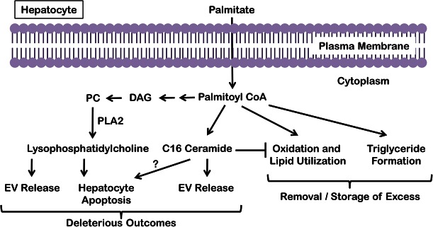
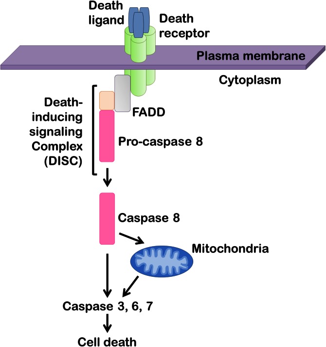
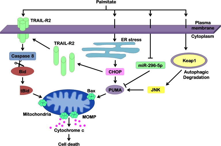
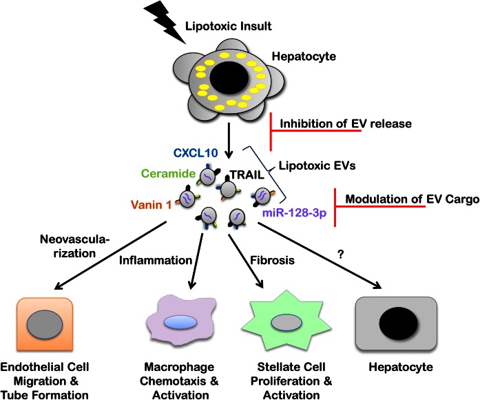
The accumulation of lipids is a histologic and biochemical hallmark of obesity-associated nonalcoholic fatty liver disease (NAFLD). A subset of NALFD patients develops progressive liver disease, termed nonalcoholic steatohepatitis, which is characterized by hepatocellular apoptosis and innate immune system-mediated inflammation. These responses are orchestrated by signaling pathways that can be activated by lipids, directly or indirectly. In this review, we discuss palmitate- and lysophosphatidylcholine (LPC)-induced upregulation of p53-upregulated modulator of apoptosis and cell-surface expression of the death receptor TNF-related apoptosis-inducing ligand receptor 2. Next, we review the activation of stress-induced kinases, mixed lineage kinase 3, and c-Jun N-terminal kinase, and the activation of endoplasmic reticulum stress response and its downstream proapoptotic effector, CAAT/enhancer binding homologous protein, by palmitate and LPC. Moreover, the activation of these stress signaling pathways is linked to the release of proinflammatory, proangiogenic, and profibrotic extracellular vesicles by stressed hepatocytes. This review discusses the signaling pathways induced by lethal and sublethal lipid overload that contribute to the pathogenesis of NAFLD.

Keywords: apoptosis; cell death; cell signaling; exosomes; extracellular vesicles; fatty acids; lipotoxicity; lysophosphatidylcholine; nonalcoholic fatty liver disease; nonalcoholic steatohepatitis; steatohepatitis.


Figures

Fig. 1.
Fig. 1.
Fate of palmitate in hepatocytes. Excess palmitate delivered to hepatocytes is converted to palmitoyl-CoA and can induce the formation of ceramide via the de novo pathway at the ER and LPC via PLA2 action on phosphatidylcholine (PC), which in turn is derived from diacylglycerol (DAG). Though palmitate can activate the proapoptotic machinery, palmitate itself and palmitate-derived LPC and ceramide all lead to the release of EVs from hepatocytes. Palmitate is buffered in hepatocytes by either oxidation or conversion to triglyceride, both of which mitigate palmitate-induced lipotoxicity.
Fig. 2.
Fig. 2.
Death receptor-mediated apoptosis. Upon binding of a ligand to the extracellular portion of the death receptor, the receptor intracellular domain recruits adaptor proteins, such as Fas-associated protein with death domain (FADD) and pro-caspase 8, to form a signaling platform termed the death-inducing signaling complex. Caspase 8 then undergoes proteolytic autoactivation, resulting in direct or indirect (via mitochondria) activation of caspases 3, 6, and 7 that execute the final steps of cellular demolition.
Fig. 3.
Fig. 3.
Apoptotic signaling networks in hepatocyte lipotoxicity. Hepatocyte treatment with palmitate results in ligand-independent clustering and activation of the TRAIL-R2, causing caspase-dependent cell death. Active caspase 8 cleaves Bid into its truncated form (tBid), which translocates to mitochondria to promote release of proapoptotic factors, such as cytochrome c. Palmitate also induces ER stress, which upregulates proapoptotic PUMA and TRAIL-R2 via transcription factor CHOP. Increased levels of PUMA facilitate hepatocyte apoptosis via the mitochondrial pathway. Palmitate has also been found to cause cellular depletion of miR-296-5p, a microRNA that, via complementary binding to the 3′-UTR of PUMA mRNA, causes its degradation. Loss of miR-296-5p during lipotoxicity removes this break on PUMA posttranscriptional regulation, thereby increasing cellular levels of PUMA. Finally, palmitate induces autophagic degradation of Keap1, resulting in JNK activation and PUMA upregulation. Along with other proapoptotic inputs, PUMA sensitizes the hepatocyte to Bax activation and MOMP culminating in cell death.
Fig. 4.
Fig. 4.
EVs in lipotoxic signaling and therapeutic opportunities. Hepatocyte lipotoxicity promotes EV release. Recent in vitro and in vivo studies have defined multiple roles of lipotoxic EVs in NASH pathogenesis through cell-to-cell communication via various cargoes. CXCL10 and ceramide-enriched EVs mediate monocyte/macrophage chemotaxis to the liver, while TRAIL-enriched EVs contribute to macrophage activation resulting in the sterile inflammatory response observed in a nutrient excess mouse model of NASH. Vanin 1-enriched EVs mediate endothelial cell migration and tube formation in vitro and neovascularization in a NASH mouse model, while miR-128-3p-laden EVs enhance HSC proliferation and activation in vitro. Inhibition of EV release and modulation of EV cargo to interrupt deleterious target cell responses are opportunities for EV-based therapies.

References

    1. Unger R. H., Clark G. O., Scherer P. E., and Orci L.. 2010. Lipid homeostasis, lipotoxicity and the metabolic syndrome. Biochim. Biophys. Acta. 1801: 209–214. - PubMed
    1. Rinella M. E. 2015. Nonalcoholic fatty liver disease: a systematic review. JAMA. 313: 2263–2273. - PubMed
    1. Puri P., Baillie R. A., Wiest M. M., Mirshahi F., Choudhury J., Cheung O., Sargeant C., Contos M. J., and Sanyal A. J.. 2007. A lipidomic analysis of nonalcoholic fatty liver disease. Hepatology. 46: 1081–1090. - PubMed
    1. Gorden D. L., Ivanova P. T., Myers D. S., McIntyre J. O., VanSaun M. N., Wright J. K., Matrisian L. M., and Brown H. A.. 2011. Increased diacylglycerols characterize hepatic lipid changes in progression of human nonalcoholic fatty liver disease; comparison to a murine model. PLoS One. 6: e22775. - PMC - PubMed
    1. Gorden D. L., Myers D. S., Ivanova P. T., Fahy E., Maurya M. R., Gupta S., Min J., Spann N. J., McDonald J. G., Kelly S. L., et al. . 2015. Biomarkers of NAFLD progression: a lipidomics approach to an epidemic. J. Lipid Res. 56: 722–736. - PMC - PubMed

Publication types

MeSH terms