. 2016 Jul;8(5):892-904.
doi: 10.1080/19420862.2016.1171432. Epub 2016 Apr 6.

Simultaneous blockade of VEGF and Dll4 by HD105, a bispecific antibody, inhibits tumor progression and angiogenesis

Affiliations

Simultaneous blockade of VEGF and Dll4 by HD105, a bispecific antibody, inhibits tumor progression and angiogenesis

Dongheon Lee et al. MAbs. 2016 Jul.

Abstract

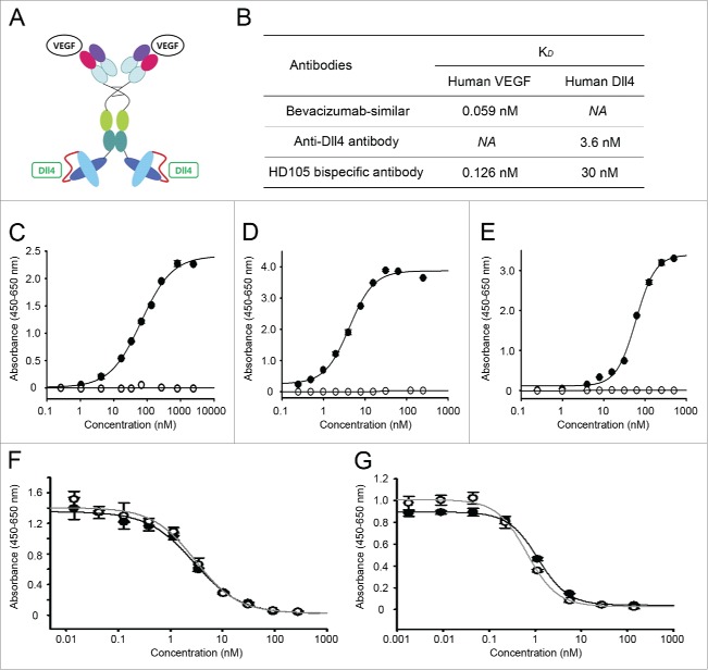
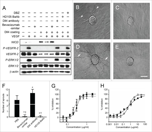
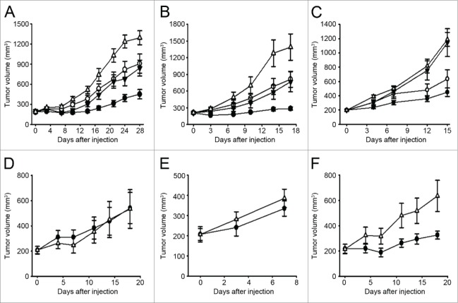
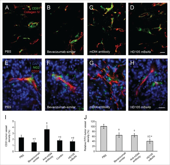
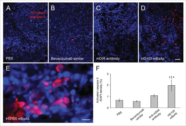
Several angiogenesis inhibitors targeting the vascular endothelial growth factor (VEGF) signaling pathway have been approved for cancer treatment. However, VEGF inhibitors alone were shown to promote tumor invasion and metastasis by increasing intratumoral hypoxia in some preclinical and clinical studies. Emerging reports suggest that Delta-like ligand 4 (Dll4) is a promising target of angiogenesis inhibition to augment the effects of VEGF inhibitors. To evaluate the effects of simultaneous blockade against VEGF and Dll4, we developed a bispecific antibody, HD105, targeting VEGF and Dll4. The HD105 bispecific antibody, which is composed of an anti-VEGF antibody (bevacizumab-similar) backbone C-terminally linked with a Dll4-targeting single-chain variable fragment, showed potent binding affinities against VEGF (KD: 1.3 nM) and Dll4 (KD: 30 nM). In addition, the HD105 bispecific antibody competitively inhibited the binding of ligands to their receptors, i.e., VEGF to VEGFR2 (EC50: 2.84 ± 0.41 nM) and Dll4 to Notch1 (EC50: 1.14 ± 0.06 nM). Using in vitro cell-based assays, we found that HD105 effectively blocked both the VEGF/VEGFR2 and Dll4/Notch1 signaling pathways in endothelial cells, resulting in a conspicuous inhibition of endothelial cell proliferation and sprouting. HD105 also suppressed Dll4-induced Notch1-dependent activation of the luciferase gene. In vivo xenograft studies demonstrated that HD105 more efficiently inhibited the tumor progression of human A549 lung and SCH gastric cancers than an anti-VEGF antibody or anti-Dll4 antibody alone. In conclusion, HD105 may be a novel therapeutic bispecific antibody for cancer treatment.

Keywords: Anti-angiogenesis; VEGF; anti-cancer; biologics; delta-like ligand; therapeutic antibody.


Figures

Figure 1.
Figure 1.
Simultaneous binding to VEGF and Dll4 by HD105 bispecific antibody leads to effective blockade of VEGF/VEGFR2 and Dll4/Notch1 interactions. The HD105 bispecific antibody was constructed of the C-terminal of the anti-VEGF (bevacizumab-similar) IgG backbone linked with a single-chain Fv targeting Dll4 (A). The binding affinity of the HD105 bispecific antibody against human VEGF or human Dll4 was determined by Biacore assays (B) and ELISAs (C, D). The KD values of each antibody against VEGF or Dll4 are summarized in Table (B). The HD105 bispecific antibody (closed circle) dose-dependently bound to human VEGF (C) or Dll4 (D). In addition, the HD105 bispecific antibody simultaneously bound to each antigen, human VEGF and human Dll4, in dual-antigen capture ELISAs (E). The anti-Dll4 antibody (open circle in C) or the anti-VEGF (bevacizumab-similar) antibody (open circle in D, E) was used as negative control. Competitive ELISAs demonstrated that the HD105 bispecific antibody inhibited the interaction between VEGF/VEGFR2 (F) or Dll4/Notch1 (G) in a dose-dependent manner. The EC50 (half maximal effective concentration) values of the anti-VEGF (bevacizumab-similar) antibody (open circle) and HD105 bispecific antibody (closed circle) for VEGF/VEGFR2 inhibition were 2.98 ± 0.5 nM and 2.84 ± 0.41 nM, respectively (F). The EC50 values of the anti-Dll4 antibody (open circle) and HD105 bispecific antibody (closed circle) were 0.65 ± 0.06 nM and 1.14 ± 0.06 nM, respectively (G).
Figure 2.
Figure 2.
Blockade of both VEGF/VEGFR2 and Dll4/Notch1 signaling pathways by HD105 bispecific antibody leads to inhibition of each signaling-induced cellular response. The HD105 bispecific antibody inhibited both the VEGF/VEGFR2 and the Dll4/Notch1 signaling pathways in HUVECs (A). The VEGF/VEGFR2 signaling pathway was monitored by the activation of VEGFR2 and ERK (phosphorylation). The Dll4/Notch1 signaling pathway was monitored by the generation of NICD (Notch-induced intracellular domain). HUVEC sprouting assays were performed in a fibrin gel in the presence of PBS (B), anti-VEGF (bevacizumab-similar) antibody (C), anti-Dll4 antibody (D), or HD105 bispecific antibody (E). Representative images show sprouting tip cells of HUVECs from the beads under basal media (B, arrowheads) and more sprouting under anti-Dll4 antibody treatment (D, arrows) but much less sprouting under anti-VEGF antibody (C) or HD105 bispecific antibody treatment (E). Scale bar (B-E), 150 μm. The bar graph (F) shows the measurement of sprouting HUVECs at 225 μm from beads (n = 20 beads/group, mean ± SE). *, P < 0.05 versus PBS. , P < 0.05vs. anti-Dll4 antibody. The HD105 bispecific antibody inhibited VEGF-dependent HUVEC proliferation (G) and Dll4-induced Notch-1-dependent activation of luciferase in SKOV-3-RBP-J Κ luciferase cells (H) in a dose-dependent manner. The IC50 values of the anti-VEGF (bevacizumab-similar) antibody (open circle) and HD105 bispecific antibody (closed circle) on HUVEC proliferation were 1.49 ± 0.04 nM and 1.58 ± 0.08 nM, respectively (G). The IC50 values of the HD105 bispecific antibody (closed circle) and the anti-Dll4 antibody (open circle) on luciferase activation were determined to be 0.62 ± 0.23 nM and 0.58 ± 0.03 nM, respectively (H).
Figure 3.
Figure 3.
Suppression of tumor progression in several cancer xenograft models by HD105 bispecific antibody. Human A549 lung cancer (A) or human SCH gastric cancer (B, C) was subcutaneously implanted into nude mice. After tumors were grown to an average volume of 150–200 mm3, PBS (open triangle), anti-VEGF (bevacizumab-similar) antibody (2.5 mg/kg, open circle), anti-mouse Dll4 antibody (2.5 mg/kg, closed triangle), or mouse HD105 bispecific antibody (3.25 mg/kg, closed circle) was intraperitoneally injected twice (A549) or once (SCH) per week (A, B). Tumor volume was calculated by the formula width × length × 0.52. The dose dependency of the mouse HD105 bispecific antibody was evaluated in human SCH gastric cancer xenograft model (C). PBS (open triangle) or mouse HD105 bispecific antibody (0.361 mg/kg, closed triangle; 1.083 mg/kg, open circle; 3.25 mg/kg, closed circle) was intraperitoneally injected once per week. The response to mouse HD105 bispecific antibody (6.5 mg/kg, once per week, closed circle) was also determined using other human gastric cancer xenograft models, including MKN-74 (D), SNU-5 (E), and SNU-16 (F). Tumor progression was not inhibited by the mouse HD105 bispecific antibody in MKN-74 and SNU-5 but was inhibited in SNU-16 similarly to SCH.
Figure 4.
Figure 4.
Suppression of tumor angiogenesis in cancer xenograft models by HD105 bispecific antibody. Fluorescence micrographs compare the vasculature of A549 human lung cancer tissues in xenograft mice after treatment with PBS (A), anti-VEGF (bevacizumab-similar) antibody (B), anti-mouse Dll4 antibody (C), or mouse HD105 bispecific antibody (D). Scale bar (A-D), 50 μm. The tumor vasculature was stained for CD31 immunoreactivity (green), and the vascular basement was stained for type IV collagen (red). Tumor vessels were decreased after treatment with anti-VEGF (bevacizumab-similar) antibody or mouse HD105 bispecific antibody, whereas tumor vessels were markedly increased after treatment with anti-mouse Dll4 antibody compared to PBS. Higher-resolution images compare the phenotype changes of tumor vessels in detail after PBS (E), anti-VEGF (bevacizumab-similar) antibody (F), anti-mouse Dll4 antibody (G), or mouse HD105 bispecific antibody treatment (H). Scale bar (E-H), 20 μm. The tumor vasculature was stained for CD31 immunoreactivity (red), and the perivascular pericyte was stained for NG2 (green). The nuclei of the tumor tissues were stained by DAPI (4′,6-diamidino-2-phenylindole). Tumor vessels after treatment with anti-mouse Dll4 antibody were conspicuously thinner and more branched than the tumor vessels of other groups. Bar graph (I) measuring tumor vessel density of A549 tumor tissues in xenograft mice confirms the conspicuous increase of tumor vessels after anti-mouse Dll4 antibody treatment but decreases after anti-VEGF (bevacizumab-similar) antibody, mouse HD105 bispecific antibody, or combination treatment with anti-mouse Dll4 antibody and anti-VEGF (bevacizumab-similar) antibody. †, P < 0.05 versus PBS. *, P < 0.05vs. anti-Dll4 antibody. However, the functional tumor vessels in SCH gastric cancer tissues assessed by intravenous FITC-labeled Lycopersicon esculentum (Tomato) lectin staining were significantly decreased after treatment with anti-VEGF (bevacizumab-similar) antibody as well as anti-mouse Dll4 antibody (J). †, P < 0.05 versus PBS. ‡, < 0.05vs. anti-VEGF (bevacizumab-similar) antibody. *, P < 0.05 versus anti-Dll4 antibody. Functional tumor vessels were more decreased after treatment with mouse HD105 bispecific antibody compared to the other groups.
Figure 5.
Figure 5.
Increase in apoptotic tumor cells in cancer xenograft models treated with HD105 bispecific antibody. Fluorescence micrographs show apoptotic cells stained for activated caspase-3 antibody (red) in SCH human gastric cancer tissues in xenograft mice after treatment with PBS (A), anti-VEGF (bevacizumab-similar) antibody (B), anti-mouse Dll4 antibody (C), and mouse HD105 bispecific antibody (D and E). Scale bar (A-D), 50 μm; (E), 20 μm. Nuclei of the tumor tissues were stained by DAPI (4′,6-diamidino-2-phenylindole, blue). The higher-resolution image confirms that activated caspase-3 antibody was stained in the cytoplasm of the apoptotic cells after mouse HD105 bispecific antibody treatment (E). The bar graph (F) measuring the cell density of apoptotic cells in SCH cancer tissues confirms the significant increase in apoptotic cells after mouse HD105 bispecific antibody treatment. *, P < 0.05vs. PBS. ‡, < 0.05 versus anti-VEGF (bevacizumab-similar)) antibody. *, P < 0.05vs. anti-Dll4 antibody.
Figure 6.
Figure 6.
Purification and stability of HD105 bispecific antibody. The HD105 bispecific antibody was produced by CHO cells and then purified by several chromatographic steps. The purity of the antibody in each purification step was analyzed by SEC-HPLC (A). The stability of the purified HD105 bispecific antibody (20 mg/ml) was monitored by SEC-HPLC (B) and SDS-PAGE (C) after 4 weeks' incubation at 4°C, 25°C, or 40°C. The binding affinity against each target of the HD105 bispecific antibody was also monitored by DACE analysis (D) after 4 weeks' incubation at 4°C, 25°C, or 40°C.

Similar articles

Cited by

References

    1. Neufeld G, Cohen T, Gengrinovitch S, Poltorak Z. Vascular endothelial growth factor (VEGF) and its receptors. FASEB J 1999; 13:9-22; PMID:9872925 - PubMed
    1. Meadows KL, Hurwitz HI. Anti-VEGF therapies in the clinic. Cold Spring Harb Perspect Med 2012; 2:a006577; PMID:23028128; http://dx.doi.org/10.1101/cshperspect.a006577 - DOI - PMC - PubMed
    1. Casak SJ, Fashoyin-Aje I, Lemery SJ, Zhang L, Jin R, Li H, Zhao L, Zhao H, Zhang H, Chen H, et al.. FDA Approval Summary: Ramucirumab for Gastric Cancer. Clin Cancer Res 2015; 21:3372-6; PMID:26048277; http://dx.doi.org/10.1158/1078-0432.CCR-15-0600 - DOI - PubMed
    1. Trichonas G, Kaiser PK. Aflibercept for the treatment of age-related macular degeneration. Ophthalmol Ther. 2013; 2:89-98; PMID:25135809; http://dx.doi.org/10.1007/s40123-013-0015-2 - DOI - PMC - PubMed
    1. Bergers G, Hanahan D. Modes of resistance to anti-angiogenic therapy. Nat Rev Cancer 2008; 8:592-603; PMID:18650835; http://dx.doi.org/10.1038/nrc2442 - DOI - PMC - PubMed

MeSH terms

Substances