

Triptolide Modulates TREM-1 Signal Pathway to Inhibit the Inflammatory Response in Rheumatoid Arthritis
- PMID: 27049384
- PMCID: PMC4848954
- DOI: 10.3390/ijms17040498
Triptolide Modulates TREM-1 Signal Pathway to Inhibit the Inflammatory Response in Rheumatoid Arthritis
Abstract
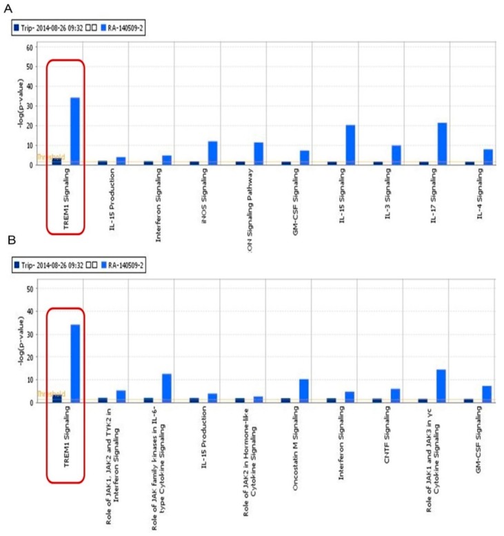
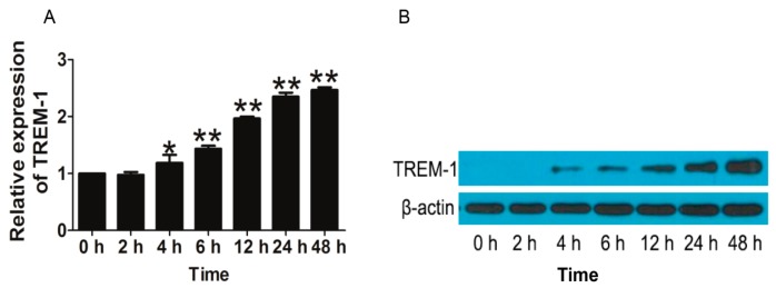
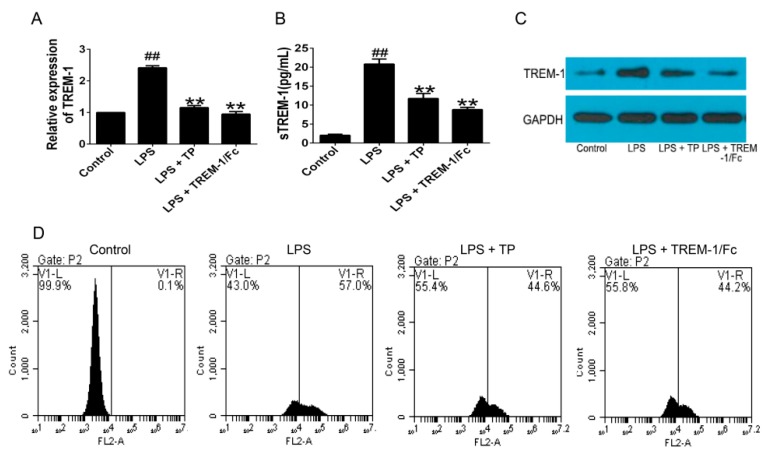
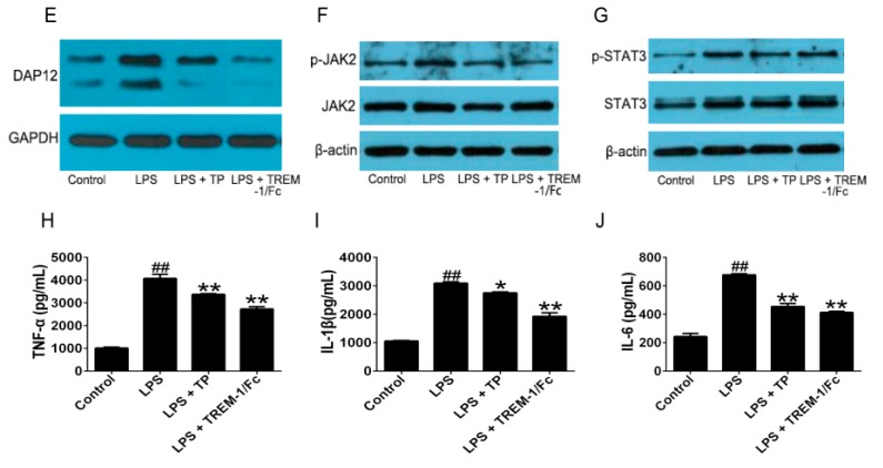
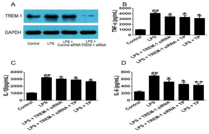
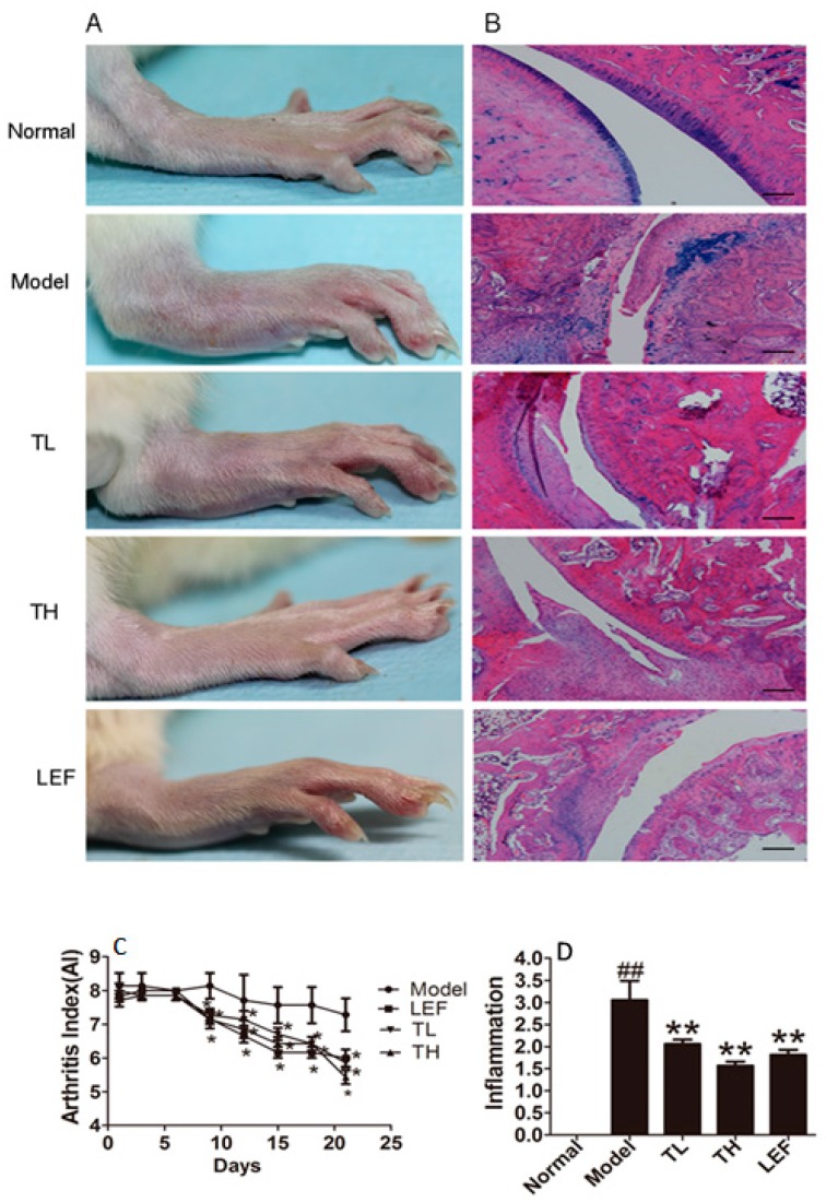
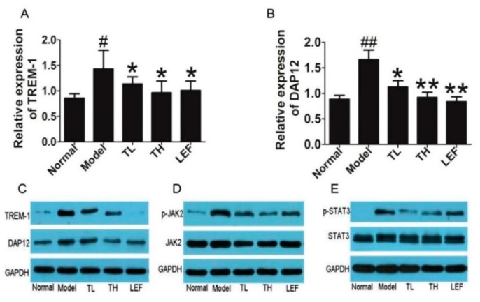
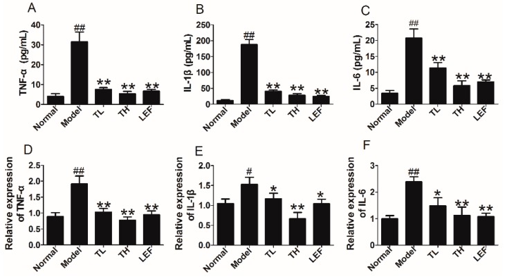
Triptolide (TP), an active component isolated from Tripterygiumwilfordii Hook F, has therapeutic potential against rheumatoid arthritis (RA). However, the underlying molecular mechanism has not been fully elucidated. The aim of this study is to investigate the mechanisms of TP acting on RA by combining bioinformatics analysis with experiment validation. The human protein targets of TP and the human genes of RA were found in the PubChem database and NCBI, respectively. These two dataset were then imported into Ingenuity Pathway Analysis (IPA) software online, and then the molecular network of TP on RA could be set up and analyzed. After that, both in vitro and in vivo experiments were done to further verify the prediction. The results indicated that the main canonical signal pathways of TP protein targets networks were mainly centered on cytokine and cellular immune signaling, and triggering receptors expressed on myeloid cells (TREM)-1 signaling was searched to be the top one shared signaling pathway and involved in the cytokine and cellular immune signaling. Further in vitro experiments indicated that TP not only remarkably lowered the levels of TREM-1 and DNAX-associated protein (DAP)12, but also significantly suppressed the activation of janus activating kinase (JAK)2 and signal transducers and activators of transcription (STAT)3. The expression of tumor necrosis factor (TNF)-α, interleukin (IL)-1β and IL-6 in lipopolysaccharides (LPS)-stimulated U937 cells also decreased after treatment with TP. Furthermore, TREM-1 knockdown was able to interfere with the inhibition effects of TP on these cytokines production. In vivo experiments showed that TP not only significantly inhibited the TREM-1 mRNA and DAP12 mRNA expression, and activation of JAK2 and STAT3 in ankle of rats with collagen-induced arthritis (CIA), but also remarkably decreased production of TNF-α, IL-1β and IL-6 in serum and joint. These findings demonstrated that TP could modulate the TREM1 signal pathway to inhibit the inflammatory response in RA.
Keywords: TREM-1 signal pathway; Triptolide; bioinformatics analysis; rheumatoid arthritis.
Figures
Similar articles
-
Triptolide inhibits the migration and invasion of rheumatoid fibroblast-like synoviocytes by blocking the activation of the JNK MAPK pathway.Int Immunopharmacol. 2016 Dec;41:8-16. doi: 10.1016/j.intimp.2016.10.005. Epub 2016 Oct 27. Int Immunopharmacol. 2016. PMID: 27816728
-
Prediction of triptolide targets in rheumatoid arthritis using network pharmacology and molecular docking.Int Immunopharmacol. 2020 Mar;80:106179. doi: 10.1016/j.intimp.2019.106179. Epub 2020 Jan 20. Int Immunopharmacol. 2020. PMID: 31972422
-
Quantitative proteomic analysis of circulating exosomes reveals the mechanism by which Triptolide protects against collagen-induced arthritis.Immun Inflamm Dis. 2024 Jun;12(6):e1322. doi: 10.1002/iid3.1322. Immun Inflamm Dis. 2024. PMID: 38888462 Free PMC article.
-
Therapeutic targets of thunder god vine (Tripterygium wilfordii hook) in rheumatoid arthritis (Review).Mol Med Rep. 2020 Jun;21(6):2303-2310. doi: 10.3892/mmr.2020.11052. Epub 2020 Apr 2. Mol Med Rep. 2020. PMID: 32323812 Review.
-
The role and mechanism of triptolide, a potential new DMARD, in the treatment of rheumatoid arthritis.Ageing Res Rev. 2025 Feb;104:102643. doi: 10.1016/j.arr.2024.102643. Epub 2024 Dec 24. Ageing Res Rev. 2025. PMID: 39722411 Review.
Cited by
-
The Anti-Inflammatory and Immunomodulatory Activities of Natural Products to Control Autoimmune Inflammation.Int J Mol Sci. 2022 Dec 21;24(1):95. doi: 10.3390/ijms24010095. Int J Mol Sci. 2022. PMID: 36613560 Free PMC article. Review.
-
Identification of peculiar gene expression profile in peripheral blood mononuclear cells (PBMC) of celiac patients on gluten free diet.PLoS One. 2018 May 24;13(5):e0197915. doi: 10.1371/journal.pone.0197915. eCollection 2018. PLoS One. 2018. PMID: 29795662 Free PMC article.
-
Therapeutic Effect of Modulating TREM-1 via Anti-inflammation and Autophagy in Parkinson's Disease.Front Neurosci. 2019 Aug 2;13:769. doi: 10.3389/fnins.2019.00769. eCollection 2019. Front Neurosci. 2019. PMID: 31440123 Free PMC article.
-
Identification of Novel Core Genes Involved in Malignant Transformation of Inflamed Colon Tissue Using a Computational Biology Approach and Verification in Murine Models.Int J Mol Sci. 2023 Feb 21;24(5):4311. doi: 10.3390/ijms24054311. Int J Mol Sci. 2023. PMID: 36901742 Free PMC article.
-
Yupingfeng San Inhibits NLRP3 Inflammasome to Attenuate the Inflammatory Response in Asthma Mice.Front Pharmacol. 2017 Dec 22;8:944. doi: 10.3389/fphar.2017.00944. eCollection 2017. Front Pharmacol. 2017. PMID: 29311942 Free PMC article.
References
Publication types
MeSH terms
Substances
LinkOut - more resources
Full Text Sources
Other Literature Sources
Medical
Miscellaneous
