. 2016 Jun;24(6):1126-1134.
doi: 10.1038/mt.2016.64. Epub 2016 Apr 6.

Allospecific Tregs Expanded After Anergization Remain Suppressive in Inflammatory Conditions but Lack Expression of Gut-homing Molecules

Affiliations

Allospecific Tregs Expanded After Anergization Remain Suppressive in Inflammatory Conditions but Lack Expression of Gut-homing Molecules

Eleni Kotsiou et al. Mol Ther. 2016 Jun.

Abstract

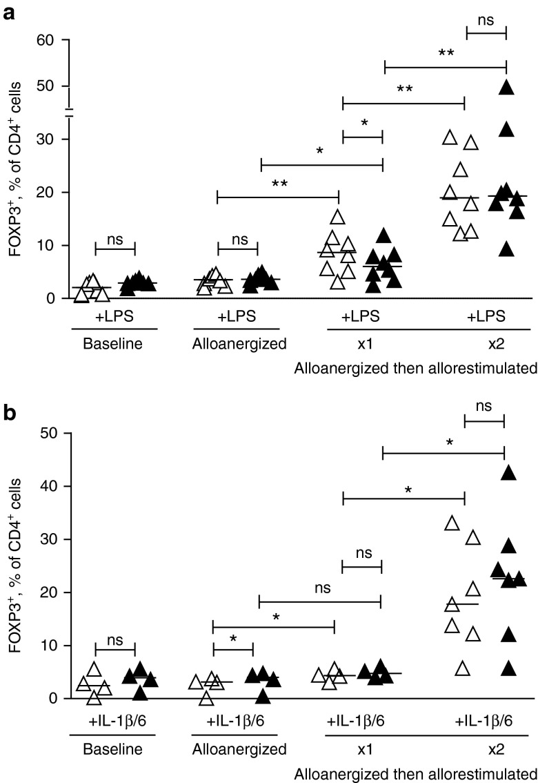
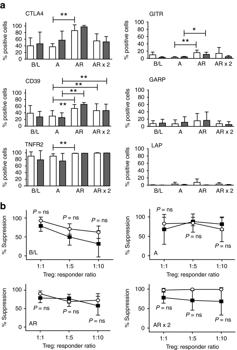
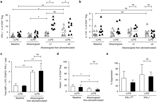
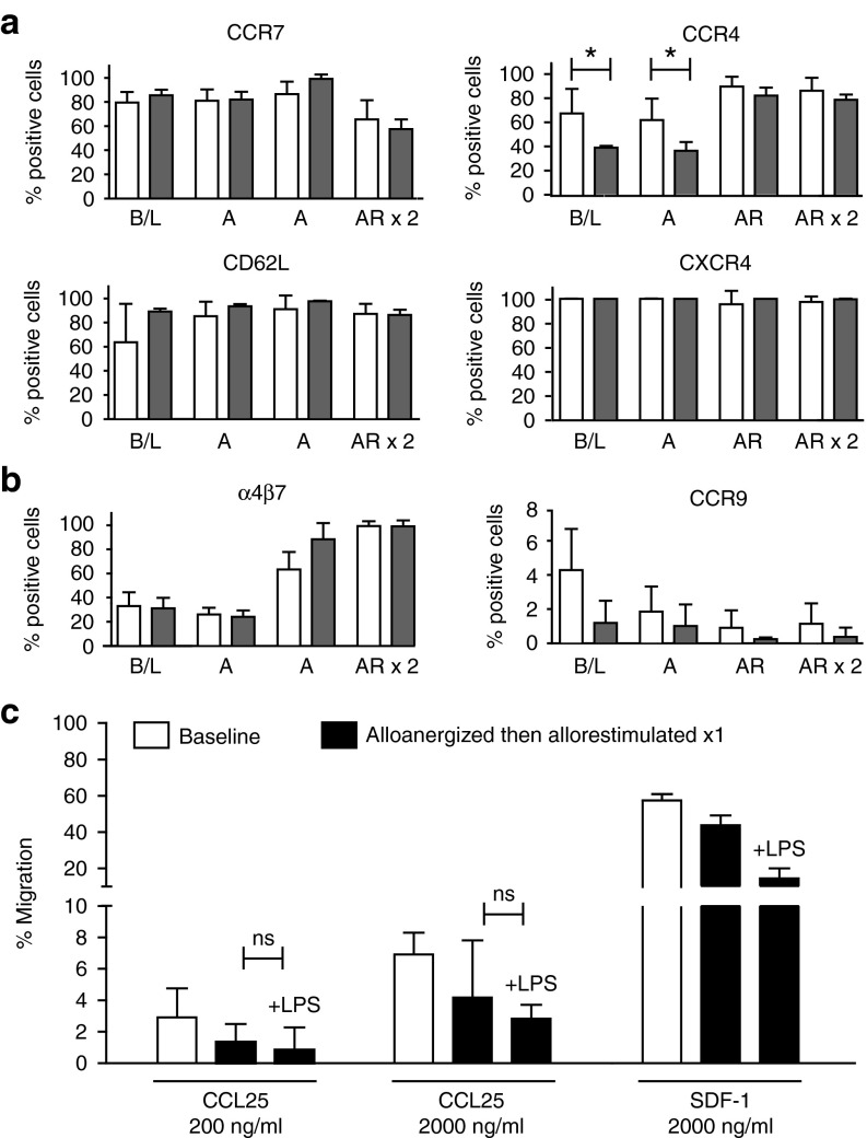
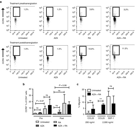
Cell therapy with antigen-specific regulatory T-cells (Treg) has great potential to selectively control unwanted immune responses after allogeneic stem-cell or solid organ transplantation and in autoimmune diseases. Ex vivo allostimulation with costimulatory blockade (alloanergization) of human T-cells expands populations of alloantigen-specific Treg, providing a cellular strategy to control donor T-cell alloresponses causing graft-versus-host disease after allogeneic hematopoietic stem-cell transplantation. Crucially, it is not known if Treg expanded in this way are stable in proinflammatory conditions encountered after transplantation, or if they possess capacity to migrate to key target organs. Using an in vitro model to functionally characterize human Treg expanded after alloanergization, we now show that these cells remain potently allosuppressive in the presence of relevant exogenous inflammatory signals. Expanded allospecific Treg retained expression of molecules conferring migratory capacity to several organs but small intestine-specific chemotaxis was markedly impaired, in keeping with the preponderance of gut graft-versus-host disease in previous clinical studies using this strategy. Importantly, impaired gut-specific chemotaxis could be partially corrected by pharmacological treatment. These findings will facilitate more effective application of this cellular approach to limit T-cell alloresponses after hematopoietic stem-cell transplantation and the wider application of the strategy to other clinical settings.


Figures

Figure 1
Figure 1
Expansion of CD4+ regulatory T-cells (Treg) after alloanergization is maintained in proinflammatory conditions. (a) FOXP3+ Treg, expressed as percentage of total CD4+ cells, after alloanergization and repeated exposure to alloantigen in the absence or presence of lipopolysaccharide (LPS). Graph depicts results from eight different HLA-mismatched stimulator-responder pairs. P values are for two-tailed student's t-test. Horizontal lines are medians. *P < 0.05, **P < 0.01, ns, not significant. (b) FOXP3+ Treg, expressed as percentage of total CD4+ cells, after alloanergization and repeated exposure to alloantigen in the absence or presence of IL-1β and Il-6. Graph depicts results from seven different HLA-mismatched stimulator-responder pairs. P values are for two-tailed student's t-test. Horizontal lines are medians. *P < 0.05, **P < 0.01, ns, not significant.
Figure 2
Figure 2
Lipopolysaccharide (LPS) does not impair allosuppressive phenotype or function of CD4+ regulatory T-cells (Treg) expanded after alloanergization. (a) Phenotype of expanded Treg after alloanergization and repeated exposure to alloantigen in the absence of presence of LPS. Bar charts show mean ±SD) frequencies of CTLA-4+, CD39+, TNFR2+, GITR+, GARP+, and LAP+ cells expressed as a proportion of CD4+ FOXP3+ Treg at baseline (B/L), after alloanergization (A) and after subsequent allorestimulation (AR). Data are for five to nine HLA-mismatched stimulator-responder pairs. *P < 0.05, **P < 0.01. (b) Allosuppressive function of CD4+ Treg at B/L, after alloanergization (A) and subsequent AR in the absence or presence of LPS. Mean percentage suppression (±SD) of alloproliferative responses of untreated responder cells by CD4+ Treg are shown. Data are for three to six HLA-mismatched stimulator-responder pairs. P values are for two-tailed t-tests, ns; nonsignificant.
Figure 3
Figure 3
Regulatory T-cells (Treg) expanded after alloanergization in lipopolysaccharide (LPS)-rich conditions are enriched with allosuppressive IFN-γ-secreting cells. (a) Frequencies of IFN-γ+ cells expressed as percentage of CD4+ Treg at baseline, after alloanergization and after allorestimulation in the absence or presence of LPS. Horizontal lines represent median values. P values are for two-tailed t-test. Results are shown for eight HLA-mismatched stimulator-responder pairs. *P < 0.05, **P < 0.01. (b) Frequencies of IL-17A+ cells expressed as percentage of Treg at baseline, after alloanergization and after allorestimulation. Horizontal lines represent median values. P values are for two-tailed t-test. Results are shown for eight HLA-mismatched stimulator- responder pairs. ns, not significant. (c) IFN-γ+ Treg following alloanergization and allorestimulation upregulate T-bet. Bar chart summarizes mean (±SD) median fluorescence intensity (MFI) of T-bet CD4+FOXP3+IFN-γ+ Treg at baseline and after alloanergization and allorestimulation. Results for three independent experiments are shown. P values are for two-tailed t-test. **P < 0.01, ns, not significant. (d) IFN-γ+ Treg following alloanergization and allorestimulation are predominantly heliosneg. Mean (±SD) frequencies of Helios+ cells expressed as a percentage of CD4+FOXP3+IFN-γ+ Treg at baseline and after alloanergization and subsequent allorestimulation. Results are for three independent experiments. P values are for two-tailed t-test. *P < 0.05, ns, not significant. (e) Allosuppressive capacity of IFN-γ+ and IFN-γneg Treg subpopulations after alloanergization and subsequent allorestimulation of healthy donor peripheral blood mononuclear cells (PBMCs) in the presence or absence of LPS. Mean percentage suppression (±SD) of alloproliferative responses of untreated responder cells are shown by purified Treg from allorestimulated alloanergized PBMCs at a ratio of 1:10 Treg: Responder cells. Results are from four independent HLA-mismatched stimulator-responder pairs. Ns, not significant.
Figure 4
Figure 4
Regulatory T-cells (Treg) expanded after alloanergization and allorestimulation display differential expression of molecules conferring organ-specific migratory capacity. (a) Mean frequencies (±SD) of cells expressing CCR7, CD62L, CCR4 and CXCR4 expressed as a proportion of Treg at baseline (B/L) and after alloanergization (A) and subsequent allorestimulation (AR) in the absence (white bars) or presence (gray bars) of lipopolysaccharide (LPS) (n = 3–9). *P<0.05, (b) Mean frequencies (±SD) of cells expressing α4β7 and CCR9 expressed as a proportion of Treg at B/L and after A and subsequent AR in the absence (white bars) or presence (gray bars) of lipopolysaccharide (LPS) (n = 3–9). (c) CCL25 and SDF-1-specific chemotaxis of CD4+ Treg from baseline and after alloanergization and restimulation in the absence or presence of LPS. Bar charts depict mean (±SD) data from three different stimulator-responder pairs. ns, not significant.
Figure 5
Figure 5
Pharmacological treatment can partially correct defective CCR9-specific chemotaxis of regulatory T-cells (Treg) expanded after alloanergization. (a) Azacitidine (AZA) and all-trans retinoic acid (RA) treatment prior to or after alloanergization increases the proportion of expanded Treg expressing CCR9. Illustrative dot pots are shown depicting CCR9 expression on CD4+ FOXP3+ Tregs after alloanergization with and without treatment with AZA, RA or both. Representative data are shown from one out of six experiments. (b) Mean frequencies (±SD) of cells expressing CCR9 expressed as a proportion of Treg after alloanergization without and with treatment with AZA, RA or both. ns, not significant. Individual P values where there was a trend to statistical significance (P > 0.05 < 0.10) are also shown. (c) CCL25 and stromal cell-derived factor 1 (SDF-1)-specific chemotaxis of CD4+ Treg from AZA/RA treated or untreated alloanergized PBMCs. Bar charts depict mean (±SD) data from three different stimulator-responder pairs. **P < 0.01, ns, not significant.

Similar articles

References

    1. Edinger, M, Hoffmann, P, Ermann, J, Drago, K, Fathman, CG, Strober, S et al. (2003). CD4+[TY: Please check and correct online + as superscript + throughout references) CD25+ regulatory T cells preserve graft-versus-tumor activity while inhibiting graft-versus-host disease after bone marrow transplantation. Nat Med 9: 1144–1150. - PubMed
    1. Xia, G, He, J, Zhang, Z and Leventhal, JR (2006). Targeting acute allograft rejection by immunotherapy with ex vivo-expanded natural CD4+ CD25+ regulatory T cells. Transplantation 82: 1749–1755. - PubMed
    1. Tang, Q, Henriksen, KJ, Bi, M, Finger, EB, Szot, G, Ye, J et al. (2004). In vitro-expanded antigen-specific regulatory T cells suppress autoimmune diabetes. J Exp Med 199: 1455–1465. - PMC - PubMed
    1. Rubtsov, YP, Niec, RE, Josefowicz, S, Li, L, Darce, J, Mathis, D et al. (2010). Stability of the regulatory T cell lineage in vivo. Science 329: 1667–1671. - PMC - PubMed
    1. Issa, F, Hester, J, Milward, K and Wood, KJ (2012). Homing of regulatory T cells to human skin is important for the prevention of alloimmune-mediated pathology in an in vivo cellular therapy model. PLoS One 7: e53331. - PMC - PubMed

Publication types

MeSH terms

LinkOut - more resources