. 2016 Apr 11;29(4):536-547.
doi: 10.1016/j.ccell.2016.03.001. Epub 2016 Mar 31.

N-Myc Drives Neuroendocrine Prostate Cancer Initiated from Human Prostate Epithelial Cells

Affiliations

N-Myc Drives Neuroendocrine Prostate Cancer Initiated from Human Prostate Epithelial Cells

John K Lee et al. Cancer Cell. .

Abstract

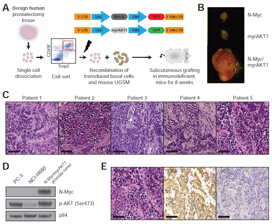
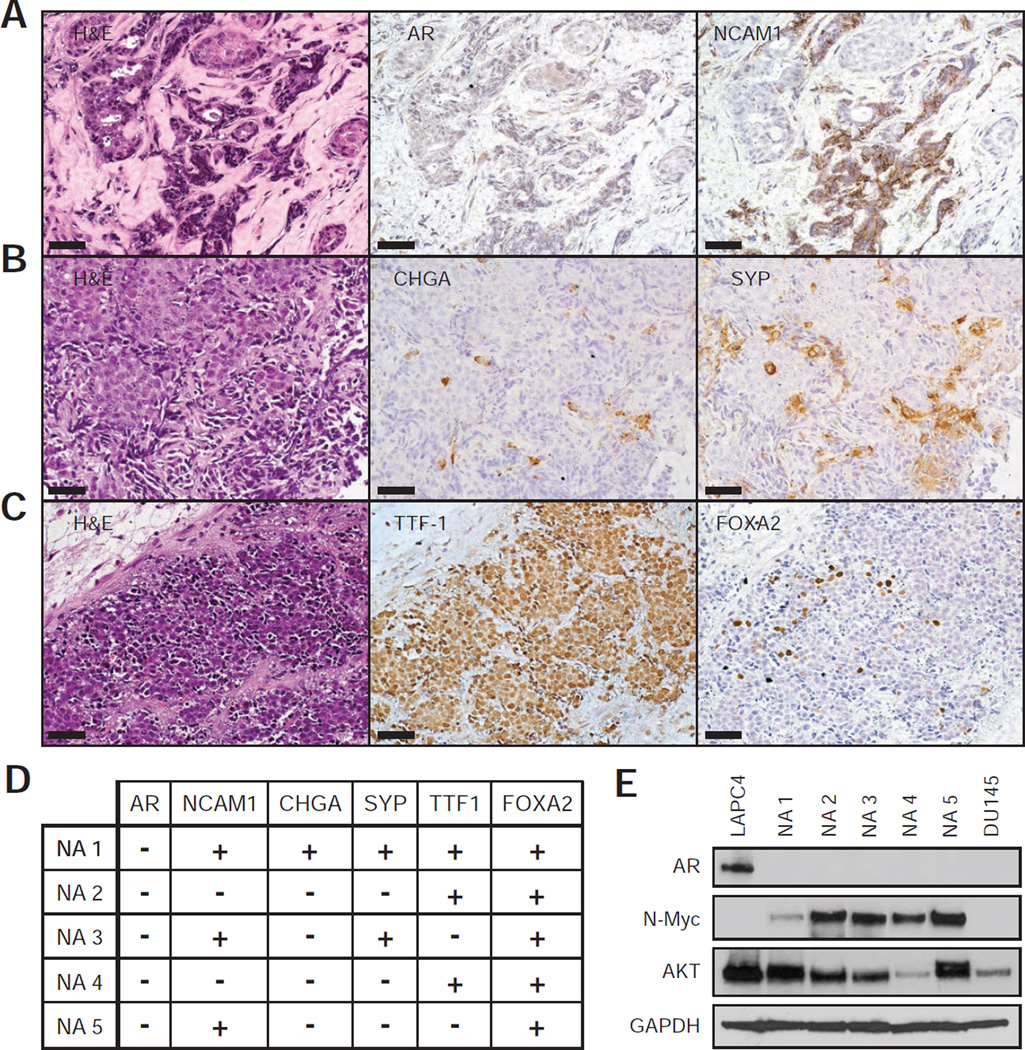
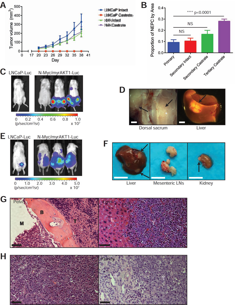
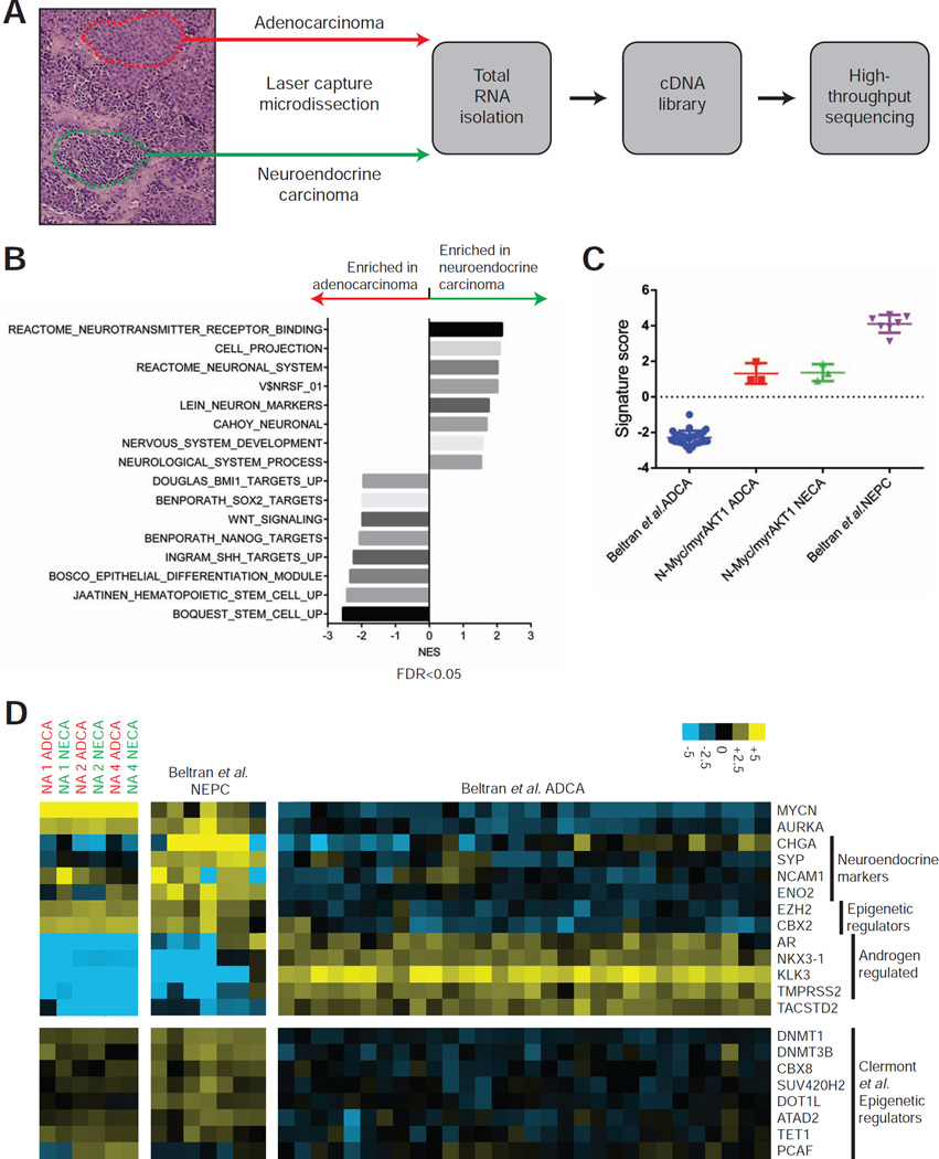
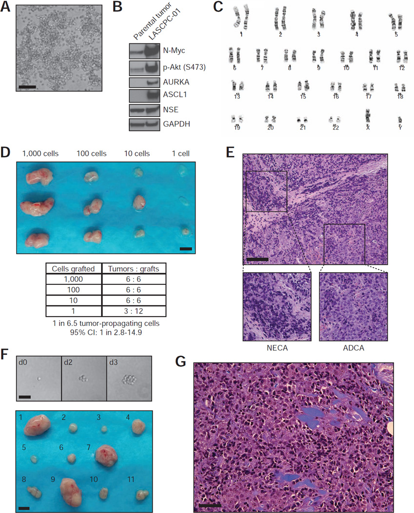
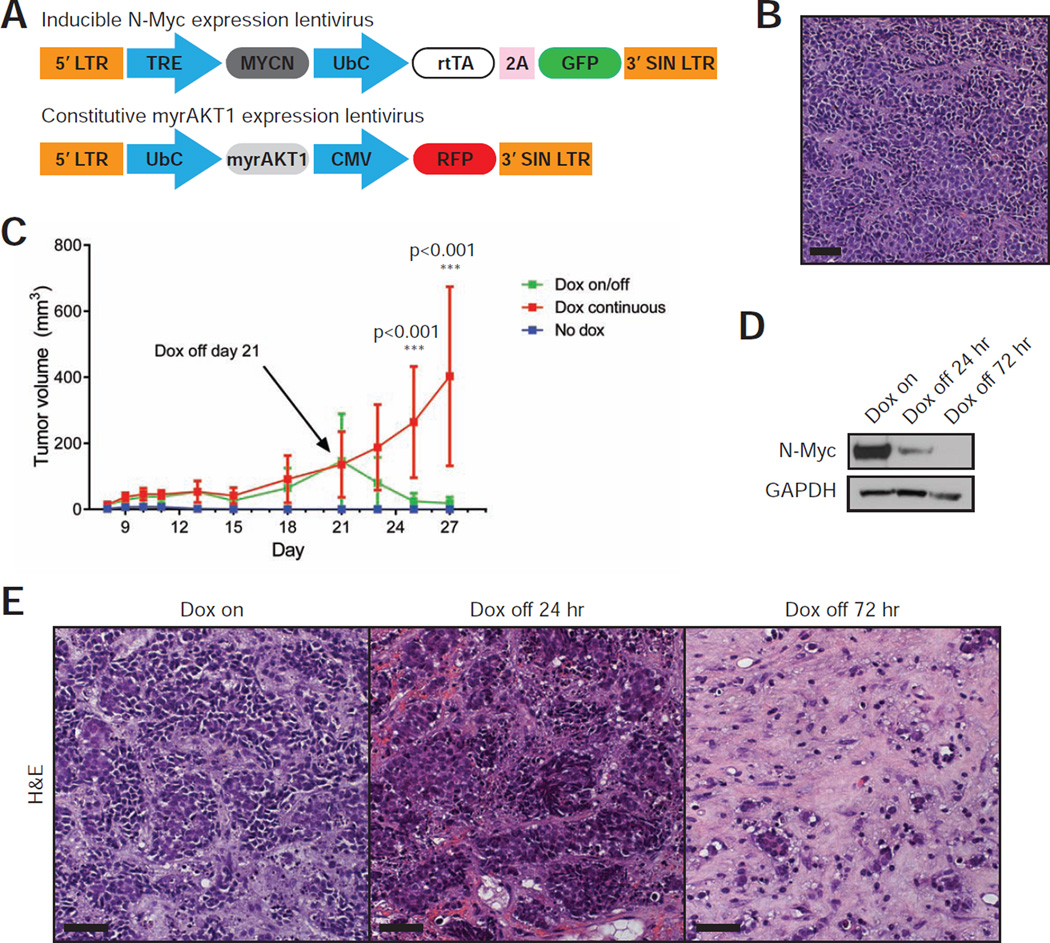
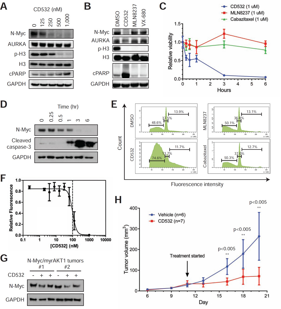
MYCN amplification and overexpression are common in neuroendocrine prostate cancer (NEPC). However, the impact of aberrant N-Myc expression in prostate tumorigenesis and the cellular origin of NEPC have not been established. We define N-Myc and activated AKT1 as oncogenic components sufficient to transform human prostate epithelial cells to prostate adenocarcinoma and NEPC with phenotypic and molecular features of aggressive, late-stage human disease. We directly show that prostate adenocarcinoma and NEPC can arise from a common epithelial clone. Further, N-Myc is required for tumor maintenance, and destabilization of N-Myc through Aurora A kinase inhibition reduces tumor burden. Our findings establish N-Myc as a driver of NEPC and a target for therapeutic intervention.


Figures

Figure 1
Figure 1. N-Myc and myrAKT1 initiate NEPC from human prostate basal epithelial cells
(A) Schematic of a human prostate regeneration and transformation assay (UbC=human ubiquitin C promoter, CMV=cytomegalovirus promoter, SIN=self-inactivating). The red square outlines the Trop2+CD49fhi basal epithelial cell population. (B) Grafts transduced with N-Myc, myrAKT1, and N-Myc/myrAKT1 harvested after 8 weeks (scale bar=2 mm). (C) H&E-stained sections of N-Myc/myrAKT1 tumors derived from individual patient prostatectomy samples (scale bar=100 µm). (D) Immunoblot of the human NEPC cell lines PC-3 and NCI-H660 and an N-Myc/myrAKT1 tumor with antibodies against N-Myc, p-AKT (Ser473), and p84 as a loading control. (E) H&E and immunohistochemical stains of an N-Myc/myrAKT1 tumor for CK8 and p63 (scale bar=100 µm). See also Figure S1 and Table S1.
Figure 2
Figure 2. Prostate tumors initiated by N-Myc and myrAKT1 lack AR expression and exhibit neuroendocrine markers
(A–C) Photomicrographs of N-Myc/myrAKT1 tumor sections containing regions of neuroendocrine carcinoma with H&E stains and immunohistochemical staining for AR and NCAM1, CHGA and SYP, and TTF-1 and FOXA2 (scale bar=100 µm). (D) Summary of the immunohistochemical staining for neuroendocrine markers in the regenerated tumors derived from five independent patient prostate samples. Positive staining represents visible staining in at least 5% of the tumor cells. (E) Immunoblot of the AR-positive human prostate cancer cell line LNCaP, five different N-Myc/myrAKT1 (NA) tumors, and the AR-null human prostate cancer cell line DU145 with antibodies against AR, N-Myc, AKT, and GAPDH as a loading control.
Figure 3
Figure 3. N-Myc/myrAKT1 prostate tumors are castration-resistant and metastasize widely
(A) LNCaP and transplanted N-Myc/myrAKT1 (NA) tumor volumes +/− SD (n=4 for each condition) in intact and surgically castrate mice over time. (B) Average percentage of neuroendocrine carcinoma identified by smart image segmentation +/− SEM in sections of primary, secondary, and tertiary N-Myc/myrAKT1 prostate tumors. p-values were calculated from an one-way analysis of variance. (C) Bioluminescent imaging of mice 21 days after tail vein injection with the LNCaP-Luc or N-Myc/myrAKT1-Luc cell lines (signal intensity is represented by radiance, p/sec/cm2/sr). (D) Gross tumor deposits marked by closed arrows localized to the sacrum and liver in N-Myc/myrAKT1-Luc injected mice (scale bar=2 mm). (E) Bioluminescent imaging of mice 74 days after orthotopic injection with the LNCaP-Luc or N-Myc/myrAKT1-Luc cell lines. (F) Gross metastatic tumors marked by closed arrows involving the liver, mesenteric lymph nodes (LNs), and kidney of N-Myc/myrAKT1-Luc mice (scale bar=1 cm). (G) H&E-stained sections of metastatic N-Myc/myrAKT1-Luc tumors in the pelvis and liver with M=marrow space, B=bone, L=liver, and T=tumor (scale bar=100 µm). (H) H&E-stained sections of metastatic N-Myc/myrAKT1-Luc tumors in a mesenteric LN and kidney with K=kidney and T=tumor (scale bar=100 µm). See also Figure S2.
Figure 4
Figure 4. Transcriptome profiling of the N-Myc/myrAKT1 tumors demonstrates similarity to human NEPC
(A) Schematic of the laser capture microdissection of matched regions of adenocarcinoma and neuroendocrine carcinoma in an N-Myc/myrAKT1 tumor and the workflow for whole transcriptome shotgun sequencing. (B) Gene set enrichment analysis for genes differentially expressed (>4-fold) in the adenocarcinoma or neuroendocrine carcinoma from the N-Myc/myrAKT1 tumor derived from Patient 1. (C) Neuroendocrine prostate cancer signature scores +/− SD for Beltran et al. neuroendocrine prostate cancer (NEPC, n=7), N-Myc/myrAKT1 adenocarcinoma (NA ADCA, n=3), N-Myc/myrAKT1 neuroendocrine carcinoma (NA NECA, n=3), and Beltran et al. adenocarcinoma (ADCA, n=30). (D) Heatmap of a selection of genes in NA ADCA, NA NECA, Beltran et al. NEPC, and Beltran et al. ADCA samples (contrast=+/−25). See also Figure S3 and Tables S2–S4.
Figure 5
Figure 5. Establishment of a human NEPC cell line LASCPC-01 with cancer stem cell-like features
(A) Photomicrograph of the LASCPC-01 cell line growing in suspension (scale bar=100 µm). (B) Immunoblot analysis of the parental N-Myc/myrAKT1 tumor from which LASCPC-01 was derived and the LASCPC-01 cell line with antibodies against N-Myc, p-AKT (Ser473), AURKA, ASCL1, NSE, and GAPDH as a loading control. (C) Conventional karyotyping of the LASCPC-01 cell. (D) Gross tumors generated from the subcutaneous xenotransplantation of serially diluted LASCPC-01 cells after five weeks (scale bar=5 mm). (E) Representative H&E-stained section of LASCPC-01 xenograft tumors with regions of neuroendocrine carcinoma (NECA) and adenocarcinoma (ADCA) (black scale bar=200 µm, white scale bar=100 µm). (F) Top panel, photomicrographs of LASCPC-1 cells in culture after single cell sorting, deposition, and culture (scale bar=50 µm). Bottom panel, gross tumors from the subcutaneous xenotransplantation of eleven clonal LASCPC-01 sublines after 4 weeks (scale bar=5 mm). (G) Representative H&E-stained section of a xenograft tumor derived from a clonal LASCPC-01 subline (scale bar=100 µm). See also Figure S4.
Figure 6
Figure 6. N-Myc expression is required for tumor maintenance in the N-Myc/myrAKTl tumors
(A) Lentiviral constructs used for doxycycline-inducible expression of N-Myc and constitutive expression of myrAKT1 (TRE=tetracycline response element, rtTA=reverse tetracycline-controlled transactivator). (B) H&E-stained section of an inducible N-Myc/myrAKT1 tumor (scale bar=100 µm). (C) Average tumor volume of passaged inducible N-Myc/myrAKT1 tumors +/− SD over time under conditions of no doxycycline (No dox, n=13), continuous doxycycline (Dox continuous, n=11), and doxycycline withdrawal on day 21 after initial doxycycline (Dox on/off, n=11). p-values were calculated from a paired t-test. (D) Immunoblot analysis of inducible N-Myc/myrAKT1 tumors after continuous doxycycline or initial doxycycline followed by withdrawal using antibodies against N-Myc and GAPDH as a loading control. (E) Representative H&E-stained sections of inducible N-Myc/myrAKT1 tumors after continuous doxycycline and after doxycycline withdrawal (scale bar=100 µm).
Figure 7
Figure 7. Therapeutic targeting of N-Myc dependence in the N-Myc/myrAKT1 model of NEPC
(A) Immunoblot analysis of LASCPC-01 cells treated with a dose range of CD532 with antibodies against N-Myc, AURKA, phosphorylated histone H3 (p-H3), histone H3, cleaved PARP (cPARP), and GAPDH as a loading control. (B) Immunoblot of LASCPC-01 treated with DMSO or 500 nM of CD532, MLN8237, or VX-680 for 3 hours with antibodies against, N-Myc, AURKA, p-H3, H3, cPARP, and GAPDH as a loading control. (C) LASCPC-01 cell viability +/− SD after 3 hours of treatment with 1 µM of CD532, MLN8237, or cabazitaxel relative to treatment with DMSO (n=6 for each condition). (D) Immunoblot of LASCPC-01 cells treated with 1 µM of CD532 over a time course with antibodies against N-Myc, cleaved caspase-3, and GAPDH as a loading control. (E) Cell cycle analysis of LASCPC-01 cells after 3 hours of treatment with DMSO or 1 µM of CD532, MLN8237, or cabazitaxel. Quantification of the sub-G1, G1, and S/G2/M fractions is shown. (F) Dose response of CD532 +/− SD (normalized to DMSO treatment only) at 48 hours using the CellTiter-Glo cell viability assay in LASCPC-01 cells. (G) Immunoblot analysis of N-Myc/myrAKT1 tumors after treatment with vehicle or CD532 for 24 hours with antibodies against N-Myc and GAPDH as a loading control. (H) Average tumor volume of LASCPC-01 subcutaneous xenografts +/− SD with vehicle (n=6) or CD532 (n=7) treatment initiated on day 11. p-values were calculated from a paired t-test. See also Figure S5 and Table S5.

Comment in

References

    1. Beltran H. The N-myc Oncogene: Maximizing its Targets, Regulation, and Therapeutic Potential. Molecular cancer research : MCR. 2014;12:815–822. - PubMed
    1. Beltran H, Rickman DS, Park K, Chae SS, Sboner A, MacDonald TY, Wang Y, Sheikh KL, Terry S, Tagawa ST, et al. Molecular characterization of neuroendocrine prostate cancer and identification of new drug targets. Cancer discovery. 2011;1:487–495. - PMC - PubMed
    1. Beltran H, Tomlins S, Aparicio A, Arora V, Rickman D, Ayala G, Huang J, True L, Gleave ME, Soule H, et al. Aggressive variants of castration-resistant prostate cancer. Clin Cancer Res. 2014;20:2846–2850. - PMC - PubMed
    1. Berman-Booty LD, Knudsen KE. Models of neuroendocrine prostate cancer. Endocrine-related cancer. 2015;22:R33–R49. - PMC - PubMed
    1. Boutros PC, Fraser M, Harding NJ, de Borja R, Trudel D, Lalonde E, Meng A, Hennings-Yeomans PH, McPherson A, Sabelnykova VY, et al. Spatial genomic heterogeneity within localized, multifocal prostate cancer. Nature genetics. 2015 - PubMed

Publication types

MeSH terms