. 2016 Apr 11;29(4):508-522.
doi: 10.1016/j.ccell.2016.03.002. Epub 2016 Mar 31.

Medulloblastoma Genotype Dictates Blood Brain Barrier Phenotype

Affiliations

Medulloblastoma Genotype Dictates Blood Brain Barrier Phenotype

Timothy N Phoenix et al. Cancer Cell. .

Abstract

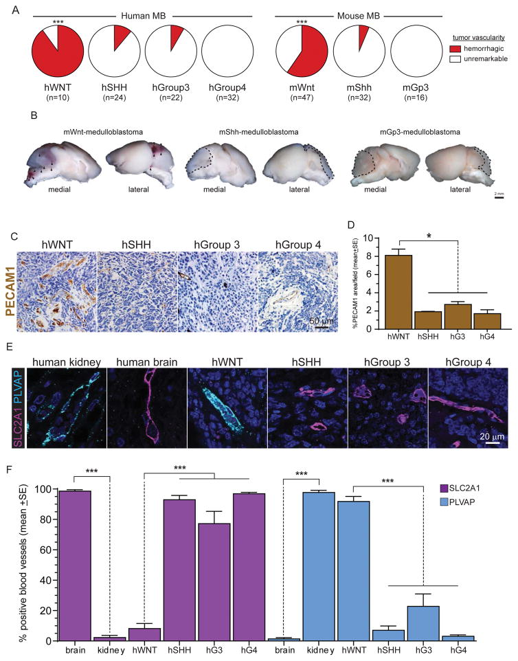
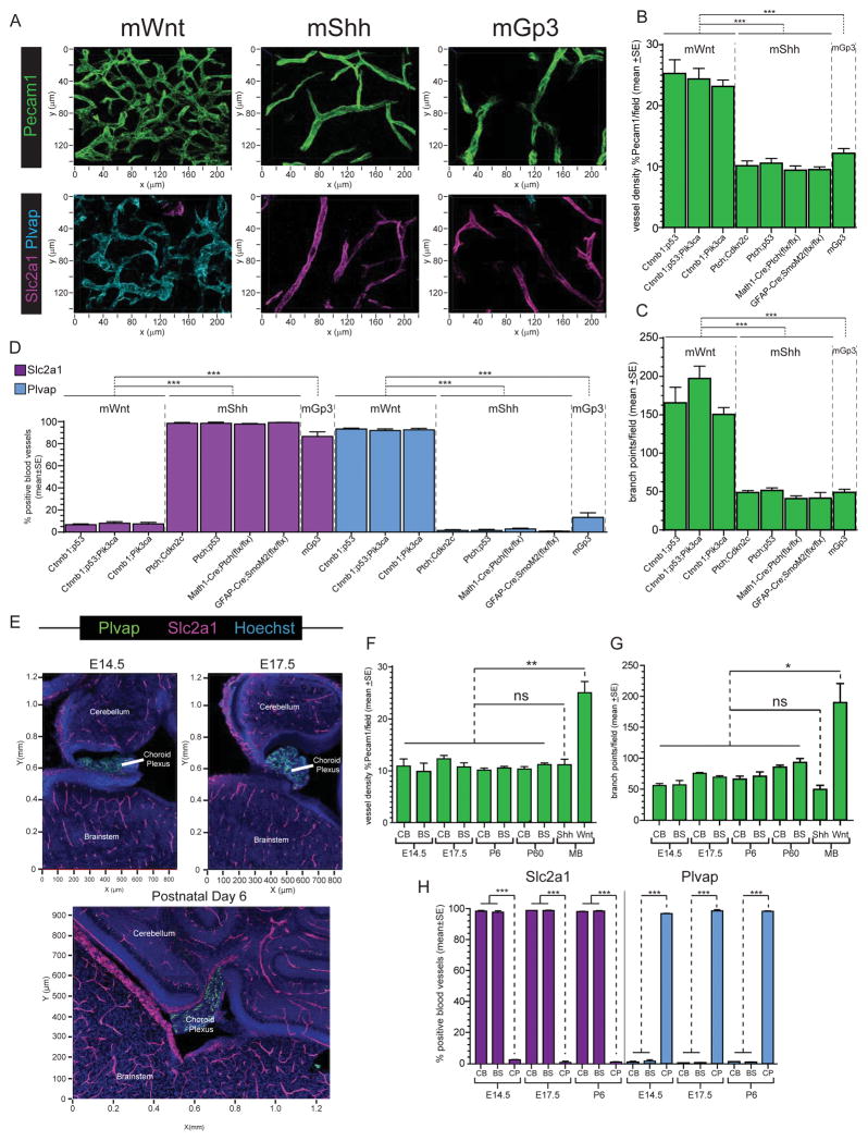
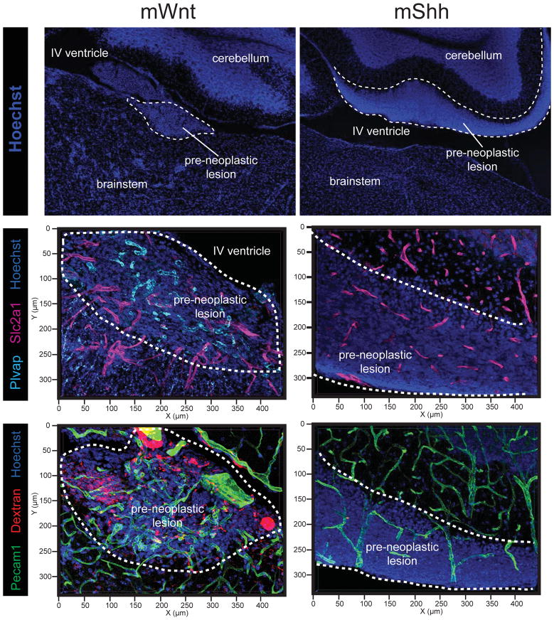
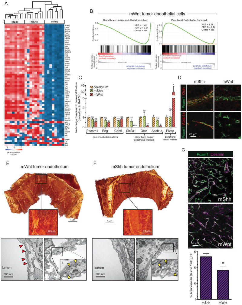
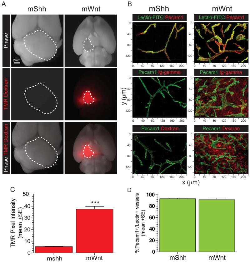
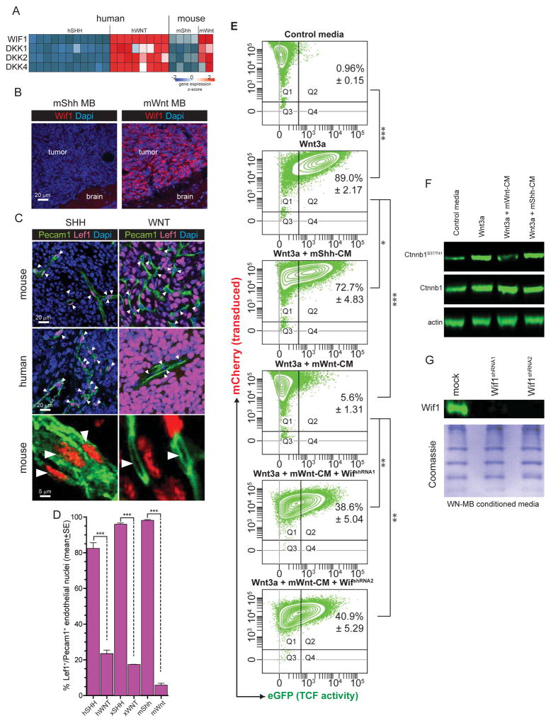
The childhood brain tumor, medulloblastoma, includes four subtypes with very different prognoses. Here, we show that paracrine signals driven by mutant β-catenin in WNT-medulloblastoma, an essentially curable form of the disease, induce an aberrant fenestrated vasculature that permits the accumulation of high levels of intra-tumoral chemotherapy and a robust therapeutic response. In contrast, SHH-medulloblastoma, a less curable disease subtype, contains an intact blood brain barrier, rendering this tumor impermeable and resistant to chemotherapy. The medulloblastoma-endothelial cell paracrine axis can be manipulated in vivo, altering chemotherapy permeability and clinical response. Thus, medulloblastoma genotype dictates tumor vessel phenotype, explaining in part the disparate prognoses among medulloblastoma subtypes and suggesting an approach to enhance the chemoresponsiveness of other brain tumors.


Figures

Figure 1
Figure 1. Human and mouse medulloblastoma vessel phenotype
A. Frequency of intra-operative haemorrhages detected in human and mouse medulloblastomas. B. Gross preparations of mouse medulloblastomas demonstrating numerous haemorrhages (arrows) in mWnt-medulloblastoma. Photomicrographs (C) and quantification (D) of PECAM1 immunohistochemical staining of human medulloblastomas. Co-immunofluorescence (E) and quantification (F) of SLC2A1 and PLVAP expression in human kidney, brain and medulloblastoma. *=P<0.05, ***=P<0.0005, Mann-Whitney. See also Figure S1.
Figure 2
Figure 2. BBB immunophenotype in mouse medulloblastoma and developing hindbrain
A. Confocal immunofluorescence of Pecam1 (top) and co-immunofluorescence of Slc2a1 and Plvap (bottom) in mWnt-, mShh, and mGroup 3-medulloblastomas. Quantification of tumour vessel density (B), tortuosity (C), and Slc2a1/Plvap expression (D) in eight variant models of mWnt-, mShh, and mGroup 3-medulloblastoma. E. Co-immunofluorescence of Slc2a1 and Plvap in developing mouse hindbrain. Quantification of vessel density (F), tortuosity (G), and Slc2a1/Plvap expression (H) in developing mouse cerebellum (CB), brainstem (BS) and choroid plexus (CP) relative to mWnt- and mShh-medulloblastomas (MB). *=P<0.05, **=P<0.005, ***=P<0.0005, Mann-Whitney. See also Figure S2.
Figure 3
Figure 3. BBB immunophenotype and porosity in early hyperplasic precursor lesions of mWnt- and mShh-medulloblastoma
Top: Hoechst staining illustrating the anatomy and location of lesions in the developing hindbrain. Middle: co-immunofluorescence of Slc2a1 and Plvap in precursor lesions of mWnt- and mShh-medulloblastoma. Bottom: Pecam31 immunofluorescence and TMR-dextran autoflourescence demonstrating leakage of dextran into mWnt- but not mShh-medulloblastoma precursor lesions.
Figure 4
Figure 4. Molecular and ultrastructural phenotype of mWnt- and mShh-medulloblastoma endothelium
A. Unsupervised hierarchical clustering of BBB endothelial specific gene expression in endothelial cells isolated from mWnt- and mShh-medulloblastoma and normal brain. B. Gene Set Enrichment Analyses of BBB endothelium enriched (left) and peripheral endothelium enriched gene expression in mWnt-medulloblastoma endothelial cells reporting normalized enrichment score (NES) and the false discovery rate (FDR) Q value. C. Reverse-transcription quantitative polymerase chain reaction analyses of endothelial specific gene expression in endothelial cells isolated from normal brain, mWnt- and mShh-medulloblastoma. D. co-immunofluorescence of Pecam1 and Abcb1a or Ocln expression in mWnt- and mShh-medulloblastoma endothelium. Electron microscopy (EM) of endothelium in mWnt- (E) and mShh-medulloblastoma (F). In both E and top=3D scanning EM, bottom=transmission EM. View is from the interior of the vessel, bright objects are mitochondria. Note regular fenestrations in mWnt-medulloblastomas on 3D scanning EM. Red arrows=pores, yellow arrows=vesicles. G. Co-immunofluorescence (top) and quantification (bottom) of Desmin and Pecam31 expression in mWnt- and mShh-medulloblastoma. *=P<0.05, **=P<0.005, ***=P<0.0005, Mann-Whitney.
Figure 5
Figure 5. BBB function in mWnt- and mShh-medulloblastoma
A. Autofluorescence of TMR-Dextran in ex vivo whole brain preparations of mice harbouring mWnt- or mShh-medulloblastoma. Dotted lines demarcate tumours. B. confocal microscopy of Pecam1 immunofluorescence and lectin-FITC autoflourescence (top), gamma-immunoglobulin coimmunofluorescence (middle); and TMR-dextran autoflourescence; (bottom). Quantification of TMR-dextran in tissue fluid (C) and lectin-FITC in vessels (D) in mWnt- and mShh-medulloblastomas. ***=P<0.0005, Mann-Whitney.
Figure 6
Figure 6. WNT-medulloblastoma paracrine signals silence WNT signalling in neighbouring cells
A. Heat map of mRNA expression of secreted WNT inhibitors by human and mouse medulloblastoma (source data Gibson et al., 2010). B. Immunofluorescence of Wif1 expression in mouse mWnt- and mShh-medulloblastoma. Co-immunofluorescence (C) and quantification (D) of Pecam1 and Lef1 expression in mouse mWnt- and mShh-medulloblastoma, arrows=nuclei. E. Fluorescence activated cell sorting plots quantifying TCF-reporter activity (x-axis) in TCF-reporter transduced cells (y-axis). Numbers in plots denote the %±SE of reporter transduced and activated cells. F. Western blots of total and phosphorylated Ctnnb1 in reporter cells shown in (E). G. Western blots of Wif1 in mWnt-medulloblastoma cells transduced with Wif1-shRNAs. *=P<0.05, **=P<0.005, ***=P<0.0005, Mann-Whitney.
Figure 7
Figure 7. Medulloblastoma-endothelial paracrine signals can be manipulated in vivo
Kaplan-Meyer survival curves of (A) mShhWif1+Dkk1- or (B) or mWntWnt1-medulloblastomas relative to controls. C. Immunofluorescence studies of the indicated proteins and TMR-dextran leakage in mShhWif1+Dkk1- and mWntWnt1-medulloblastomas relative to controls. Quantification of Lef1+ endothelial nuclei (D), Slc2a1/Plvap expression (E) and TMR-dextran leakage (F) leakage in mShhWif1+Dkk1- and mWntWnt1-medulloblastomas relative to controls. Slc2a1/Plvap expression (G) and TMR-dextran leakage (H) in admixed tumours of mWntWnt1- and mWntcontrol-medulloblastoma cells. **=P<0.005, ***=P<0.0005, Mann-Whitney. See also Figure S3.
Figure 8
Figure 8. BBB function dictates medulloblastoma exposure and response to vincristine in vivo
A. Growth inhibition assays of mouse medulloblastoma cells following 72 hour exposure to the indicated drugs. B. Kaplan-Meyer survival curves of mShhcontrol and mWntcontrol-medulloblastomas to vincristine therapy. C. Vincristine area under the curve (AUC) plots for plasma (left), tumour tissue fluid (middle) and KPT (right) in mice harbouring the indicated medulloblastomas. D. Kaplan-Meyer survival curves of mWntcontrol- or mWntWnt7a- medulloblastomas treated with vincristine therapy. *=P<0.05, **=P<0.005, Mann-Whitney.

Comment in

References

    1. Alvarez JI, Dodelet-Devillers A, Kebir H, Ifergan I, Fabre PJ, Terouz S, Sabbagh M, Wosik K, Bourbonniere L, Bernard M, et al. The Hedgehog pathway promotes blood-brain barrier integrity and CNS immune quiescence. Science. 2011;334:1727–1731. - PubMed
    1. Armulik A, Genove G, Mae M, Nisancioglu MH, Wallgard E, Niaudet C, He L, Norlin J, Lindblom P, Strittmatter K, et al. Pericytes regulate the blood-brain barrier. Nature. 2010;468:557–561. - PubMed
    1. Atkinson JM, Shelat AA, Carcaboso AM, Kranenburg TA, Arnold LA, Boulos N, Wright K, Johnson RA, Poppleton H, Mohankumar KM, et al. An integrated in vitro and in vivo high-throughput screen identifies treatment leads for ependymoma. Cancer Cell. 2011;20:384–399. - PMC - PubMed
    1. Avula S, Mallucci C, Kumar R, Pizer B. Posterior fossa syndrome following brain tumour resection: review of pathophysiology and a new hypothesis on its pathogenesis. Child's nervous system : ChNS : official journal of the International Society for Pediatric Neurosurgery. 2015;31:1859–1867. - PubMed
    1. Biechele TL, Adams AM, Moon RT. Transcription-based reporters of Wnt/beta-catenin signaling. Cold Spring Harb Protoc. 2009;2009 pdb prot5223. - PubMed

Publication types

MeSH terms