. 2016 Apr 6:7:11215.
doi: 10.1038/ncomms11215.

Circular RNA profiling reveals an abundant circHIPK3 that regulates cell growth by sponging multiple miRNAs

Affiliations

Circular RNA profiling reveals an abundant circHIPK3 that regulates cell growth by sponging multiple miRNAs

Qiupeng Zheng et al. Nat Commun. .

Abstract

Circular RNAs (circRNAs) represent a class of widespread and diverse endogenous RNAs that may regulate gene expression in eukaryotes. However, the regulation and function of human circRNAs remain largely unknown. Here we generate ribosomal-depleted RNA sequencing data from six normal tissues and seven cancers, and detect at least 27,000 circRNA candidates. Many of these circRNAs are differently expressed between the normal and cancerous tissues. We further characterize one abundant circRNA derived from Exon2 of the HIPK3 gene, termed circHIPK3. The silencing of circHIPK3 but not HIPK3 mRNA significantly inhibits human cell growth. Via a luciferase screening assay, circHIPK3 is observed to sponge to 9 miRNAs with 18 potential binding sites. Specifically, we show that circHIPK3 directly binds to miR-124 and inhibits miR-124 activity. Our results provide evidence that circular RNA produced from precursor mRNA may have a regulatory role in human cells.


Figures
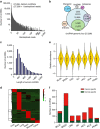
Figure 1
Figure 1. Profiling of circular RNAs in human normal and cancerous tissues.
(a) The number of circRNAs and back-spliced reads identified in six human normal tissues and seven human cancerous tissues. (b) Genomic origin of human circRNAs. (c) The length distribution for exonic circRNAs (n=20,533, only known spliced length was considered). (d) Clustered heatmap for tissue-specific circRNAs from six human normal tissues, with rows representing circRNAs and columns representing tissues. The circRNAs were classified according to the Pearson correlation. The numerical data represented log10-transformed mean SRPBM of two replicates. (e) Violin plot of relative abundance of circRNAs in seven cancer tissues compared with the paired normal tissues. Data are expressed as the log2 foldchange of SRPBM. The white dot represents the median. (f) Numbers of specific circRNAs identified in seven cancerous and matched normal tissues. Cancer-specific circRNAs are shown in red. Normal specific circRNAs are shown in green.
Figure 2
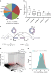
Figure 2. The characteristics of circular RNA abundance in human cells.
(a) Number of circRNAs produced from one gene (20,530 circRNAs from 5,955 host genes). (b) The box plots describe the comparison of the levels of most abundantly expressed circRNA isoform (circGene) and other circRNAs (circGene.x) from one gene locus (n=3,687). The ends of the boxes define the 25th and 75th centiles, a line indicates the median, and bars define the 5th and 95th centiles. The first five circRNAs were presented. (c) Schematic illustration of the methodology to estimate either the circular ratio at the 5′ end (5′ CR) or at 3′ end (3′ CR) for circRNAs. Ci and ci represent the back-spliced junctions (support for circRNA) and number of reads spanning these junctions, respectively; Li and li represented linear spliced junctions and the number of reads spanning these junctions, respectively. Solid squares, exons; broken lines, linear spliced junctions; arc lines, back-spliced junctions. (d) Multidimensional scaling screen for highly abundant circRNAs (n=27,293, three outlines were not shown). Red dots and black dots represented highly abundant circRNAs and low-abundance circRNAs, respectively. circHIPK3 is highlighted in blue. The cut-off for highly abundant circRNAs is shown in grey. The pseudocount of 1 was added to SRPBM to avoid log10-transform issues. Thus, the cut-off of log10-transformed SRPBM is 0.3010. (e) A density plot of the flanking intron length of highly abundant circRNAs (red) and low-abundance circRNAs (green).
Figure 3
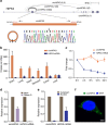
Figure 3. Characterization of circHIPK3 RNA in human cells.
(a) The genomic loci of five circRNAs in HIPK3 gene. The supported unique reads were presented. The expression of circHIPK3 was validated by RT–PCR followed by sanger sequencing. Arrows represent divergent primers binding to the genome region of circHIPK3. (b) Absolute quantification for circHIPK3 and HIPK3 mRNA in six human normal tissues. (c) qRT–PCR for the abundance of circHIPK3 and HIPK3 mRNA in HeLa cells treated with Actinomycin D at the indicated time points. (d) qRT–PCR for the abundance of circHIPK3 and HIPK3 mRNA in HeLa cells treated with RNase R. The amount of circHIPK3 and HIPK3 mRNA were normalized to the value measured in the mock treatment. (e) qRT–PCR data indicating the abundance of circHIPK3 and HIPK3 mRNA in either the cytoplasm or nucleus of HeLa cells. The amounts of circHIPK3 and HIPK3 mRNA were normalized to the value measured in the cytoplasm. Data in (ce) are the means±s.e.m. of three experiments. (f) RNA fluorescence in situ hybridization for circHIPK3. Nuclei were stained with 4,6-diamidino-2-phenylindole (DAPI). Scale bar, 5 μm.
Figure 4
Figure 4. circHIPK3 is derived from HIPK3 exon2 due to long flanking introns.
(a) Schematics showing that the genomic regions of HIPK3 Exon2 contain flanking Alu repeats and long introns. Alu elements and long introns were deleted using CRISPR/Cas9 systems. Primers flanking the gRNA recognition were used to detect deletions. (b) Schematic diagram of circHIPK3 expression vectors with various genomic sequences for circHIPK3 recapitulation (#1–4). The genomic region of circHIPK3 was cloned into the pcDNA3.0 vector with the upstream and downstream intron sequences, which included Alu elements (#1). Deletions were introduced into the HIPK3 expression plasmid. (#2–4). (c,d) Northern blot and qRT–PCR for circHIPK3 in cells transfected with the expression plasmids (#1–4). (e,f) Normal PCR and qRT–PCR data for CRISPR/Cas9-mediated genomic deletions in HEK-293 T cells. Two pairs of gRNAs (gRNA1+gRNA2, gRNA3+gRNA4) were used to mediate the deletion of the proximal Alu elements. M: 100 bp DNA ladder. (g) Normal PCR and qRT–PCR data for large deletion mediated by four sets of paired gRNAs. The primer site was designed outside the deleted region. For the control gRNA, the expected product was not shown because of too large PCR product. For the loading control, a genomic region from Actin gene loci was amplifed in eg. Data in dg are the means±s.e.m. of three experiments. *P<0.05, **P<0.01 (Student's t-test).
Figure 5
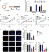
Figure 5. Silencing of circHIPK3 RNA inhibits human cell proliferation.
(a) Schematic illustration showing three targeted siRNAs. Si-HIPK3 targets the HIPK3 linear transcript, si-circHIPK3 targets the back-splice junction of circHIPK3, and si-both targets both the linear and circular species. (b) qRT–PCR for circHIPK3 and HIPK3 mRNA in HEK-293 T cells treated with three siRNAs as described above. Data are the means±s.e.m. of three experiments. (ce) Proliferation of HuH-7, HCT-116 and HeLa cells transfected with the above three siRNAs assessed using a CCK-8 kit at the indicated days. Data in ce are the means±s.e.m. of three experiments. (f) DNA synthesis assessed using an EdU (5-ethynyl-2'-deoxyuridine) assay in HuH-7 cells transfected with the above three siRNAs for 48 h. Cells were fluorescently stained with EdU (red). Nuclei were stained with DAPI (blue). Micrographs represent at least three experiments. Scale bar, 200 μm. (g) Quantitative EdU assay data from f and Supplementary Fig. 7. Data in dg are the means±s.e.m. of three experiments. *P<0.05, **P<0.01 (Student's t-test).
Figure 6
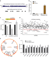
Figure 6. circHIPK3 serves as a sponge for multiple miRNAs in human cells.
(a) Schematic illustration showing that the conservation across 100 vertebrate species and AGO2 binding sites in circHIPK3 genomic region. (b) Ago2 RNA immunoprecipitation (RIP) assay for the amount of circHIPK3 in HEK-293 T cells stably expressing Flag-AGO2 or Flag-GFP. Data are the means±s.e.m. of three experiments. (c) The entire circHIPK3 sequence (red) was cloned into the downstream region of the luciferase gene, denoted LUC-cHIPK3. Luciferase reporter assay for the luciferase activity of LUC-cHIPK3 in HEK-293 T cells co-transfected with siRNA against circHIPK3 or circHIPK3 expressing vector. Data are the means±s.e.m. of three experiments. (d) Luciferase reporter assay for the luciferase activity of LUC-cHIPK3 in HEK-293 T cells transfected with a library of 424 miRNA mimics to identify miRNAs that were able to bind to the circHIPK3 sequence. Nine miRNAs that inhibited luciferase activity by 30% are indicated by red dots. (e) A schematic drawing showing the putative binding sites of the miRNAs associated with circHIPK3. (f) Luciferase reporter assay for the luciferase activity of LUC-cHIPK3 or LUC-cHIPK3-mutant in HEK-293 T cells co-transfected with miRNA mimics. Data are the means±s.e.m. of three experiments.
Figure 7
Figure 7. circHIPK3 sponges with miR-124 and inhibits its activity.
(a) Proliferation assessed using a CCK-8 kit in HEK-293 T cells transfected with nine miRNA mimics or control RNA (20 nM). (b) qRT–PCR analysis of circHIPK3 level in the streptavidin captured fractions from the HEK-293 T cell lysates after transfection with 3′-end biotinylated miR-124 or control RNA (NC). (c) Co-localization between miR-124 and circHIPK3 was observed (arrowheads) by RNA in situ hybridization in HeLa cells after co-transfection with circHIPK3 and miR-124 expressing vectors. Nuclei were stained with DAPI. Scale bar, 5μm. (d) qRT–PCR analysis of IL6R and DLX2 expression in HEK-293 T cells after transfected with si-cHIPK3, miR-124 mimics or miR-124 with circHIPK3 expressing vector (p-cHIPK3). (e) Proliferation assessed using a CCK-8 kit in cells transfected with circHIPK3 or miR-124 (10 nM) as indicated. Data in a,b,d are the means±s.e.m. of three experiments. (f) qRT–PCR for the abundance of circHIPK3 relative to ACTB and miR-124 relative RNU6B in six human normal tissues. The correlation between circHIPK3 and miR-124 is also shown. *P<0.05, **P<0.01 (Student's t-test).

References

    1. Jeck W. R. & Sharpless N. E. Detecting and characterizing circular RNAs. Nat. Biotechnol. 32, 453–461 (2014). - PMC - PubMed
    1. Lasda E. & Parker R. Circular RNAs: diversity of form and function. RNA 20, 1829–1842 (2014). - PMC - PubMed
    1. Nigro J. M. et al.. Scrambled exons. Cell 64, 607–613 (1991). - PubMed
    1. Cocquerelle C., Mascrez B., Hetuin D. & Bailleul B. Mis-splicing yields circular RNA molecules. FASEB J. 7, 155–160 (1993). - PubMed
    1. Zaphiropoulos P. G. Circular RNAs from transcripts of the rat cytochrome P450 2C24 gene: correlation with exon skipping. Proc. Natl Acad. Sci. USA 93, 6536–6541 (1996). - PMC - PubMed

Publication types

MeSH terms