. 2016 Nov 1;33(21):1919-1935.
doi: 10.1089/neu.2015.4348. Epub 2016 May 16.

Endoplasmic Reticulum Stress and Disrupted Neurogenesis in the Brain Are Associated with Cognitive Impairment and Depressive-Like Behavior after Spinal Cord Injury

Affiliations

Endoplasmic Reticulum Stress and Disrupted Neurogenesis in the Brain Are Associated with Cognitive Impairment and Depressive-Like Behavior after Spinal Cord Injury

Junfang Wu et al. J Neurotrauma. .

Abstract

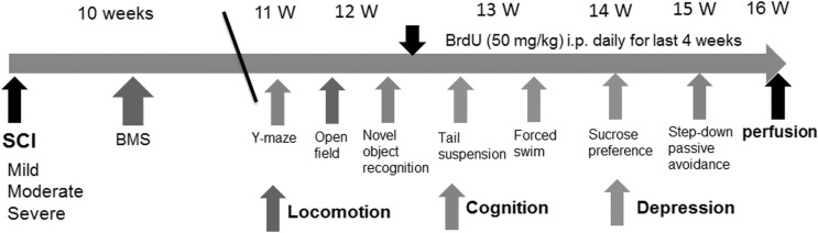
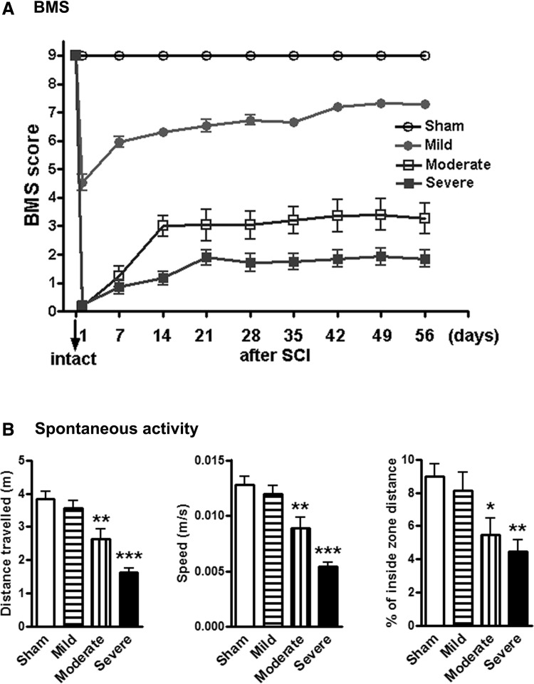
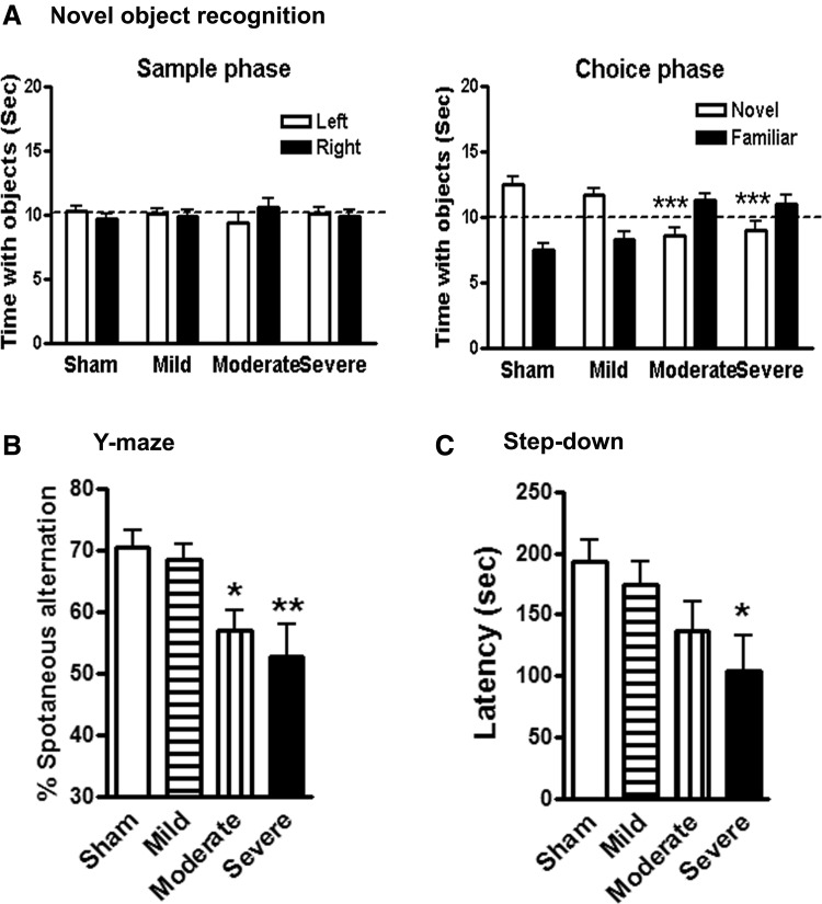
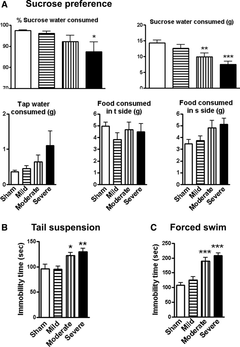
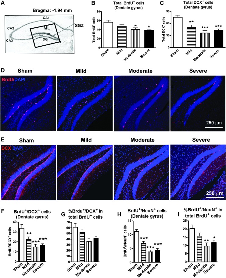
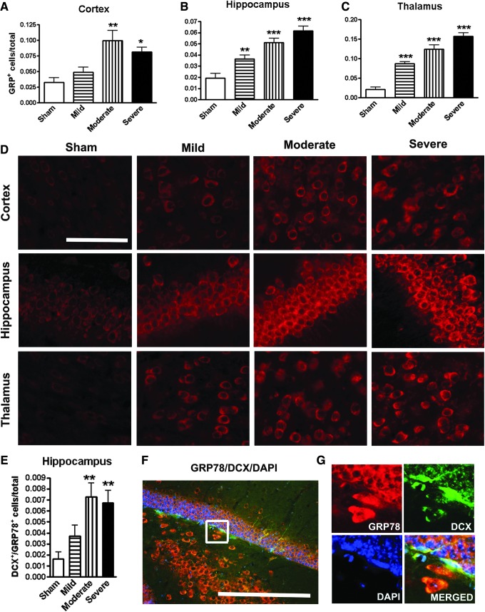
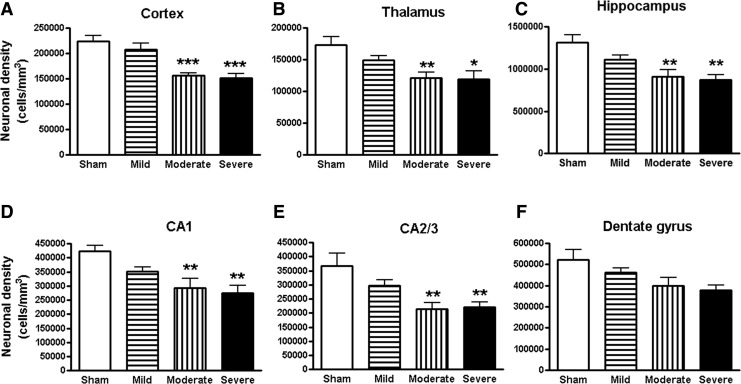
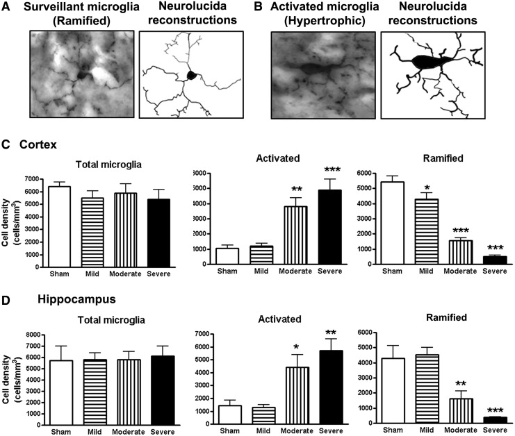
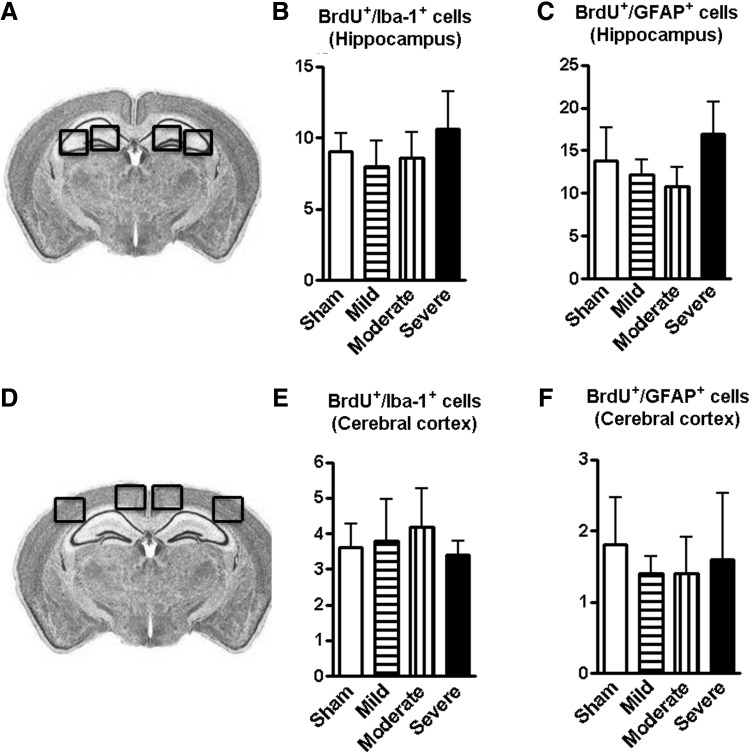
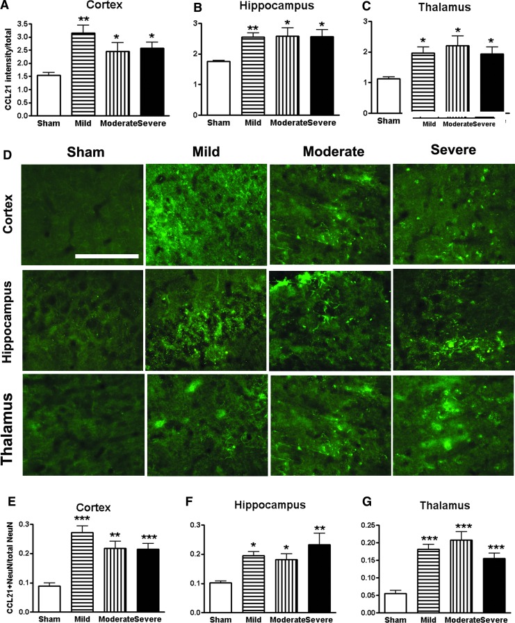
Clinical and experimental studies show that spinal cord injury (SCI) can cause cognitive impairment and depression that can significantly impact outcomes. Thus, identifying mechanisms responsible for these less well-examined, important SCI consequences may provide targets for more effective therapeutic intervention. To determine whether cognitive and depressive-like changes correlate with injury severity, we exposed mice to sham, mild, moderate, or severe SCI using the Infinite Horizon Spinal Cord Impactor and evaluated performance on a variety of neurobehavioral tests that are less dependent on locomotion. Cognitive impairment in Y-maze, novel objective recognition, and step-down fear conditioning tasks were increased in moderate- and severe-injury mice that also displayed depressive-like behavior as quantified in the sucrose preference, tail suspension, and forced swim tests. Bromo-deoxyuridine incorporation with immunohistochemistry revealed that SCI led to a long-term reduction in the number of newly-generated immature neurons in the hippocampal dentate gyrus, accompanied by evidence of greater neuronal endoplasmic reticulum (ER) stress. Stereological analysis demonstrated that moderate/severe SCI reduced neuronal survival and increased the number of activated microglia chronically in the cerebral cortex and hippocampus. The potent microglial activator cysteine-cysteine chemokine ligand 21 (CCL21) was elevated in the brain sites after SCI in association with increased microglial activation. These findings indicate that SCI causes chronic neuroinflammation that contributes to neuronal loss, impaired hippocampal neurogenesis and increased neuronal ER stress in important brain regions associated with cognitive decline and physiological depression. Accumulation of CCL21 in brain may subserve a pathophysiological role in cognitive changes and depression after SCI.

Keywords: ER stress; adult neurogenesis; brain; cognition/depression; spinal cord injury.


Conflict of interest statement

Author Disclosure Statement No competing financial interests exist.

Figures

<b>FIG. 1.</b>
FIG. 1.
A timeline of the in vivo experimental design. A battery of behavioral tests were evaluated according to the schedule outlined and compared following sham, mild-level (30 kDyne), moderate-level (60 kDyne), or severe-level (100 kDyne) spinal cord injury (SCI). Assessment includes motor function (Basso Mouse Scale [BMS], open field spontaneous activity), cognition (Y-maze, novel object recognition, step-down passive avoidance), and depression (sucrose preference, tail suspension and forced swim tests). ip, intraperitoneal.
<b>FIG. 2.</b>
FIG. 2.
Spinal cord injury (SCI) causes impairments of motor function in an injury-severity dependent manner. (A) Hindlimb locomotor function was evaluated using the Basso Mouse Scale (BMS) score. SCI reduced BMS scores in an injury-severity dependent manner. (B) Determination of spontaneous locomotor activity. SCI resulted in a significant reduced distance traveled (left panel), walking speed (middle panel), and percent of inside zone distance (right panel), compared with sham mice.
<b>FIG. 3.</b>
FIG. 3.
Spinal cord injury (SCI) causes learning and memory impairments. (A) Novel object recognition test. Mice from sham or mild groups spent more time than chance (10 sec) with the novel object 24 h after training (sample phase), indicating intact memory. SCI mice with moderate or severe injury spent significantly less time with the novel object. (B) Y-maze spontaneous alteration test. Sham mice showed approximately 70% spontaneous alteration, indicative of functional working memory. Moderate or severe SCI caused a significant reduction of spontaneous alteration, compared with sham animals. (C) Step-down passive avoidance (SDPA) test. During SDPA conditioning trials, all groups exhibited similar short retention times before stepping down onto the platform and receiving a shock. During testing for memory of the aversive experiences 24 h later, SCI mice showed significantly reduced latency to step-down from the platform, compared with sham mice. n = 11–15/group. *p < 0.05, **p < 0.01, ***p < 0.001 vs. sham group.
<b>FIG. 4.</b>
FIG. 4.
Spinal cord injury (SCI) mice display depressive-like behavior. (A) Sucrose preference (SP) test. The SP is calculated by divided consumption of sweetened water (0.5% saccharine) by total consumption of water (sweetened water plus plain water). The food preference also is calculated as a control to demonstrate that mice do not show a place preference. SCI mice showed significantly reduced sweet water consumption without a change in food consumption. (B) Tail-suspension test: SCI resulted in significantly increases in immobility times, compared with Sham group. (C) Forced swim test: Rodent develops depression-like status demonstrated by immobility in unescapable water cylinder. Depression-like effect is increased with the severity. n = 11–15/group. *p < 0.05, **p < 0.01, ***p < 0.001 vs. sham group.
<b>FIG. 5.</b>
FIG. 5.
The hippocampal neurogenesis is impaired following spinal cord injury (SCI). (A) Approximate positions of the hippocampal anatomical structures are shown in an atlas overlay. The insert shows the dentate gyrus (DG) area for digital images captured at 20× magnification, which included granular layer (GL), subgranular zone (SGZ), and hilus. (B) Proliferating cells stained by bromo-deoxyuridine (BrdU) were significantly decreased in the moderate and severe SCI groups, compared with sham mice. *p < 0.05 vs. sham group. n = 8–9. (C) Immature neurons stained by doublecortin (DCX) were significantly decreased in the mild, moderate, and severe SCI groups, compared with sham-operated group. **p < 0.01, ***p < 0.001 vs. sham group. n = 8–9. (D and E) Representative images showed BrdU+ and DCX+ cells along with nuclear staining (4',6-diamidino-2-phenylindole, blue) in the hippocampal DG sub-region in sham, mild, moderate, and severe SCI mice. Scale bar = 250 μm. (F–I) Number of co-localized BrdU+/DCX+ and BrdU+/NeuN+ cells showed a significant decrease in proliferating immature (F) and mature (H) neurons in mice with mild, moderate, and severe SCI. The percentage of BrdU+/DCX+ in total proliferating cells (BrdU+) did not reach the significance while the percentage of proliferating mature neurons (BrdU+/NeuN+) was significantly decreased in the moderate and severe SCI groups, compared with the sham-operated group. *p < 0.05, **p < 0.01, ***p < 0.001 vs. sham group. n = 8–9. Color image is available online at www.liebertpub.com/neu
<b>FIG. 6.</b>
FIG. 6.
Spinal cord injury (SCI) increases neuronal endoplasmic reticulum (ER) stress in the brain. (A–C) Quantification of ER stress marker 78-kDa glucose regulation protein (GRP78)+ cells showed significantly increased in the cortex, hippocampus, and thalamus in an injury severity–dependent manner. (D) Representative images of immunohistochemistry staining for GRP78 in the key brain regions from sham, mild, moderate, and severe SCI animals. (E) Quantification of GRP78+/ doublecortin (DCX)+ double-labeling cells in hippocampal DG. (F) A representative image for GRP78+/DCX+ cells from moderate SCI mice. (G) High-magnification image of GRP78 co-labeling with DCX from the insert in F. All scale bars are 50 μm. *p < 0.05, **p < 0.01, ***p < 0.001 vs. sham group. n = 6–7. Color image is available online at www.liebertpub.com/neu
<b>FIG. 7.</b>
FIG. 7.
Spinal cord injury (SCI) mediated chronic neurodegeneration in the brain. Unbiased stereological assessment of neuronal density was performed on cresyl-violet–stained section in the cortex, thalamus, and CA1, CA2/3, and dentate gyrus sub-regions of hippocampus. Surviving neurons were significantly reduced in cortex (A), thalamus (B), hippocampus (C), and CA1 (D), CA2/3 (E) sub-regions of hippocampus in both the moderate and severe SCI groups. The survival neurons in the DG (F) were not significantly different. *p < 0.05, **p < 0.01, ***p < 0.001 vs. sham group. n = 5–6.
<b>FIG. 8.</b>
FIG. 8.
Chronic spinal cord injury (SCI) increased activated microglial phenotypes in the brain at 16 weeks post-lesion. (A and B) Representative Iba-1 immunohistochemistry images displaying surveillant (ramified morphology) or activated (hypertrophic morphology) microglial phenotypes and the corresponding Neurolucida reconstructions. (C and D) Unbiased stereological quantitative assessment in the cortex (C) and hippocampus (D) revealed significant increased numbers of activated microglia displaying a hypertrophic cellular morphology and reduced numbers of surveillant microglia displaying the ramified cellular morphology in moderate/severe SCI mice, compared with sham mice. No significant differences were observed in the number of total microglia across the groups. *p < 0.05, **p < 0.01, ***p < 0.001, SCI vs. sham groups, n = 5–6.
<b>FIG. 9.</b>
FIG. 9.
Spinal cord injury (SCI) does not affect gliogenesis in the hippocampus and cerebral cortex. (A and B) Illustration of the hippocampal and cerebral cortical anatomical regions used for image analysis. (C–F) Numbers of co-localized bromo-deoxyuridine (BrdU)+/Iba-1+ and BrdU+/ glial fibrillary acidic protein + cells in the hippocampus and cortex did not change among all of SCI groups. n = 5–6.
<b>FIG. 10.</b>
FIG. 10.
Spinal cord injury (SCI) triggers CCL21 accumulation in the brain regions at 16 weeks post-injury. (A–C) Quantification of CCL21 immunointensity showed significantly increased in the cortex, hippocampus, and thalamus in all SCI groups. (D) Representative images of immunohistochemistry staining for CCL21 in the key brain regions from sham, mild, moderate, and severe SCI animals. (E–G) The proportion of CCL21+/NeuN+ double-labeling cells in total number of neurons was significantly increased in SCI groups. Scale bar is 50 μm. *p < 0.05, **p < 0.01, ***p < 0.001 vs. sham group. n = 5–6. Color image is available online at www.liebertpub.com/neu

Similar articles

Cited by

References

    1. Davidoff G.N., Roth E.J., and Richards J.S. (1992). Cognitive deficits in spinal cord injury: epidemiology and outcome. Arch. Phys. Med. Rehabil. 73, 275–284 - PubMed
    1. Jegede A.B., Rosado-Rivera D., Bauman W.A., Cardozo C.P., Sano M., Moyer J.M., Brooks M., and Wecht J.M. (2010). Cognitive performance in hypotensive persons with spinal cord injury. Clin. Auton. Res. 20, 3–9 - PMC - PubMed
    1. Lazzaro I., Tran Y., Wijesuriya N., and Craig A. (2013). Central correlates of impaired information processing in people with spinal cord injury. J. Clin. Neurophysiol. 30, 59–65 - PubMed
    1. Dowler R.N., Harrington D.L., Haaland K.Y., Swanda R.M., Fee F., and Fiedler K. (1997). Profiles of cognitive functioning in chronic spinal cord injury and the role of moderating variables. J. Int. Neuropsychol. Soc. 3, 464–472 - PubMed
    1. Jensen M.P., Kuehn C.M., Amtmann D., and Cardenas D.D. (2007). Symptom burden in persons with spinal cord injury. Arch. Phys. Med. Rehabil. 88, 638–645 - PMC - PubMed

Publication types

LinkOut - more resources