. 2016 Apr 12;15(2):229-37.
doi: 10.1016/j.celrep.2016.03.024. Epub 2016 Mar 31.

Differential Mitochondrial Requirements for Radially and Non-radially Migrating Cortical Neurons: Implications for Mitochondrial Disorders

Affiliations

Differential Mitochondrial Requirements for Radially and Non-radially Migrating Cortical Neurons: Implications for Mitochondrial Disorders

Erika G Lin-Hendel et al. Cell Rep. .

Abstract

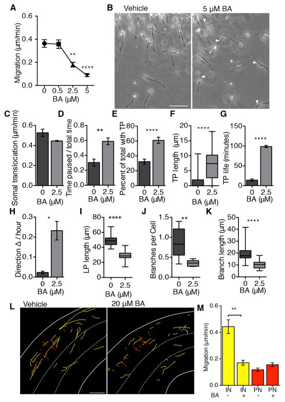
Mitochondrial dysfunction has been increasingly linked to neurodevelopmental disorders such as intellectual disability, childhood epilepsy, and autism spectrum disorder, conditions also associated with cortical GABAergic interneuron dysfunction. Although interneurons have some of the highest metabolic demands in the postnatal brain, the importance of mitochondria during interneuron development is unknown. We find that interneuron migration from the basal forebrain to the neocortex is highly sensitive to perturbations in oxidative phosphorylation. Both pharmacologic and genetic inhibition of adenine nucleotide transferase 1 (Ant1) disrupts the non-radial migration of interneurons, but not the radial migration of cortical projection neurons. The selective dependence of cortical interneuron migration on oxidative phosphorylation may be a mechanistic pathway upon which multiple developmental and metabolic pathologies converge.


Figures

Figure 1
Figure 1. Mitochondrial localization in migrating neurons
(A) Schemata of interneuron (IN; 1–3) morphologies displayed during migration. (B) Confocal immunofluorescence (IF) images of mitochondria in migrating INs in vitro displaying varying localization patterns according to morphology. Cytosol (GFP), mitochondria (Tom20), nuclei (DAPI). Scale bar = 10 μm. (C) Quantification of clustering of mitochondria in subcellular locations. Region I: trailing process (TP); II: overlapping nucleus (Nuc); III: 5 μm anterior to nucleus (5 μm AN); IV: cytoplasmic bleb (bleb); V: leading process (LP); VI: leading process tip (LPT). Clustering varied markedly between IN morphologies. IN morphology 1 clustered in III, *p = 0.039. IN morphology 2, clustered in IV, *p = 0.02, while IN morphology 3 clustered in I, and II, *p = 0.0343, ***p = 0.0002 Bars represent median with 25–75th percentiles ± min/max value of percent total mitochondrial area (%TMA) normalized to region’s percent of total cell area (%TCA). n = 15 cells each type; Freidman’s test with Dunn’s correction. (D) Schemata of pyramidal neuron (PN) migration morphology. (E) Confocal IF images of mitochondria in representative migrating PN. Cytosol (GFP), mitochondria (MitoDsRed), nuclei (DAPI). Scale bar = 10 μm. (F) Quantification of clustering mitochondria in migrating PNs. Region I: TP; II: Nuc; III: 5 μm AN; IV: cytoplasmic bleb (bleb); V: LP; VI: leading process tip LPT. *p = 0.0327, **p = 0.0064, ****p < 0.0001. n = 15 cells; Freidman’s test with Dunn’s correction. (G) Time-lapse imaging of a migrating Dlx5/6CIG IN in vitro (cytosol, GFP; mitochondria, Mitotracker® Red CMXRos) show intracellular movement of mitochondria. 1 frame=10 minutes. Scale bar = 10 μm. (See also Movie S2) (H) Quantification of INs displaying a extensive movement of mitochondrial through the cell versus those where the mitochondria remain confined to one compartment. The majority of INs showed this movement of mitochondria throughout the cell during migration. p < 0.0001, unpaired t-test, n = 5 independent cultures, 200 cells. Values represent mean ± SEM (I) Live time-lapse imaging of a migrating PN (cytosol, GFP; mitochondria, MitoDsRed). 1 frame = 10 minutes. Scale bar = 10 μm. (See also Movie S3) (J) Quantification of PNs movement of mitochondrial through the cell versus those where the mitochondria remain confined to one compartment. Intracellular movement of mitochondria through PNs was essentially not observed. p = 0.002, Mann-Whitney, n = 6 independent cultures, 82 cells. Values represent mean ± SEM.
Figure 2
Figure 2. Pharmacological inhibition of OXPHOS reduces IN migration
(A) MGE explants were cultured in high glucose +/− BA, an inhibitor of OXPHOS. Inhibiting OXPHOS decreased IN migration. (BA concentration: 0 μM and 0.5 μM = 0.36 ± 0.4; 2.5 μM = 0.18 ± 0.02, 5 μM = 0.09 ± 0.01; all in μm/min ± SEM; 0 vs. 2.5 or 5, p ≤ 0.001, n = 5 independent cultures, >150 cells each, ANOVA test). (B) Representative phase image of BA treated MGE INs. Arrows identify elongated trailing processes. Scale bar = 50 μm. (C) Somal translocation was calculated for each migrating IN, while slightly slower this did not reach significance. (BA concentration: 0 μM = 0.53 ± 0.04; 2.5 μM = 0.45 ± 0.01; all in μm/min ± SEM; p ≤ 0.075, n = 5 independent cultures, >150 cells each, unpaired t-test test, Welch’s correction) (D) The time a migrating neuron was paused versus moving was also calculated. In the presence of BA the neurons spend significantly more time paused compared to control cells. (BA concentration: 0 μM = 0.30 ± 0.05; 2.5 μM = 0.59 ± 0.05. All in total time paused/imaging period ± SEM; p ≤ 0.002, n = 5 independent cultures, >150 cells each, unpaired t-test test, Welch’s correction) (E) In addition to decreased migration, inhibition of OXPHOS increased the frequency (p <0.001, unpaired t-test with Welch’s correction, mean ± SEM), (F) length (p < 0.0001, Mann Whitney, median with interquartile range ± min/max), and (G) life of trailing processes (p < 0.0001, Mann-Whitney). For each, n = 5 independent cultures, 100 cells each. (H) OXPHOS inhibition also causes increases in direction changes (the mean number of direction changes/hour ± SEM was 0.024 ± 0.006 for 0 μM BA; and 0.23 ± 0.05 for 2.5 μM BA, p < 0.0001; n = 5 independent cultures, 100 cells each, unpaired t-test with Welch’s correction). (I–K) Leading process length, branches/cell, and brach length were all significantly reduced in migrating INs treated with BA when compared to controls (p < 0.0001, p < 0.001, p < 0.0001 respectively, Mann Whitney) (L) Sample migration paths of INs (yellow) and PNs (red) in E16 brain slices treated with vehicle or 20 μM BA (See also Movie S5). CP = Cortical plate, IZ = Intermediate Zone, VZ = Ventricular Zone. Scale bar = 150 μm. (M) IN migration rates decreased in slices treated with BA (p = 0.0023, n = 6 individuals, 20 INs each) whereas PNs were unaffected (p > 0.05, n = 6 individuals, ≥15 PNs in each). Unpaired t-test with Welch’s correction, mean ± SEM.
Figure 3
Figure 3. Interneuron migration is reduced in embryonic Ant1 mutants
(A) At E13.5 migrating INs (labeled by Calbindin immunohistochemistry) have not traveled as far in Ant1−/− brains compared to Ant+/+ brains. The white arrow indicates the leading migrating INs. Ant1+/+ served as the standardized control distance of 1.0 ± 0.02 with Ant1−/− IN migration showing on average 0.85 ± 0.02 relative distance units ± SEM or a 15% reduction, p < 0.0001. Scale bar = 200 μm. (B) Quantification of relative distance of leading cells of migrating INs. ****p < 0.0001, n = 5 individuals, 25 cells each, Mann-Whitney test. Values represent median with interquartile range ± min/max. (C) Quantification of relative INs in cortex; Ant1+/+ = 1.0 ± 0.06; Ant1−/− = 0.7 ± 0.04; normalized INs in cortex ± SEM, p = 0.004 **p = 0.004, n = 5, unpaired t-test with Welch’s correction. (D) Ant1−/− INs in the cortex have increased trailing process (TP) length. Ant1+/+: 1.1 μm ± 0.3; Ant1−/−: 5.2 μm ± 1.1 (± SEM), *p = 0.022, n = 5 individuals, 25 cells each, unpaired t-test with Welch’s correction. (E) Proliferation in germinal ventricular zone (VZ), indicated by Ki67 immunostaing was not impacted by loss of Ant1. Medial ganglionic eminence (MGE) and pallium (Ctx). Ant1+/+ vs. Ant1−/− for each region, p = 0.286 for MGE, p = 0.309 for Ctx; n = 5 individuals, unpaired t-test with Welch’s correction. (F) Loss of Ant1 did not increase cell death, indicated by Caspase 3, in the MGE and the ventral and dorsal pallium (V/D Ctx). Ant1+/+ vs. Ant1−/− for each region, p > 0.99, n = 5 individuals, Kruskal-Wallis, Dunn’s correction. Values represent median, 25th–75th percentile ± min/max (G) INs in cortex of Ant1−/− mice displayed abnormal leading process orientation. Arrowheads = misaligned INs. Scale bar = 75μm. (H) Quantification of cortical IN leading process orientation into quadrants: Q1, dorsal; Q2, pial; Q3, ventral; Q4, ventricular orientation. The average percent of IN oriented in Q1 for Ant1+/+ was 80 ± 4 and 50 ± 4; Ant1−/− (± SEM), ****p = 0.0001, **p = 0.0069, n = 5, ANOVA with Sidak’s correction
Figure 4
Figure 4. Abnormal migration by Ant1−/− interneurons
(A) MGE explants were cultured in vitro for 16 hrs. Ant1−/− INs did not migrate out of the explant as far as controls. (B) Quantification of relative distance of IN migration from explant showing a statistically significant difference in the distance migrated between Ant1−/− and Ant1−/− INs., Ant1+/+ is calculated as the normalized distance (1.0 ± 0.03) and Ant1−/− was on average approximately 40% reduced (0.61 ± 0.08); normalized distance ± SEM **p = 0.0034, n = 6, 50 cells each genotype, unpaired t-test with Welch’s correction. Scale bar = 250 μm. (C) Quantification of IN migration rates of wildtype and mutant INs. Ant1−/− IN migration rates had slower migration rates (****p < 0.0001), and were more sensitive to BA treatment compared to Ant1+/+(****p < 0.001). Migration rates Ant1+/+: 0.42 ± 0.01; Ant1−/−: 0.24 ± 0.01; μm ± SEM, n = 5, 150 cells each condition, ANOVA with Bonferonni’s correction. (D) Examples of migration path of GFP+ INs in slice culture (also see Movie S7). Dots = start; lines = paths. Scale bars, 150 μm. (E) Ant1−/− INs in slices display decreased migration rates relative to control. The relative migration rate was compared between Ant1+/+: 0.94 ± 0.04 and Ant1−/−: 0.65 ± 0.04; relative migration rate ± SEM, **** p < 0.0001, n = 5, unpaired t-test with Welch’s correction. (F) Ant1−/− INs display increased direction changes. Ant1+/+: 0.07 ± 0.01; Ant1−/−: 0.25 ± 0.02; direction changes per cell/hr ± SEM, p < 0.0001, n = 5, unpaired t-test with Welch’s correction. (G) Frequency plot of IN direction changes; dark grey = Ant1+/+, light grey = Ant1−/−. ****p < 0.0001, ***p = 0.0006, n = 5, two-way ANOVA with Sidak’s correction.
Figure 5
Figure 5. Loss of Ant1 causes shift in centrosome position
(A) Sample images of centrosome position in INs show mislocalized centrosome in Ant1−/− INs and BA treated INs (C) in vitro. Gamma-tubulin = red, nucleus = blue, cytoplasm = green. Scale bar = 15 μm. (B, D) Scatter plot of centrosome score of Ant−/− and BA treated INs (Grey lines = average nuclear length). Ant1+/+: 0.06 ± 0.01; Ant1−/−: 0.02 ± 0.01; centrosome score ± SEM; p = 0.0005, n= 75 cells each genotype, Mann-Whitney test, median with interquartile range ± min/max.

Similar articles

Cited by

References

    1. Adeva-Andany M, Lopez-Ojen M, Funcasta-Calderon R, Ameneiros-Rodriguez E, Donapetry-Garcia C, Vila-Altesor M, Rodriguez-Seijas J. Comprehensive review on lactate metabolism in human health. Mitochondrion. 2014;17C:76–100. - PubMed
    1. Bellion A, Baudoin JP, Alvarez C, Bornens M, Metin C. Nucleokinesis in tangentially migrating neurons comprises two alternating phases: forward migration of the Golgi/centrosome associated with centrosome splitting and myosin contraction at the rear. The Journal of neuroscience: the official journal of the Society for Neuroscience. 2005;25:5691–5699. - PMC - PubMed
    1. Benson DL, Watkins FH, Steward O, Banker G. Characterization of GABAergic neurons in hippocampal cell cultures. Journal of neurocytology. 1994;23:279–295. - PubMed
    1. Bertholet AM, Millet AM, Guillermin O, Daloyau M, Davezac N, Miquel MC, Belenguer P. OPA1 loss of function affects in vitro neuronal maturation. Brain: a journal of neurology. 2013;136:1518–1533. - PubMed
    1. Cheng A, Wan R, Yang JL, Kamimura N, Son TG, Ouyang X, Luo Y, Okun E, Mattson MP. Involvement of PGC-1alpha in the formation and maintenance of neuronal dendritic spines. Nature communications. 2012;3:1250. - PMC - PubMed

Publication types

Substances