. 2016 Apr 19;113(16):4320-5.
doi: 10.1073/pnas.1519858113. Epub 2016 Apr 5.

Metabolomics-assisted proteomics identifies succinylation and SIRT5 as important regulators of cardiac function

Affiliations

Metabolomics-assisted proteomics identifies succinylation and SIRT5 as important regulators of cardiac function

Sushabhan Sadhukhan et al. Proc Natl Acad Sci U S A. .

Abstract

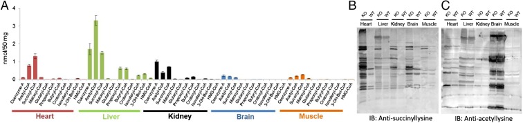
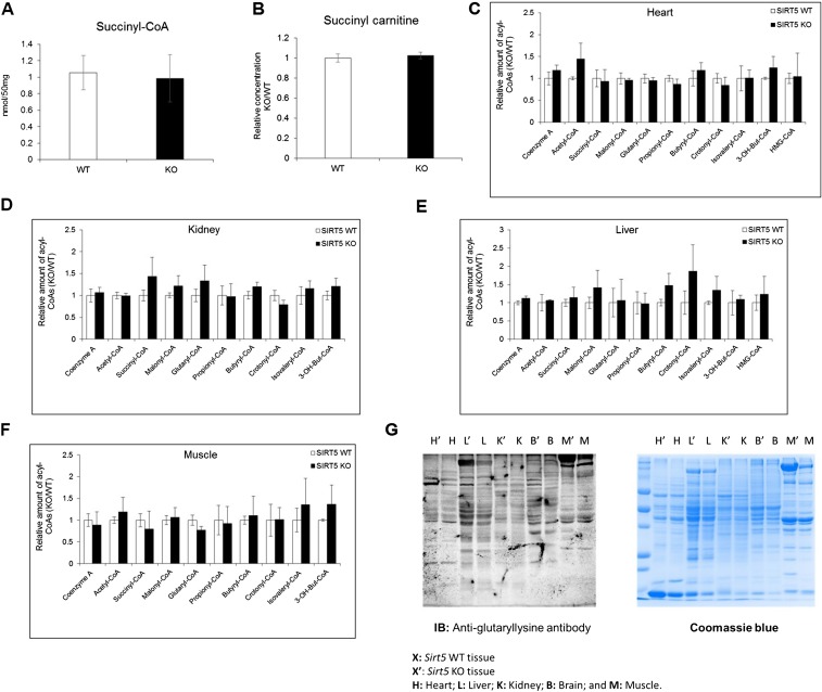
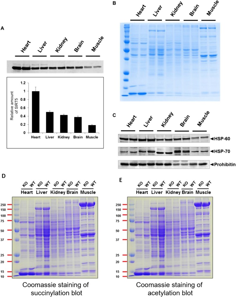
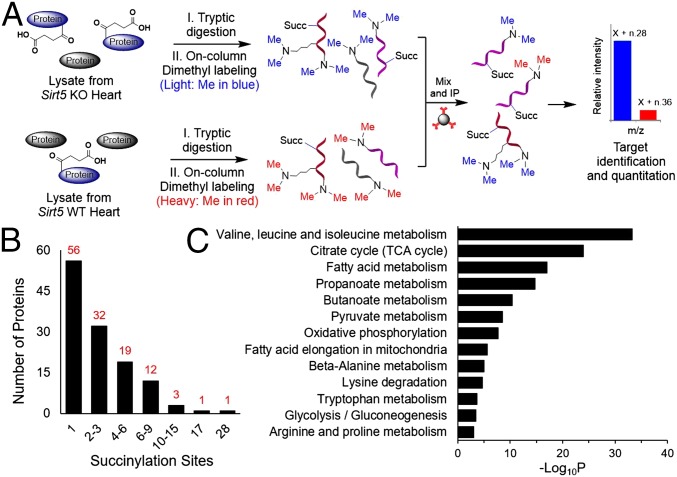
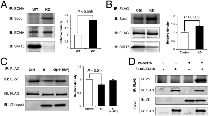
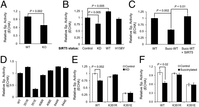
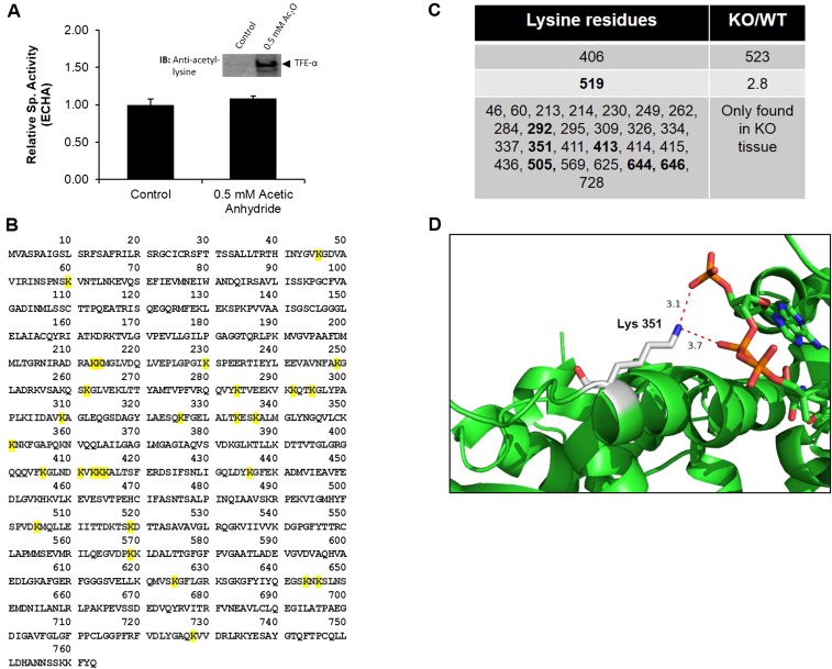
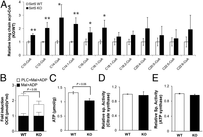
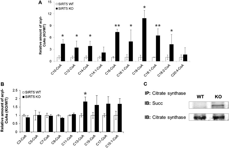
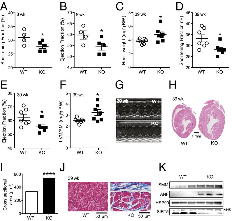
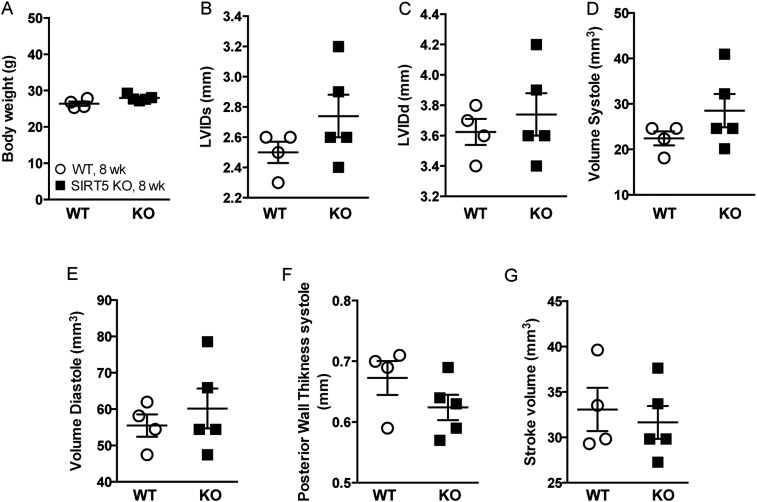
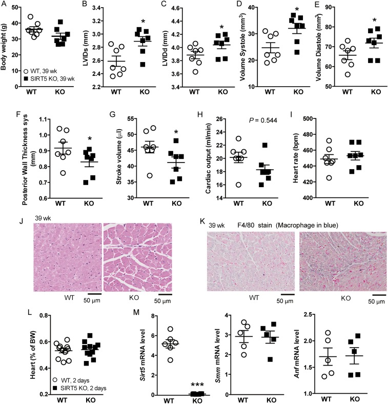
Cellular metabolites, such as acyl-CoA, can modify proteins, leading to protein posttranslational modifications (PTMs). One such PTM is lysine succinylation, which is regulated by sirtuin 5 (SIRT5). Although numerous proteins are modified by lysine succinylation, the physiological significance of lysine succinylation and SIRT5 remains elusive. Here, by profiling acyl-CoA molecules in various mouse tissues, we have discovered that different tissues have different acyl-CoA profiles and that succinyl-CoA is the most abundant acyl-CoA molecule in the heart. This interesting observation has prompted us to examine protein lysine succinylation in different mouse tissues in the presence and absence of SIRT5. Protein lysine succinylation predominantly accumulates in the heart whenSirt5is deleted. Using proteomic studies, we have identified many cardiac proteins regulated by SIRT5. Our data suggest that ECHA, a protein involved in fatty acid oxidation, is a major enzyme that is regulated by SIRT5 and affects heart function.Sirt5knockout (KO) mice have lower ECHA activity, increased long-chain acyl-CoAs, and decreased ATP in the heart under fasting conditions.Sirt5KO mice develop hypertrophic cardiomyopathy, as evident from the increased heart weight relative to body weight, as well as reduced shortening and ejection fractions. These findings establish that regulating heart metabolism and function is a major physiological function of lysine succinylation and SIRT5.

Keywords: desuccinylation; fatty acid metabolism; hypertrophic cardiomyopathy; lysine succinylation; sirtuin.


Conflict of interest statement

The authors declare no conflict of interest.

Figures

Fig. 1.
Fig. 1.
Protein lysine succinylation occurs to the greatest extent in the heart. (A) Profiling of short-chain CoAs among different tissues from Sirt5 WT mice using LC-MS/MS (mean ± SEM, n = 3 mice). (B and C) Western blot of different tissue lysates (25 μg each) against (B) antisuccinyllysine antibody and (C) against antiacetyllysine antibody. Sirt5 KO heart has the highest succinylation level. Coomassie-stained gels (loading control) are shown in Fig. S2 D and E.
Fig. S1.
Fig. S1.
(A) Concentration of succinyl-CoA in mouse hearts from WT and Sirt5 KO animals. (B) Relative level of succinyl-carnitine in mouse hearts from WT and Sirt5 KO animals. (CF) Relative level of short-chain acyl-CoAs in mouse heart, kidney, liver, and muscle from WT and Sirt5 KO animals after 30 min of endurance exercise. The metabolomics data are provided as mean ± SEM, n = 3 per genotype. (G) Western blot of different tissue lysates (25 μg each) from Sirt5 KO (designated by the prime sign) and WT mouse tissues against antiglutaryllysine antibody. B, brain; H, heart; K, kidney; L, liver; M, muscle.
Fig. S2.
Fig. S2.
(A) SIRT5 Western blot for different tissue lysates (25 μg each) from Sirt5 WT mouse tissues showing that the heart has the highest amount of SIRT5. The experiment was done with tissues from two different mice (duplicate). (B) Coomassie-stained gel showing equal loading of total protein. (C) Prohibitin, HSP-60, and HSP-70 were used as a loading control. Because the readings with different markers were not consistent (e.g., in the brain lysate the Hsp70 level was very high but prohibitin level was very low), we used the total protein amount to normalize the results in A. (D and E) Coomassie-stained gels (loading control) for Western blot of different tissue lysates (25 μg each) against antisuccinyllysine antibody (D) and antiacetyllysine antibody (E).
Fig. 2.
Fig. 2.
Workflow of the dimethyl-labeling strategy for the succinylome analysis. (A) One milligram of total protein from Sirt5 KO and WT heart was separately digested with trypsin and labeled with light and heavy dimethyl groups, respectively. The isotopically labeled peptides were mixed together and immunoprecipitated with antisuccinyllysine antibody. Succinyl-lysine peptides were then analyzed by nano LC-MS/MS. (B) Distribution of number of lysine succinylation sites per protein. (C) Metabolic pathways enriched with lysine succinylated proteins.
Fig. 3.
Fig. 3.
Lack of SIRT5 leads to hypersuccinylation on ECHA. (A) ECHA was immunoprecipitated from Sirt5 WT and KO mouse heart using ECHA-specific antibody. Sirt5 KO mouse heart had increased succinylation on endogenous ECHA. (B) Flag-ECHA expressed in HEK-293T Sirt5 KD cells showed increased succinylation compared with Flag-ECHA from control KD cells. (C) Overexpression of WT SIRT5, but not catalytically inactive SIRT5-H158Y, decreased the succinylation level of ECHA. Quantitative representation of relative density of succinylation (mean ± SEM, n = 3) is shown for AC. (D) Flag-ECHA and V5-tagged SIRT5 were co-overexpressed in HEK-293T cells. Immunoprecipitation of Flag-ECHA pulled down V5-tagged SIRT5.
Fig. 4.
Fig. 4.
SIRT5 increases ECHA activity by desuccinylation. (A) ECHA activity was higher in Sirt5 WT mouse hearts than in Sirt5 KO mice. (B) Flag-ECHA expressed in HEK-293T control, Sirt5 KD, and SIRT5 (WT or H158Y) overexpressing cells showed activities consistent with the hypothesis that SIRT5 increases ECHA activity by desuccinylation. (C) Recombinant ECHA and ECHB (coexpressed and purified in E. coli) could be nonenzymatically succinylated, which decreased the ECHA activity. Incubation with SIRT5 and NAD restored ECHA activity. (D) K351 is the only residue that decreases ECHA activity when mutated to E. (E) Neither K351R nor K351E show any change in activity when purified from HEK-293T control or Sirt5 KD cells. (F) Unlike WT, K351R and K351E mutant ECHA does not lose any additional activity when incubated with succinyl-CoA. Data shown as mean ± SEM, n = 3.
Fig. S3.
Fig. S3.
(A) Recombinant ECHA and ECHB complex was nonenzymatically acetylated with 0.5 mM acetic anhydride for 15 min at room temperature. Data are shown as mean ± SEM, n = 3. ECHA did not show any significant change in activity after chemical acetylation. (Inset) Western blot against antiacetyllysine antibody shows acetylation on ECHA after treating with 0.5 mM acetic anhydride. (B) Succinylated lysine residues of ECHA identified from our proteomics analysis are highlighted in yellow. (C) Fold change of succinyl-lysine–containing peptides (KO/WT) calculated from the peak area of the peptides. (D) The interactions between Lys351 ε-N (denoted by blue) and the negatively charged CoA phosphate groups (oxygen is denoted by red) are shown in the X-ray crystal structure of M. tuberculosis ECHA in complex with free CoA bound at the hydratase active sites (PDB ID code 4B3J). The figure was generated using PyMOL. Sequence alignment of ECHA showed Lys351 in mice is aligned with Ala-312 in M. tuberculosis. Hence, Ala-312 in ECHA of M. tuberculosis was mutated to Lys using PyMOL.
Fig. 5.
Fig. 5.
SIRT5 deficiency leads to accumulation of long-chain CoAs and decreased cardiac ATP levels. (A) Relative levels of long-chain CoA thioesters in Sirt5 KO hearts compared with WT (after 30 min of exercise, **P < 0.05, *P < 0.1). (B) Normalized fatty acid oxidation was significantly reduced in permeabilized Sirt5 KO heart tissue. Mitochondrial respiration in response to palmitoyl-l-carnitine (PLC) was monitored. Malate (2 mM) and ADP (2.5 mM) were used as a pretreatment. (C) Cardiac ATP levels were measured in Sirt5 WT and KO mice after 24 h of fasting. (D) Enzymatic activity of citrate synthase was measured in heart extracts from Sirt5 WT and KO mice. (E) Complex V activities were measured from Sirt5 WT and KO mice heart mitochondria. All data shown as mean ± SEM, n = 3 per genotype.
Fig. S4.
Fig. S4.
(A) Relative level of long-chain CoA thioesters in Sirt5 KO hearts compared with WT after 24 h of fasting (mean ± SEM, n = 3 per genotype, **P < 0.05, *P < 0.1). (B) Relative level of odd-chain acyl-CoAs in Sirt5 KO hearts compared with WT after 30 min of exercise (mean ± SEM, n = 3 per genotype, *P < 0.1). (C) Citrate synthase from Sirt5 KO mouse heart had increased succinylation.
Fig. 6.
Fig. 6.
SIRT5 deficiency causes hypertrophic cardiomyopathy. (A and B) Shortening fraction (A) and ejection fraction (B) were reduced in Sirt5 KO mice (n = 4 and 5 for Sirt5 WT and KO respectively, 8-week-old males). (C) Normalized heart weight of Sirt5 WT and KO male mice (n = 7 per genotype). (D–F) The shortening fraction (D), and ejection fraction (E) were significantly reduced whereas left ventricular mass to body mass (LVM/BM, F) was significantly increased in hearts of Sirt5 KO mice. (G) Representative M-mode images of echocardiography showing cardiac dysfunction in Sirt5 KO mice. (H and I) H&E staining of heart cross-sections (H) and quantification of cardiomyocytes cross-sectional areas (n = 100 per genotype, I) showing cardiac hypertrophy in the Sirt5 KO mice. The two black boxes in H indicate the localization of the images that are shown in larger magnification in Fig. S6J. (J) Masson’s trichrome stain in cross-sections of the heart showing increased fibrosis in Sirt5 KO hearts. (K) Evaluation of SMM, ANP, HSP90, and SIRT5 protein levels in Sirt5 WT and KO mouse hearts. Experiments in C–J were performed with hearts of 39-week-old male mice. All graphs shown as mean ± SEM, *P < 0.05, ****P < 0.0001.
Fig. S5.
Fig. S5.
(A) Body weight of Sirt5 WT and KO male mice (n = 4–5 per genotype, 8-wk-old males). (B) Left ventricular internal dimension in systole (LVIDs), (C) left ventricular internal dimension in diastole (LVIDd), (D) volume in systole, (E) volume in systole, (F) posterior wall thickness, and (G) stroke volume did not altered significantly in Sirt5 KO male mice. All graphs are shown as mean ± SEM.
Fig. S6.
Fig. S6.
(A) Body weight of Sirt5 WT and KO male mice (n = 7 per genotype, 39-wk-old males). (BH) Left ventricular internal dimension in systole (LVIDs, B), left ventricular internal dimension in diastole (LVIDd, C), and volume in systole (D) and diastole (E) were significantly increased whereas posterior wall thickness (F), stroke volume (G), and cardiac output (H) were reduced in Sirt5 KO male mice. (I) Heart rate did not show clear differences between the genotypes. (J) Representative zoomed-in (black boxes in Fig. 6H) image of H&E staining of heart cross-sections (39-wk-old males). (K) F4/80 staining in cross-sectional heart showing increased macrophage infiltration in Sirt5 KO heart (39-wk-old males). (L) Normalized heart weight of WT and Sirt5 KO pups (n = 11 per genotypes, 2-d-old pups) does not show any difference. (M) mRNA levels of Sirt5, Smm, and Anf as markers of cardiac dysfunction were evaluated by qRT-PCR (n = 5 per genotype, 2-d-old pups). All graphs are shown as mean ± SEM, ***P < 0.001, *P < 0.05.

References

    1. Du J, et al. Sirt5 is a NAD-dependent protein lysine demalonylase and desuccinylase. Science. 2011;334(6057):806–809. - PMC - PubMed
    1. Lin H, Su X, He B. Protein lysine acylation and cysteine succination by intermediates of energy metabolism. ACS Chem Biol. 2012;7(6):947–960. - PMC - PubMed
    1. Tan M, et al. Identification of 67 histone marks and histone lysine crotonylation as a new type of histone modification. Cell. 2011;146(6):1016–1028. - PMC - PubMed
    1. Zhang Z, et al. Identification of lysine succinylation as a new post-translational modification. Nat Chem Biol. 2011;7(1):58–63. - PMC - PubMed
    1. Chen Y, et al. Lysine propionylation and butyrylation are novel post-translational modifications in histones. Mol Cell Proteomics. 2007;6(5):812–819. - PMC - PubMed

Publication types