Case Reports
. 2016 Apr;31(4):635-40.
doi: 10.3346/jkms.2016.31.4.635. Epub 2016 Mar 10.

A Case Report of a Middle East Respiratory Syndrome Survivor with Kidney Biopsy Results

Affiliations
Case Reports

A Case Report of a Middle East Respiratory Syndrome Survivor with Kidney Biopsy Results

Ran-Hui Cha et al. J Korean Med Sci. 2016 Apr.

Abstract

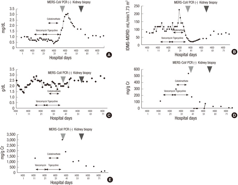
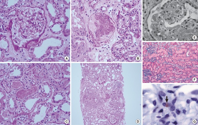
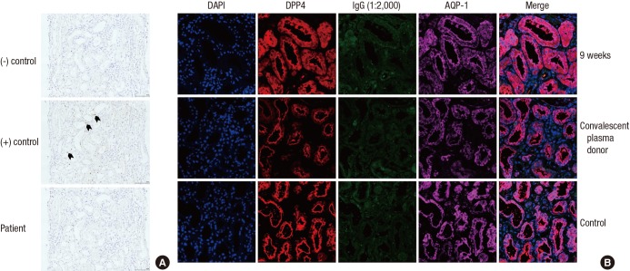
A 68-year old man diagnosed with Middle East Respiratory Syndrome-Coronavirus (MERS-CoV) presented with multiple pneumonic infiltrations on his chest X-ray, and the patient was placed on a mechanical ventilator because of progressive respiratory failure. Urinary protein excretion steadily increased for a microalbumin to creatinine ratio of 538.4 mg/g Cr and a protein to creatinine ratio of 3,025.8 mg/g Cr. The isotope dilution mass spectrometry traceable serum creatinine level increased to 3.0 mg/dL. We performed a kidney biopsy 8 weeks after the onset of symptoms. Acute tubular necrosis was the main finding, and proteinaceous cast formation and acute tubulointerstitial nephritis were found. There were no electron dense deposits observed with electron microscopy. We could not verify the virus itself by in situ hybridization and confocal microscopy (MERS-CoV co-stained with dipeptidyl peptidase 4). The viremic status, urinary virus excretion, and timely kidney biopsy results should be investigated with thorough precautions to reveal the direct effects of MERS-CoV with respect to renal complications.

Keywords: Acute Tubulointerstitial Nephritis; Kidney Tubular Necrosis, Acute; Middle East Respiratory Syndrome-Coronavirus; Renal Pathology.


Conflict of interest statement

DISCLOSURE: The authors have no potential conflicts of interest to disclose.

Figures

Fig. 1
Fig. 1
The progression of renal function, albumin, and urinary protein excretion. (A, B) The IDMS traceable serum creatinine and IDMS-MDRD estimated glomerular filtration rate. (C) The progression of serum albumin level. (D, E) The microalbumin to creatinine ratio and protein to creatinine ratio from randomly collected urine. The polymerase chain reaction of Middle East Respiratory Syndrome-Coronavirus from the respiratory tract specimens was negative on hospital day 37; the patient underwent a kidney biopsy on hospital day 55. IDMS, isotope dilution mass spectrometry; HOD, hospital day; MDRD, modification of diet in renal disease.
Fig. 2
Fig. 2
Light microscopic findings of kidney tissues (H&E staining) (17,18). (A) Glomerular pathology was not observed. (B) Acute tubular necrosis showing denuded tubular epithelial cells. (C) Some tubules showed protenaceous cast formation. (D) Acute tubulointerstitial nephritis showing numerous inflammatory cells in tubules and the interstitium (A, B, C: magnification ☓ 200, D: magnification ☓ 40). (E) Proximal tubules with vacuolar degenerative alterations (magnification ☓ 400) (17). (F) Moderate necrosis and detachment of tubular epithelium (magnification ☓ 100) (18). (G) Viral antigen detected in the glomerular macrophage (arrow) (magnification ☓ 1000) (18).
Fig. 3
Fig. 3
The detection of Middle East Respiratory Syndrome-Coronavirus: in situ hybridization and confocal microscopy. (A) Middle East Respiratory Syndrome-Coronavirus (MERS-CoV) was not evident in the in-situ hybridization method (magnification ☓ 200). The first and the second rows indicate negative and positive control (black arrow). The third row shows results from patients. There was no brown punctuate dot in the nucleus and/or cytoplasm. (B) The kidney tissue samples were co-stained for MERS-CoV (human IgG; green) and dipeptidyl peptidase 4 (DPP 4; red). DPP 4 was primarily stained in proximal tubules (aquaporin-1; violet). The first and second rows indicate the reaction using patient serum 9 weeks after symptom onset and the serum from a convalescent plasma donor. The third row is the negative control.

References

    1. Memish ZA, Zumla AI, Al-Hakeem RF, Al-Rabeeah AA, Stephens GM. Family cluster of Middle East respiratory syndrome coronavirus infections. N Engl J Med. 2013;368:2487–2494. - PubMed
    1. Guery B, Poissy J, el Mansouf L, Séjourné C, Ettahar N, Lemaire X, Vuotto F, Goffard A, Behillil S, Enouf V, et al. MERS-CoV study group Clinical features and viral diagnosis of two cases of infection with Middle East Respiratory Syndrome coronavirus: a report of nosocomial transmission. Lancet. 2013;381:2265–2272. - PMC - PubMed
    1. Zaki AM, van Boheemen S, Bestebroer TM, Osterhaus AD, Fouchier RA. Isolation of a novel coronavirus from a man with pneumonia in Saudi Arabia. N Engl J Med. 2012;367:1814–1820. - PubMed
    1. Poissy J, Goffard A, Parmentier-Decrucq E, Favory R, Kauv M, Kipnis E, Mathieu D, van der Werf S, Guery B, MERS-CoV Biology Group Kinetics and pattern of viral excretion in biological specimens of two MERS-CoV cases. J Clin Virol. 2014;61:275–278. - PMC - PubMed
    1. Raj VS, Mou H, Smits SL, Dekkers DH, Müller MA, Dijkman R, Muth D, Demmers JA, Zaki A, Fouchier RA, et al. Dipeptidyl peptidase 4 is a functional receptor for the emerging human coronavirus-EMC. Nature. 2013;495:251–254. - PMC - PubMed

Publication types

MeSH terms