

Time-dependent low-field MRI characteristics of canine blood: an in vitro study
- PMID: 27051346
- PMCID: PMC4808634
- DOI: 10.4142/jvs.2016.17.1.103
Time-dependent low-field MRI characteristics of canine blood: an in vitro study
Abstract
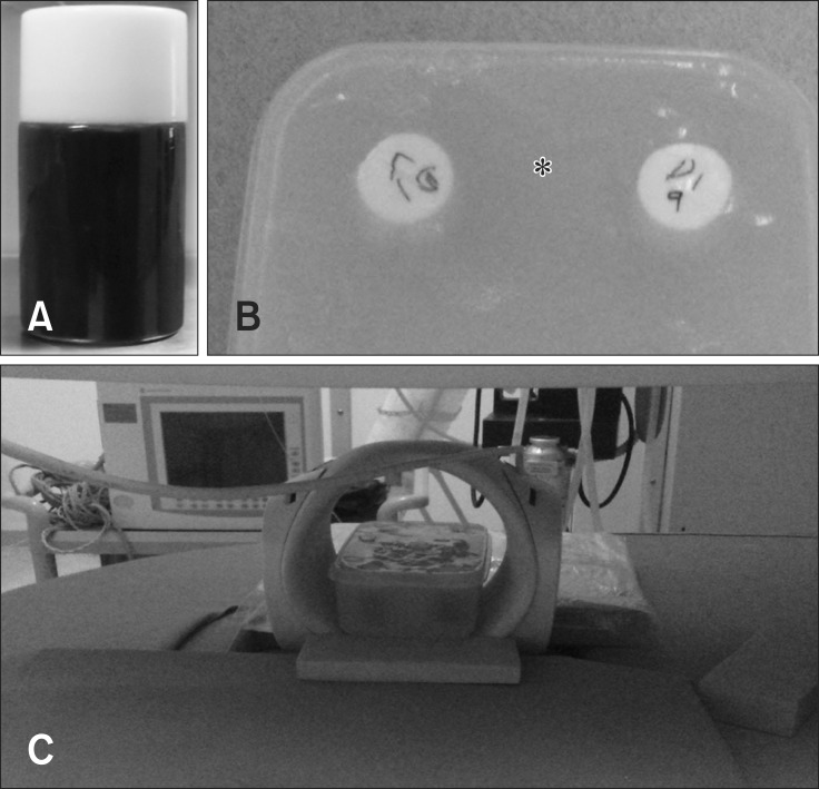
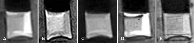
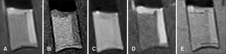
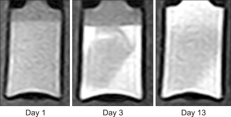
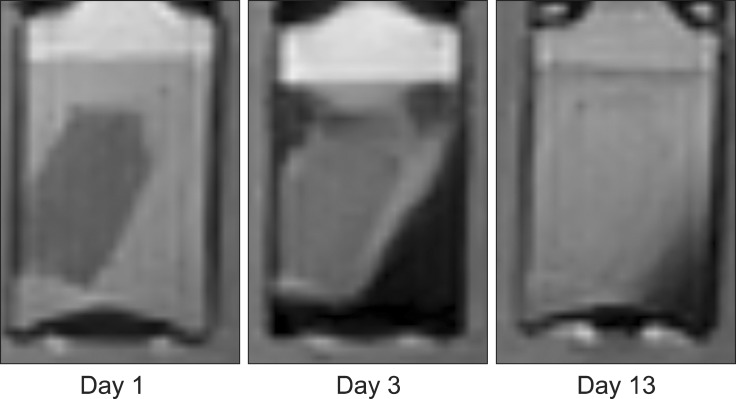
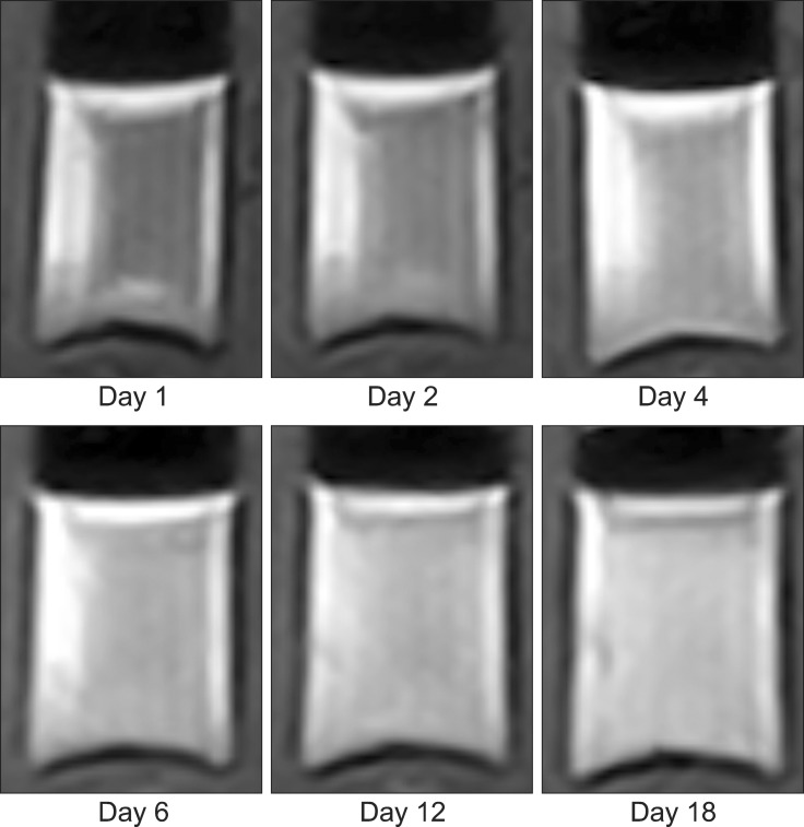
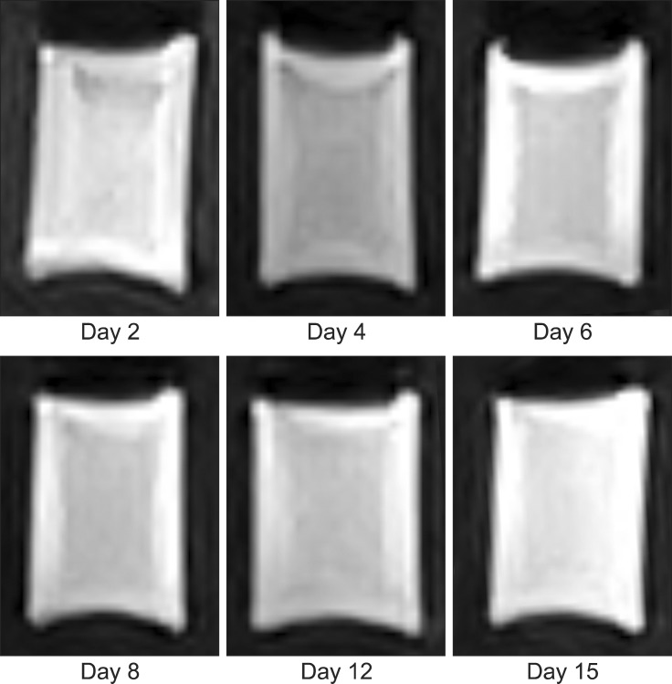
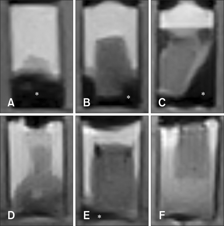
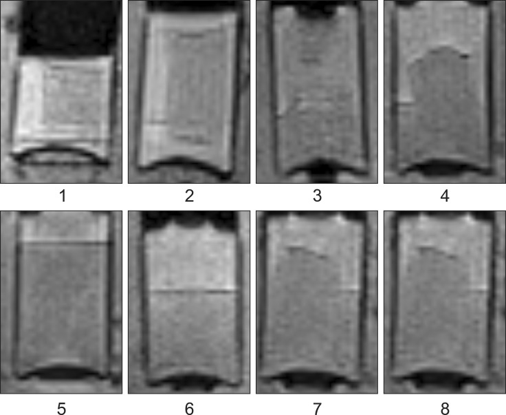
This study was conducted to assess time-sensitive magnetic resonance (MR) changes in canine blood using low-field MR. Arterial and venous blood samples were collected from eight healthy beagle dogs. Samples were placed in 5-mL tubes and imaged within 3 hours of collection at 1 day intervals from day 1 to day 30. The following sequences were used: T1-weighted (T1W), T2-weighted (T2W), fluid-attenuated inversion recovery (FLAIR), short tau inversion recovery (STIR), and T2-star gradient-echo (T2(*)-GRE). Visual comparison of the images revealed that four relatively homogenous blood clots and twelve heterogeneous blood clots developed. The margination of the clot and plasma changed significantly on day 2 and day 13. On day 2, heterogeneous blood clots were differentiated into 2 to 3 signal layers in the T2W, T1W, and especially the STIR images. Hypointense signal layers were also detected in the blood clots in STIR images, which have T2 hypo, FLAIR hypo, and T1 hyper intense signals. In all images, these signal layers remained relatively unchanged until day 13. Overall, the results suggest that hematomas are complex on low-field MRI. Accordingly, it may not be feasible to accurately characterize hemorrhages and predict clot age based on low-field MRI.
Keywords: blood; canine; in vitro; low field; magnetic resonance.
Conflict of interest statement
Figures
Similar articles
-
Chronological low field magnetic resonance appearance of canine spinal epidural hemorrhage model.J Vet Sci. 2019 Mar;20(2):e7. doi: 10.4142/jvs.2019.20.e7. Epub 2019 Mar 8. J Vet Sci. 2019. PMID: 30944530 Free PMC article.
-
Differentiation of white and red thrombus with magnetic resonance imaging: a phantom study.Chin Med J (Engl). 2012 Jun;125(11):1889-92. Chin Med J (Engl). 2012. PMID: 22884048
-
MRI of clot in cerebral venous thrombosis: high diagnostic value of susceptibility-weighted images.Stroke. 2006 Apr;37(4):991-5. doi: 10.1161/01.STR.0000206282.85610.ae. Epub 2006 Feb 16. Stroke. 2006. PMID: 16484607
-
Magnetic resonance imaging in dogs with neuroangiostrongyliasis (rat lungworm disease).Parasitology. 2021 Feb;148(2):198-205. doi: 10.1017/S0031182020001742. Epub 2020 Sep 21. Parasitology. 2021. PMID: 32951620 Free PMC article.
-
[Diagnostic imaging of hemangiomas in the brain].Brain Nerve. 2011 Jan;63(1):5-15. Brain Nerve. 2011. PMID: 21228443 Review. Japanese.
Cited by
-
MRI Findings of Early-Stage Hyperacute Hemorrhage Causing Extramedullary Compression of the Cervical Spinal Cord in a Dog with Suspected Steroid-Responsive Meningitis-Arteritis.Front Vet Sci. 2017 Sep 27;4:161. doi: 10.3389/fvets.2017.00161. eCollection 2017. Front Vet Sci. 2017. PMID: 29021984 Free PMC article.
-
Advanced Hemophilic Arthropathy: Sensitivity of Soft Tissue Discrimination With Musculoskeletal Ultrasound.J Ultrasound Med. 2018 Aug;37(8):1945-1956. doi: 10.1002/jum.14541. Epub 2018 Jan 24. J Ultrasound Med. 2018. PMID: 29363781 Free PMC article.
-
Musculoskeletal ultrasound for intra-articular bleed detection: a highly sensitive imaging modality compared with conventional magnetic resonance imaging.J Thromb Haemost. 2018 Mar;16(3):490-499. doi: 10.1111/jth.13930. Epub 2018 Jan 19. J Thromb Haemost. 2018. PMID: 29274196 Free PMC article.
References
-
- Atlas SW, Mark AS, Grossman RI, Gomori JM. Intracranial hemorrhage: gradient-echo MR imaging at 1.5 T. Comparison with spin-echo imaging and clinical applications. Radiology. 1988;168:803–807. - PubMed
-
- Bellon EM, Haacke EM, Coleman PE, Sacco DC, Steiger DA, Gangarosa RE. MR artifacts: a review. AJR Am Roentgenol. 1986;147:1271–1281. - PubMed
-
- Bradley WG., Jr MR appearance of hemorrhage in the brain. Radiology. 1993;189:15–26. - PubMed
-
- Brooks RA, Di Chiro G, Patronas N. MR imaging of cerebral hematomas at different field strengths: theory and applications. J Comput Assist Tomogr. 1989;13:194–206. - PubMed
Publication types
MeSH terms
LinkOut - more resources
Full Text Sources
Other Literature Sources
Medical
