

Fossa navicularis magna detection on cone-beam computed tomography
- PMID: 27051639
- PMCID: PMC4816771
- DOI: 10.5624/isd.2016.46.1.47
Fossa navicularis magna detection on cone-beam computed tomography
Abstract
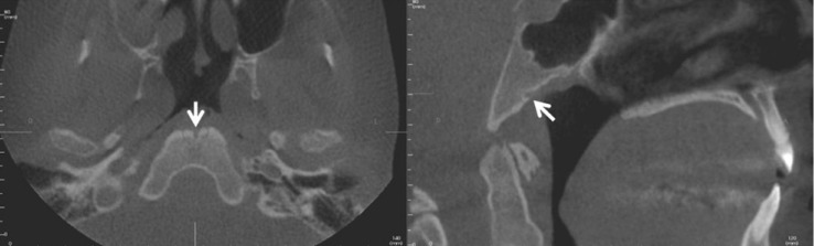
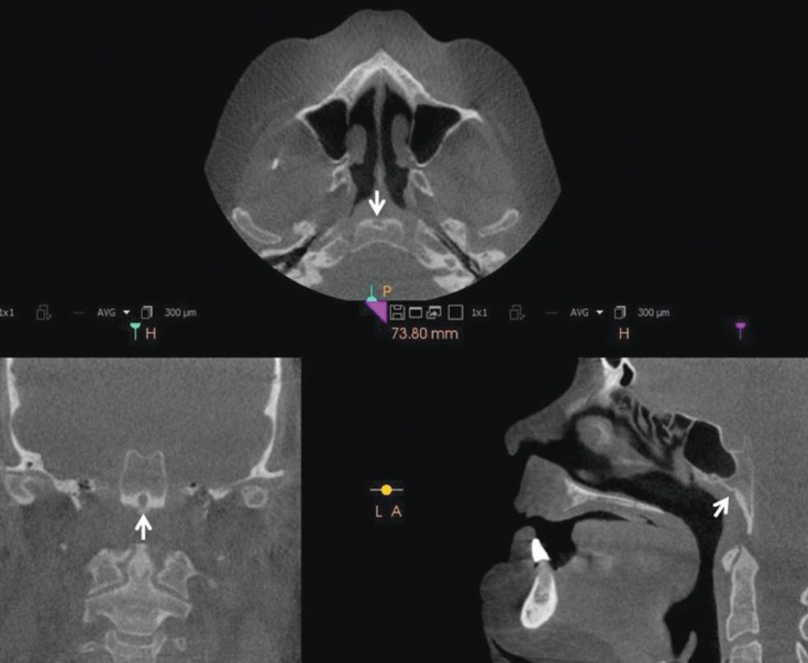
Herein, we report and discuss the detection of fossa navicularis magna, a close radiographic anatomic variant of canalis basilaris medianus of the basiocciput, as an incidental finding in cone-beam computed tomography (CBCT) imaging. The CBCT data of the patients in question were referred for the evaluation of implant sites and to rule out pathology in the maxilla and mandible. CBCT analysis showed osseous, notch-like defects on the inferior aspect of the clivus in all four cases. The appearance of fossa navicularis magna varied among the cases. In some, it was completely within the basiocciput and mimicked a small rounded, corticated, lytic defect, whereas it appeared as a notch in others. Fossa navicularis magna is an anatomical variant that occurs on the inferior aspect of the clivus. The pertinent literature on the anatomical variations occurring in this region was reviewed.
Keywords: Clivus Fossa, Posterior; Cone-Beam Computed Tomography; Multidetector Computed Tomography; Skull Base.
Figures


Similar articles
-
Evaluation of canalis basilaris medianus using cone-beam computed tomography.Imaging Sci Dent. 2016 Jun;46(2):141-4. doi: 10.5624/isd.2016.46.2.141. Epub 2016 Jun 23. Imaging Sci Dent. 2016. PMID: 27358822 Free PMC article.
-
Prevalence and morphometric features of fossa navicularis on cone beam computed tomography in Turkish population.Folia Morphol (Warsz). 2017;76(4):715-719. doi: 10.5603/FM.a2017.0030. Epub 2017 Mar 29. Folia Morphol (Warsz). 2017. PMID: 28353302
-
Prevalence of anatomical variants in the clivus: fossa navicularis magna, canalis basilaris medianus, and craniopharyngeal canal.Surg Radiol Anat. 2019 Apr;41(4):477-483. doi: 10.1007/s00276-019-02200-3. Epub 2019 Feb 6. Surg Radiol Anat. 2019. PMID: 30725217
-
Anomalies of the clivus of interest in dental practice: A systematic review.Imaging Sci Dent. 2021 Dec;51(4):351-361. doi: 10.5624/isd.20210039. Epub 2021 Aug 11. Imaging Sci Dent. 2021. PMID: 34987995 Free PMC article. Review.
-
Intracranial infection caused by spreading through the fossa naviclaris magna - a case report and review of the literature.Int J Pediatr Otorhinolaryngol. 2013 Dec;77(12):1919-21. doi: 10.1016/j.ijporl.2013.09.013. Epub 2013 Sep 23. Int J Pediatr Otorhinolaryngol. 2013. PMID: 24148862 Review.
Cited by
-
The First Histological Analysis of the Tissues Lining the Fossa Navicularis: Insights to its Etiology.Cureus. 2017 May 31;9(5):e1299. doi: 10.7759/cureus.1299. Cureus. 2017. PMID: 28680787 Free PMC article.
-
Rhinolith: An incidental radiographic finding.Imaging Sci Dent. 2021 Sep;51(3):333-336. doi: 10.5624/isd.20200126. Epub 2021 Aug 11. Imaging Sci Dent. 2021. PMID: 34621662 Free PMC article.
-
Clival canal and clival foramen development in the fetal and infant basioccipital.Childs Nerv Syst. 2017 Jul;33(7):1209-1216. doi: 10.1007/s00381-017-3466-2. Epub 2017 May 24. Childs Nerv Syst. 2017. PMID: 28540612 Free PMC article.
-
Investigation of the prevalence and main features of skull-base anomalies and characteristics of the sphenoid sinus using cone-beam computed tomography.J Korean Assoc Oral Maxillofac Surg. 2022 Aug 31;48(4):207-218. doi: 10.5125/jkaoms.2022.48.4.207. J Korean Assoc Oral Maxillofac Surg. 2022. PMID: 36043251 Free PMC article.
-
Morphological Measurement and Anatomical Variations of the Clivus Using Computed Tomography.J Neurol Surg B Skull Base. 2021 Feb 18;83(Suppl 2):e75-e82. doi: 10.1055/s-0040-1722712. eCollection 2022 Jun. J Neurol Surg B Skull Base. 2021. PMID: 35833004 Free PMC article.
References
-
- Neelakantan A, Rana AK. Benign and malignant diseases of the clivus. Clin Radiol. 2014;69:1295–1303. - PubMed
-
- Shah A, Goel A. Clival dysgenesis associated with Chiari Type 1 malformation and syringomyelia. J Clin Neurosci. 2010;17:400–401. - PubMed
-
- Segal N, Atamne E, Shelef I, Zamir S, Landau D. Intracranial infection caused by spreading through the fossa naviclaris magna - a case report and review of the literature. Int J Pediatr Otorhinolaryngol. 2013;77:1919–1921. - PubMed
-
- Prabhu SP, Zinkus T, Cheng AG, Rahbar R. Clival osteomyelitis resulting from spread of infection through the fossa navicularis magna in a child. Pediatr Radiol. 2009;39:995–998. - PubMed
-
- Collins HB., Jr Frequency and distribution of fossa pharyngea in human crania. Am J Phys Anthropol. 1927;11:101–106.
Publication types
LinkOut - more resources
Full Text Sources
Other Literature Sources
