. 2016 Apr 7:6:24048.
doi: 10.1038/srep24048.

RXR controlled regulatory networks identified in mouse brain counteract deleterious effects of Aβ oligomers

Affiliations

RXR controlled regulatory networks identified in mouse brain counteract deleterious effects of Aβ oligomers

Kyong Nyon Nam et al. Sci Rep. .

Abstract

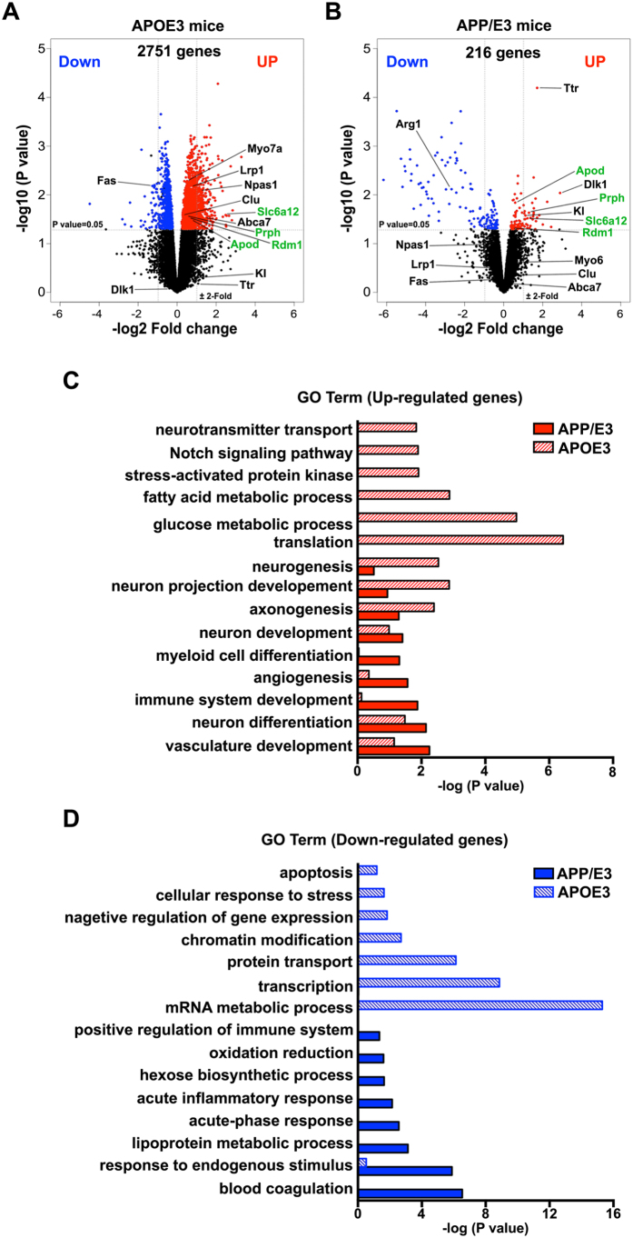
Bexarotene, a selective agonist for Retinoid X receptors (RXR) improves cognitive deficits and amyloid-β (Aβ) clearance in mice. Here we examine if the effect of bexarotene on RXR cistrome and transcriptomes depend on APOE isoform and Aβ deposition. We found bexarotene increased RXR binding to promoter regions in cortex of APOE3 mice. Rho family GTPases and Wnt signaling pathway were highly enriched in ChIP-seq and RNA-seq datasets and members of those pathways - Lrp1, Lrp5, Sfrp5 and Sema3f were validated. The effect of APOE isoform was compared in APOE3 and APOE4 mice and we found significant overlapping in affected pathways. ChIP-seq using mouse embryonic stem cells and enrichment levels of histone marks H3K4me3 and H3K27me3 revealed that, bexarotene induced epigenetic changes, consistent with increased neuronal differentiation and in correlation with changes in transcription. Comparison of transcriptome in APOE3 and APP/APOE3 mice revealed that amyloid deposition significantly affects the response to bexarotene. In primary neurons, bexarotene ameliorated the damaged dendrite complexity and loss of neurites caused by Aβ42. Finally, we show that the disruption of actin cytoskeleton induced by Aβ42 in vitro was inhibited by bexarotene treatment. Our results suggest a mechanism to establish RXR therapeutic targets with significance in neurodegeneration.


Conflict of interest statement

The authors declare no competing financial interests.

Figures

Figure 1
Figure 1. ChIP-seq data reveal changes in the distribution of RXR binding in APOE3 mice brain induced by bexarotene treatment.
APOE3 mice were treated with bexarotene or vehicle as described in Materials & Methods and ChIP-seq was performed using material from cortex. (A) Venn diagram showing the number of overlapping peaks in bexarotene (Bexa) and vehicle (Veh) treated APOE3 mice (Blue: Veh, Red: Bexa); (B) Distribution of RXR binding showing significant shift towards RXR binding in promoter regions of annotated genes in bexarotene versus vehicle treated mice. (C) Most significant functional annotation categories (GO Term Biological Process, DAVID); (D) ChIP-qPCR validation data of genes with enriched RXR binding in promoter regions. Fasn gene is used as a positive control. (E) Comparative heatmap of RXR cistrome of bexarotene treated APOE3 and APOE4 mice Shown are the most significant distinct Biological Function categories identified by IPA in each experiment. Blue represents vehicle and red represents bexarotene. Gray boxes indicate no overlapping categories. Negative log10 (P value) denotes lower significance and red color higher significance.
Figure 2
Figure 2. Bexarotene treatment significantly affects genes related to nervous system development, axon guidance and neurites extension.
RNA was isolated from cortices of the same APOE3 mice treated with bexarotene or vehicle as shown on Fig. 1. (A) Scatter plot of transcripts in vehicle and bexarotene treated APOE3 mice. Transcripts identified as significantly differentially expressed at p < 0.05 cut-off are colored in blue (down-regulated) and red (up-regulated). Up-regulated transcripts associated with neurite extension are shown in green, and down-regulated genes associated with inflammation in orange. Gene symbols of representative transcripts are shown next to the corresponding values. (B) Most significant common canonical pathways identified by ChIP-seq and RNA-seq in APOE3 mice; (C) Significantly changed levels of RNA expression of genes in categories “neuron differentiation” and “neuron projection”; statistics is by edgeR, p < 0.05, n = 4/group; (D) Validation of RNA-seq results by qPCR; APOE3 mice, n = 4/group; (E) Validation by qPCR in N2a cells: Cells were treated with 1 μM bexarotene or vehicle for 24 hours and 48 hours (details in Materials & Methods). Values are mean ± SEM. For (D,E) statistics is by t-test; n = 3, *p < 0.05, **p < 0.01, ***p <  0.001.
Figure 3
Figure 3. Bexarotene induces epigenetic changes in correlation with neuronal differentiation in ES cells.
ES cells were treated with 5 μM Bexarotene for 4 days and markers for neuronal differentiation were examined 6 days after plating (Day10, Neurons). Control cells were treated with vehicle and processed in the same way. (A) Outline of the protocol for ES cell treatment and differentiation. EBs, embryoid Bodies; (B) Genome browser view of lineage markers in ES cells for H3K4me3 identified by ChIP-seq at EBs stage or 4 days after bexarotene (Bexa, Day4) and vehicle (Veh, Day4) treatment. Note that for pluripotency markers Nanog and Oct4/Pou5f1 data are compared at EBs vs Bexa, Day4, and neuronal markers Pax6 and Syp are compared 4 days after bexarotene or vehicle treatment. (C) Bexarotene treatment increases the expression of Pax6 and Syp; (D) Comparative heatmap of significantly changed GO categories (gene lists derived from edgeR output tables) in APOE3 and APOE4 mice and epigenetic changes (analyzed ChIP-seq datasets) in bexarotene or vehicle treated ES cells (Day 4). Shown are the most significant distinct BP categories as determined by David in each experiment. Blue color indicates up-regulated genes and red indicates down-regulated genes in RNA-seq. For ChIP-seq data, Blue represents vehicle and red represents bexarotene. Gray boxes indicate no overlapping categories. Negative log10 (P value) denotes lower significance and red color higher significance; (E) Validation of RNA-seq results by qPCR in ES cells; Cells were treated with 5 μM bexarotene or vehicle for different time (24 h, Day 4 and Day 10). Values are mean ± SEM and statistics is by t-test; n = 3, *p < 0.05, **p < 0.01, ***p < 0.001.
Figure 4
Figure 4. Aβ deposition differentially affects transcriptome changes in response to bexarotene in APOE3 and APP/E3 mice.
Six month old mice of both APOE3 and APP/E3 genotypes were treated with bexarotene or vehicle (n = 4 for each condition) and RNA-seq datasets were used to determine differential gene expression. (A,B) Volcano plots for comparing transcripts in vehicle and bexarotene treated APOE3 and APP/E3 mice. Transcripts identified as significantly differentially expressed at P < 0.05 are colored in blue (down-regulated) and red (up-regulated). Gene symbols of representative genes associated with Aβ clearance and phagocytosis are indicated for both genotypes. The gene symbols in green denote common genes in both APOE3 and APP/E3 mice. (C,D) Comparison of the most significant distinct functional categories (GO Term Biological process) of differentially affected genes in both APOE3 and APP/E3 mice as identified by DAVID. (C) Up-regulated genes and (D) Down-regulated. Solid bar indicates APP/E3 mice and pattern bar indicates APOE3 mice.
Figure 5
Figure 5. Bexarotene treatment maintains dendrite complexity and prevents loss of neurites in primary neurons exposed to Aβ.
Mouse primary cortical neurons were infected with GFP expressing lentivirus at DIV0. DIV14 cells were pre-treated with 1 μM bexarotene or vehicle for 24 hours followed by 0.5 μM Aβ42 for 24 hours. (A) Digital reconstructed images of Aβ or Bexa followed by Aβ treated cells, respectively; (B) Number of Intersections. Statistics is by two-way repeated measures ANOVA with number of intersections as an independent variable, treatment group as a factor, and distance from the soma as the repeated-measure factor. There is an interaction F(171,2280) = 6.23, p < 0.0001. Total neurite length (C), Number of Bifurcations (D) and Number of branches (E) were significantly increased in the neurons treated with bexarotene. Statistics for (C–E) is by one-way ANOVA. For each treatment group, images were taken from at least 4 wells and of more than 2 GFP+ neurons per well. (F) Primary neurons were treated for 6 or 24 hours with bexarotene and mRNA level for selected genes related to categories “neuron projections” and “Wnt signaling” were examined by qPCR. Values are mean ± SEM; Statistics is by t-test; n = 3, *p < 0.05, **p < 0.01 versus vehicle group.
Figure 6
Figure 6. Bexarotene inhibits disruption of actin cytoskeleton caused by Aβ42 in N2a cells.
N2a cells were treated with vehicle or 0.5 μM bexarotene for 24 hours and then treated with 0.5 μM Aβ42 for 24 hours. Cells were stained with alexa 488 phalloidin for visualizing F-actin puncta. (A) Representative images of phalloidin staining. The white arrows point intracellular accumulation of F-actin puncta. (B) The Quantification of F-actin puncta formation. The percentage of cells with F-actin puncta formation was calculated for each condition from merged DAPI and phalloidin images from 9 pictures per condition. Values are mean ± SEM and statistics is by one-way ANOVA followed by Tukey’s post-test; n = 9, *p < 0.05, ***p < 0.001. (C) N2a cells were treated as shown on A and B and mRNA level for selected RXR targets was examined by qPCR. Values are mean ± SEM; Statistics is by t-test; n = 3, *p < 0.05, **p < 0.01 versus vehicle group; ##p < 0.01, ###p < 0.001 versus Aβ group.

Similar articles

Cited by

References

    1. Evans R. M. & Mangelsdorf D. J. Nuclear receptors, RXR, and the big bang. Cell 157, 255–266 (2014). - PMC - PubMed
    1. Skerrett R., Malm T. & Landreth G. Nuclear receptors in neurodegenerative diseases. Neurobiol Dis 72 Pt A, 104–116 (2014). - PMC - PubMed
    1. Koldamova R. P. et al. The liver X receptor ligand T0901317 decreases Amyloid-β production in vitro and in a mouse model of Alzheimer’s Disease. J Biol Chem 280, 4079–4088 (2005). - PubMed
    1. Jiang Q. et al. ApoE promotes the proteolytic degradation of Aβ. Neuron 58, 681–693 (2008). - PMC - PubMed
    1. Koldamova R., Staufenbiel M. & Lefterov I. Lack of ABCA1 considerably decreases brain ApoE level and increases amyloid deposition in APP23 mice. J Biol Chem 280, 43224–43235 (2005). - PubMed

Publication types

MeSH terms

LinkOut - more resources