. 2016 Apr 27;138(16):5392-402.
doi: 10.1021/jacs.6b01989. Epub 2016 Apr 19.

Stability of Protein-Specific Hydration Shell on Crowding

Affiliations

Stability of Protein-Specific Hydration Shell on Crowding

Kuo-Ying Huang et al. J Am Chem Soc. .

Abstract

We demonstrate that the effect of protein crowding is critically dependent on the stability of the protein's hydration shell, which can dramatically vary between different proteins. In the human eye lens, γS-crystallin (γS-WT) forms a densely packed transparent hydrogel with a high refractive index, making it an ideal system for studying the effects of protein crowding. A single point mutation generates the cataract-related variant γS-G18V, dramatically altering the optical properties of the eye lens. This system offers an opportunity to explore fundamental questions regarding the effect of protein crowding, using γS-WT and γS-G18V: (i) how do the diffusion dynamics of hydration water change as a function of protein crowding?; and (ii) upon hydrogel formation of γS-WT, has a dynamic transition occurred generating a single population of hydration water, or do populations of bulk and hydration water coexist? Using localized spin probes, we separately probe the local translational diffusivity of both surface hydration and interstitial water of γS-WT and γS-G18V in solution. Surprisingly, we find that under the influence of hydrogel formation at highly crowded γS-WT concentrations up to 500 mg/mL, the protein hydration shell remains remarkably dynamic, slowing by less than a factor of 2, if at all, compared to that in dilute protein solutions of ∼5 mg/mL. Upon self-crowding, the population of this robust surface hydration water increases, while a significant bulk-like water population coexists even at ∼500 mg/mL protein concentrations. In contrast, surface water of γS-G18V irreversibly dehydrates with moderate concentration increases or subtle alterations to the solution conditions, demonstrating that the effect of protein crowding is highly dependent on the stability of the protein-specific hydration shell. The core function of γS-crystallin in the eye lens may be precisely its capacity to preserve a robust hydration shell, whose stability is abolished by a single G18V mutation.


Figures

Figure 1.
Figure 1.
The loop 2 region where the spin labels are placed is shown for γS-WT (light green) and γS-G18V (light blue) where cysteine residues are highlighted in yellow (A) Endogenous cysteine residues. (B) Spin-labeling Cys substitution at position 28 (C) Spin-labeling Cys substitution at position 32.
Figure 2.
Figure 2.
A. CWEPR spectra of γS-WT (green) and γS-G18V (blue) at low concentration (~10mg/ml). B. Representative CWEPR spectra of γS-G18V in the monomeric state (I.), with onset of aggregation (II.), and upon aggregation (III.). C Representative CWEPR spectra of γS-WT stored below ~100 mg/ml prior to spin labeling show mobile spin labels at 5 and 270 mg/ml, respectively (IV,V). A broader line (VI.) is observed when a threshold concentration (>100 mg/ml) for γS-WT is reached prior to spin labeling, as observed for protein concentrations between 5–550 mg/ml (see Section 2 in SI).
Figure 3.
Figure 3.
A. ODNP-measured translational correlation time of surface hydration water around γS-WT (green open square), S3-A28C (orange open circle), S3-T32C (pink open triangle), and interstitial hydration water (black open square), along with G18V surface hydration (sample 1v: blue square, 2v: blue circle, 3v: blue triangle, 4v: blue upside-down triangle, and 5v: blue diamond), S3-G18V/A28C (orange left flag), and S3-G18V/T32C (pink right flag). B. Region from 0 to 350 ps zoomed in to show γS-WT variation. The surface hydration dynamics of γS-WT remain relatively invariant up to 550 mg/mL. The diffusivity of interstitial water was similar to that of bulk water at γS-WT < 200 mg/mL (reference value for bulk water given at 0 mg/mL), and slowed about two fold at > 400 mg/mL. The translational correlation time of γS-G18V in the monomeric form (blue square) was indistinguishable from that of γS-WT (green open square) in dilute solution, though the results for γS-G18V were highly sample preparation dependent, with slower water correlation times observed for samples stored at high concentrations, even after subsequent dilution.
Figure 4.
Figure 4.
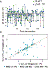
A. Comparison of the NH temperature coefficient, Δδ/ΔT, for γS-WT (green) and γS-G18V (blue). Most Δδ/ΔT values for residues in the C-terminal domain are unchanged, while, many Δδ/ΔT values for residues in the N-terminal domain differ between γS-G18V and γS-WT. Values above the dotted line at −4.6 ppb are indicative of residues involved in intramolecular hydrogen bonds, and those below ones that are hydrogen bond to solvent. B. NH temperature coefficients, Δδ/ΔT, for γS-WT (x-axis) vs. γS-G18V (y-axis). The largest deviations in the NH temperature coefficient are observed in the N-terminal domain closest to the mutation site (dark teal), with significant deviations also occurring in the latter part of the N-terminal domain (light blue). Few differences are seen in the C-terminal domain (salmon), mostly corresponding to residues in the interdomain interface. The dashed line has a slope of 1 as a guide to identify the residues for which γS-WT and γS-G18V have the same Δδ/ΔT values.
Figure 5.
Figure 5.
γS-WT and γS-G18V present subtle differences in intramolecular/solvent hydrogen bonding and surface hydrophobicity. Increased fluorescence of bis-ANS at 500 nm was observed upon binding to γS-G18V (blue) compared to γS-WT (green), consistent with an increase in exposed hydrophobic surface area for γS-G18V.

References

    1. Minton AP Biopolymers 1981, 20, 2093.
    1. Zhou H-X; Rivas G; Minton AP Annual review of biophysics 2008, 37, 375. - PMC - PubMed
    1. Miklos AC; Sarkar M; Wang Y; Pielak GJ Journal of the American Chemical Society 2011, 133, 7116. - PubMed
    1. Predeus AV; Gul S; Gopal SM; Feig M The Journal of Physical Chemistry. B 2012, 116, 8610. - PMC - PubMed
    1. Gnutt D; Gao M; Brylski O; Heyden M; Ebbinghaus S Angewandte Chemie 2015, 54, 2548–2551. - PMC - PubMed

Publication types

LinkOut - more resources