Review
. 2016 Jul 1;116(1):122-34.
doi: 10.1152/jn.00087.2016. Epub 2016 Apr 6.

Strategies for targeting primate neural circuits with viral vectors

Affiliations
Review

Strategies for targeting primate neural circuits with viral vectors

Yasmine El-Shamayleh et al. J Neurophysiol. .

Abstract

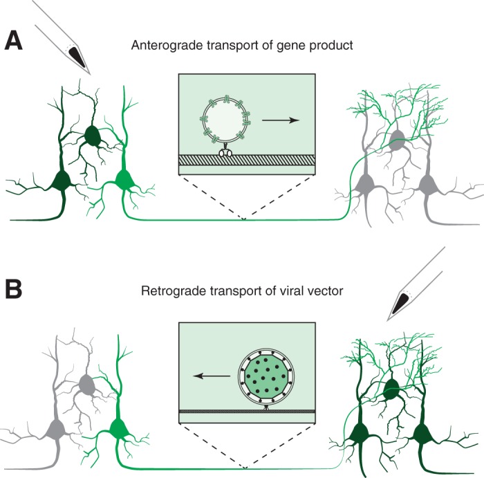
Understanding how the brain works requires understanding how different types of neurons contribute to circuit function and organism behavior. Progress on this front has been accelerated by optogenetics and chemogenetics, which provide an unprecedented level of control over distinct neuronal types in small animals. In primates, however, targeting specific types of neurons with these tools remains challenging. In this review, we discuss existing and emerging strategies for directing genetic manipulations to targeted neurons in the adult primate central nervous system. We review the literature on viral vectors for gene delivery to neurons, focusing on adeno-associated viral vectors and lentiviral vectors, their tropism for different cell types, and prospects for new variants with improved efficacy and selectivity. We discuss two projection targeting approaches for probing neural circuits: anterograde projection targeting and retrograde transport of viral vectors. We conclude with an analysis of cell type-specific promoters and other nucleotide sequences that can be used in viral vectors to target neuronal types at the transcriptional level.

Keywords: gene therapy; optogenetics; primate; targeting; viral vector.


Figures

Fig. 1.
Fig. 1.
Viral vector tropism in primate neocortex. Coronal sections of visual area V1 of a rhesus macaque. Tissue was stained for SMI-32 (red) and with DAPI (blue), and transduced neurons are green. A: injection of LV-CaMKIIa-ArchT-GFP titered at 1.8 × 10⁷ infectious units/ml. B: injection of AAV9-hSyn-ChR2-eYFP titered at 2.75 × 10¹³ genomic copies/ml. A total of 5 μl of each viral vector was injected (1 μl injected at each of 5 sites, spaced 500 μm apart). Scale bars, 500 μm. The sparse labeling is consistent with some studies (Gerits et al. 2015) but not others (Diester et al. 2011).
Fig. 2.
Fig. 2.
Illustration of projection targeting approaches. A: anterograde projection targeting. A viral vector is injected into a source area (left) where neuronal cell bodies are transduced. Gene products are trafficked along axons (center) to their terminations in a recipient area (right). B: retrograde projection targeting. A viral vector is injected into a recipient area (right). Virions are retrogradely transported along axons to cell bodies (left) where genes are expressed. In both scenarios, some neurons are transduced (light and dark green) whereas others are not transduced (gray). The subset of transduced neurons available for selective manipulation are highlighted (light green).
Fig. 3.
Fig. 3.
Anterograde projection targeting of V1 axons projecting to the SC (for details of the AAV1-hSyn-ChR2-mCherry vector injection, see Jazayeri et al. 2012). In each experiment, an optical fiber and a tungsten electrode were independently lowered through a common guide tube to the surface of the SC. The tip of the electrode was positioned in the superficial layers, and the tip of the optical fiber was positioned just outside of the SC. Electrical responses to 473-nm light flashes were recorded. A: rasters and PSTHs showing entrainment of multiunit SC responses to periodic illumination (blue dashed lines). B: the position of each dot represents the receptive field location of a tested SC site. The size and color of each dot show the change in spike rate following a pulse of blue light; they are proportional to the number of spikes during the 10 ms after each light pulse divided by the number of spikes during the 10 ms preceding each light pulse. Optogenetic activation was greatest at SC sites with receptive fields that overlapped those at the V1 injection site (yellow square). C: coronal section through the left SC of the same animal showing ChR2-mCherry+ axons (red) and DAPI (blue). Scale bar, 500 μm.

References

    1. Aartsen WM, van Cleef KW, Pellissier LP, Hoek RM, Vos RM, Blits B, Ehlert EM, Balaggan KS, Ali RR, Verhaagen J, Wijnholds J. GFAP-driven GFP expression in activated mouse Müller glial cells aligning retinal blood vessels following intravitreal injection of AAV2/6 vectors. PLoS One 5: e12387, 2010. - PMC - PubMed
    1. Abbott NJ. Evidence for bulk flow of brain interstitial fluid: significance for physiology and pathology. Neurochem Int 45: 545–552, 2004. - PubMed
    1. Adamantidis AR, Zhang F, Aravanis AM, Deisseroth K, de Lecea L. Neural substrates of awakening probed with optogenetic control of hypocretin neurons. Nature 450: 420–424, 2007. - PMC - PubMed
    1. Alexander IE, Russell DW, Miller AD. Transfer of contaminants in adeno-associated virus vector stocks can mimic transduction and lead to artifactual results. Hum Gene Ther 8: 1911–1920, 1997. - PubMed
    1. Arnold CD, Gerlach D, Stelzer C, Boryn LM, Rath M, Stark A. Genome-wide quantitative enhancer activity maps identified by STARR-seq. Science 339: 1074–1077, 2013. - PubMed

Publication types