. 2016 Apr 7:6:24226.
doi: 10.1038/srep24226.

Loss-of-function Mutation in PMVK Causes Autosomal Dominant Disseminated Superficial Porokeratosis

Affiliations

Loss-of-function Mutation in PMVK Causes Autosomal Dominant Disseminated Superficial Porokeratosis

Jiuxiang Wang et al. Sci Rep. .

Abstract

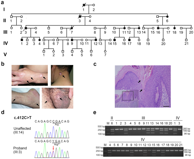
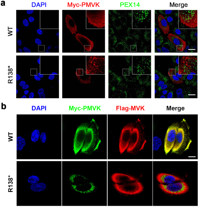
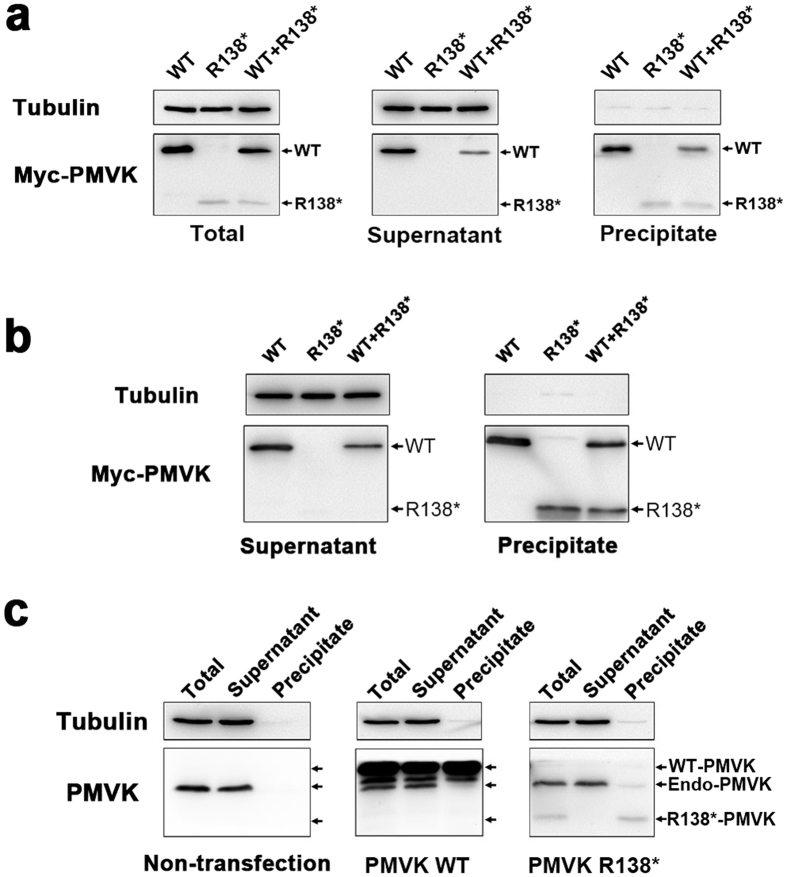
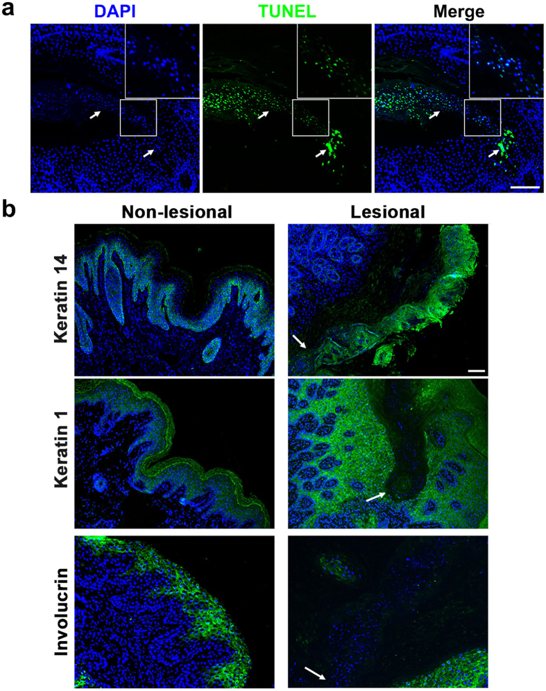
Disseminated superficial porokeratosis (DSP) is a rare keratinization disorder of the epidermis. It is characterized by keratotic lesions with an atrophic center encircled by a prominent peripheral ridge. We investigated the genetic basis of DSP in two five-generation Chinese families with members diagnosed with DSP. By whole-exome sequencing, we sequencing identified a nonsense variation c.412C > T (p.Arg138*) in the phosphomevalonate kinase gene (PMVK), which encodes a cytoplasmic enzyme catalyzing the conversion of mevalonate 5-phosphate to mevalonate 5-diphosphate in the mevalonate pathway. By co-segregation and haplotype analyses as well as exclusion testing of 500 normal control subjects, we demonstrated that this genetic variant was involved in the development of DSP in both families. We obtained further evidence from studies using HaCaT cells as models that this variant disturbed subcellular localization, expression and solubility of PMVK. We also observed apparent apoptosis in and under the cornoid lamella of PMVK-deficient lesional tissues, with incomplete differentiation of keratinocytes. Our findings suggest that PMVK is a potential novel gene involved in the pathogenesis of DSP and PMVK deficiency or abnormal keratinocyte apoptosis could lead to porokeratosis.


Conflict of interest statement

The authors declare no competing financial interests.

Figures

Figure 1
Figure 1. The pedigree tree, clinical manifestations, and mutation information of Family 1.
(a) Pedigree of Family 1. Black symbols, affected individuals; White symbols, unaffected individuals; Arrow, the proband of the family. Genomic DNA of II:6 (affected), III:14 (unaffected), and IV:1 (affected) were used for whole-exome sequencing. (b) Representative keratotic lesions of the skin. Lesions are indicated by arrows on the hand, neck, leg, and scrotum of the proband. (c) Histological examination of the lesional tissue from the proband. Typical cornoid lamellae with the absence of a granular layer below the parakeratotic column were indicated by arrows. Scale bar, 50 μm. (d) Sanger sequencing of the heterozygous c.412C > T PMVK mutation in the proband (III:3). The mutant base is marked by an arrow. The affected codon is underlined. (e) Segregation analysis of the mutation in Family 1. The mutation eliminated an Alu I site and led to two additional bands in the restriction fragment length polymorphism (RFLP) analysis.
Figure 2
Figure 2. The R138* Mutation Disturbs the Cellular Localization of PMVK.
(a) Abnormal localization of the R138* mutant PMVK in HaCaT cells. WT PMVK shows a diffuse cytoplasmic distribution, whereas mutant PMVK aggregates in the cytoplasm in a punctate pattern. Neither the WT nor the mutant PMVK co-localizes with the peroxisomal maker PEX14. Boxes on the right top show the partial enlarged details. Green, PEX14; Red, PMVK; Scale bars, 20 μm. (b) The R138* mutation destroys the co-localization of PMVK and MVK in HaCaT cells. WT PMVK showed the same dispersed distribution as MVK in cytoplasm, yet mutant PMVK did not co-localize with MVK. Green, PMVK; Red, MVK; Scale bar, 10 μm.
Figure 3
Figure 3. The Solubility of Endogenously and Exogenously Expressed WT and R138* PMVK in cells.
(a) The R138* mutant PMVK shows reduced expression and solubility in HaCaT cells. Myc-tagged WT and R138* PMVK were expressed in HaCaT cells. The total level of WT PMVK was higher than that of mutant PMVK. WT PMVK was present in both soluble (supernatant) and insoluble (precipitate) fractions, but mutant PMVK was exclusively present in the precipitate. Tubulin was served as a marker for soluble proteins and also a loading control. (b) The R138* mutation disturbs the expression and solubility of PMVK in ARPE19 cells. Myc-tagged WT and R138* PMVK were exogenously expressed in ARPE19 cells. Soluble and insoluble fractions were separated and subjected to the detection of PMVK protein levels by Western blot. (c) The solubility of endogenous PMVK is not affected by exogenously expressed WT and R138* PMVK. In non-transfected HaCaT cells, PMVK was fully soluble. When HaCaT cells were transfected with Myc-tagged WT or R138* PMVK plasmids, endogenous PMVK was still largely present in the supernatant (soluble fraction). Tubulin was used as a loading control and a marker for soluble proteins.
Figure 4
Figure 4. Abnormal Apoptosis and Differentiation of Keratinocytes in the Lesional Tissues of PMVK-deficient Individuals.
(a) Apoptosis of keratinocytes in the lesional tissues. TUNEL-positive cells were observed in and under the cornoid lamella (indicated by arrows). The enlarged images show the co-localization of TUNEL signals and nuclei. Scale bar, 50 μm. (b) Aberrant distributions of the keratinocyte differentiation markers in the lesional tissues. Antibodies against keratin 14, keratin1or involucrin were used to reveal the differentiation process from immature stratum basale cells to mature keratinocytes in stratum granulosum and stratum corneum. The nuclei of keratinocytes in the lesional and non-lesional tissues were visualized by DAPI staining. The arrows indicate the bottom of cornoid lamellae. Scale bar, 20 μm.

References

    1. Sertznig P., von Felbert V. & Megahed M. Porokeratosis: present concepts. J Eur Acad Dermatol 26, 404–412 (2012). - PubMed
    1. Online Mendelian Inheritance in Man, OMIM®. McKusick-Nathans Institute of Genetic Medicine, Johns Hopkins University (Baltimore, MD). Available at: http://www.omim.org/. (Accessed: 8th September 2015).
    1. Zhang S. et al.. Exome sequencing identifies MVK mutations in disseminated superficial actinic porokeratosis. Nat Genet 44, 1156–1160 (2012). - PubMed
    1. Cui H. et al.. Exome sequencing identifies SLC17A9 pathogenic gene in two Chinese pedigrees with disseminated superficial actinic porokeratosis. J Med Genet 51, 699–704 (2014). - PubMed
    1. Zeng K., Zhang Q., Li L., Duan Y. & Liang Y. Splicing mutation in MVK is a cause of porokeratosis of Mibelli. Arch Dermatol Res 306, 749–755 (2014). - PubMed

Publication types

MeSH terms

Substances