. 2016 Apr 7:6:24131.
doi: 10.1038/srep24131.

Concentration Dependent Ion-Protein Interaction Patterns Underlying Protein Oligomerization Behaviours

Affiliations

Concentration Dependent Ion-Protein Interaction Patterns Underlying Protein Oligomerization Behaviours

Helena Batoulis et al. Sci Rep. .

Abstract

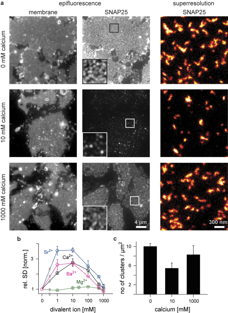
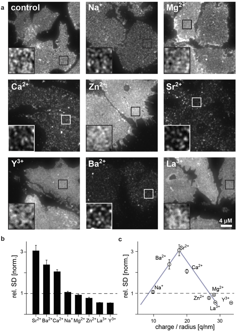
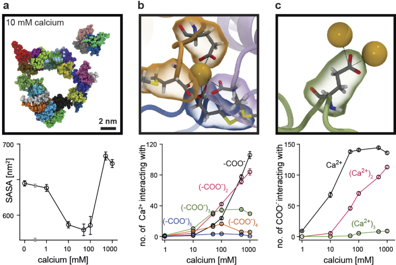
Salts and proteins comprise two of the basic molecular components of biological materials. Kosmotropic/chaotropic co-solvation and matching ion water affinities explain basic ionic effects on protein aggregation observed in simple solutions. However, it is unclear how these theories apply to proteins in complex biological environments and what the underlying ionic binding patterns are. Using the positive ion Ca(2+) and the negatively charged membrane protein SNAP25, we studied ion effects on protein oligomerization in solution, in native membranes and in molecular dynamics (MD) simulations. We find that concentration-dependent ion-induced protein oligomerization is a fundamental chemico-physical principle applying not only to soluble but also to membrane-anchored proteins in their native environment. Oligomerization is driven by the interaction of Ca(2+) ions with the carboxylate groups of aspartate and glutamate. From low up to middle concentrations, salt bridges between Ca(2+) ions and two or more protein residues lead to increasingly larger oligomers, while at high concentrations oligomers disperse due to overcharging effects. The insights provide a conceptual framework at the interface of physics, chemistry and biology to explain binding of ions to charged protein surfaces on an atomistic scale, as occurring during protein solubilisation, aggregation and oligomerization both in simple solutions and membrane systems.


Conflict of interest statement

The authors declare no competing financial interests.

Figures

Figure 1
Figure 1. SNAP25 in native plasma membrane sheets shows biphasic clustering in response to divalent ions.
PC12 plasma membrane sheets incubated at 37 °C for 10 min with the indicated concentrations of calcium, strontium, barium or magnesium chloride, fixed, immunostained for SNAP25 and analysed with epifluorescence or superresolution STED microscopy. (a) Membrane sheets incubated with 0, 10, or 1000 mM CaCl2. Epifluorescence recordings from the dye TMA-DPH (left; documenting the integrity of the membranes) and the immunostaining (middle; overviews are shown at the same, magnified insets at arbitrary scaling). Right, STED micrographs of SNAP25 immunofluorescence to which the “red hot” look up table was applied which displays increasingly brighter pixel intensities applying a colour code from black to red to yellow to white. (b) SNAP25 clustering was quantified by calculating the relative standard deviation (rel. SD) of the immunostaining pattern, normalized to the baseline condition which contained no divalent cations. Values are means ± s.e.m. (c) SNAP25 cluster density (means ± s.e.m.) resolved by superresolution STED microscopy. Cluster size was similar under all conditions (Supplementary Fig. 4).
Figure 2
Figure 2. The efficacy of mono-, di- and trivalent ions on SNAP25 clustering in the cell membrane depends on ion charge-to-radius ratio.
(a) Membrane sheets incubated at 37 °C for 10 min with 1 mM chloride salts of Na+, Mg2+, Ca2+, Zn2+, Sr2+, Y3+, Ba2+ and La3+ (sorted by atomic number), fixed, and immunostained for SNAP25. Overviews are shown at the same, magnified views at arbitrary intensity scaling. (b) The degree of SNAP25 clustering is expressed as rel. SD of the immunostaining pattern, normalized to the control condition. Ions are shown in decreasing order of their clustering efficacy. (c) Plotting values from (b) versus the ratio of ion charge-to-crystal radius shows a distribution resembling a volcano plot (grey lines). For relation to other ion properties see Supplementary Fig. 5. Values are means ± s.e.m.
Figure 3
Figure 3. Ion-protein binding patterns depend on the calcium concentration.
(a) Snapshot from the end of the MD simulation in the presence of 10 mM CaCl2 (for comparison see Supplementary Fig. 6 showing snapshots from the respective end states of all simulation runs and conditions). The solvent accessible surface area (SASA), averaged over the last 20 ns of the simulation (means ± s.e.m.) illustrating the degree of peptide clustering. (b) Example of an interaction between one calcium ion (yellow, van der Waals representation) and four carboxylate groups provided by three peptides (orange, magenta and blue). For clarity only protein residues providing a carboxylate group for Ca2+ binding are shown. The graph shows the number of interactions (averaged over the last 20 ns, means ± s.e.m.) between one calcium ion and i carboxylate groups (i = 1, 2, …, 6; indicated by different colours; for clarity i = 6 is not shown) as a function of Ca2+ concentration. (c) Example of an interaction between one glutamic acid carboxylate group and two calcium ions, at two different binding distances (see text). The graph shows how the number of interactions between one carboxylate group and i calcium ions (i = 1, 2, or 3) depends on the calcium concentration.

References

    1. Ma C. & Bloomfield V. A. Condensation of supercoiled DNA induced by MnCl2. Biophys. J. 67, 1678–1681 (1994). - PMC - PubMed
    1. Tang J. X., Wong S., Tran P. T. & Janmey P. A. Counterion induced bundle formation of rodlike polyelectrolytes. Berichte Bunsenges. Für Phys. Chem. 100, 796–806 (1996).
    1. Alies B., Hureau C. & Faller P. The role of metal ions in amyloid formation: general principles from model peptides. Metallomics 5, 183 (2013). - PubMed
    1. Breydo L. & Uversky V. N. Role of metal ions in aggregation of intrinsically disordered proteins in neurodegenerative diseases. Metallomics 3, 1163–1180 (2011). - PubMed
    1. Salinas B. A. et al. Understanding and modulating opalescence and viscosity in a monoclonal antibody formulation. J. Pharm. Sci. 99, 82–93 (2010). - PMC - PubMed

Publication types

MeSH terms

LinkOut - more resources