Comment
. 2016 Jun;23(6):548-56.
doi: 10.1038/gt.2016.31. Epub 2016 Apr 7.

Reduced retinal transduction and enhanced transgene-directed immunogenicity with intravitreal delivery of rAAV following posterior vitrectomy in dogs

Affiliations
Comment

Reduced retinal transduction and enhanced transgene-directed immunogenicity with intravitreal delivery of rAAV following posterior vitrectomy in dogs

R F Boyd et al. Gene Ther. 2016 Jun.

Abstract

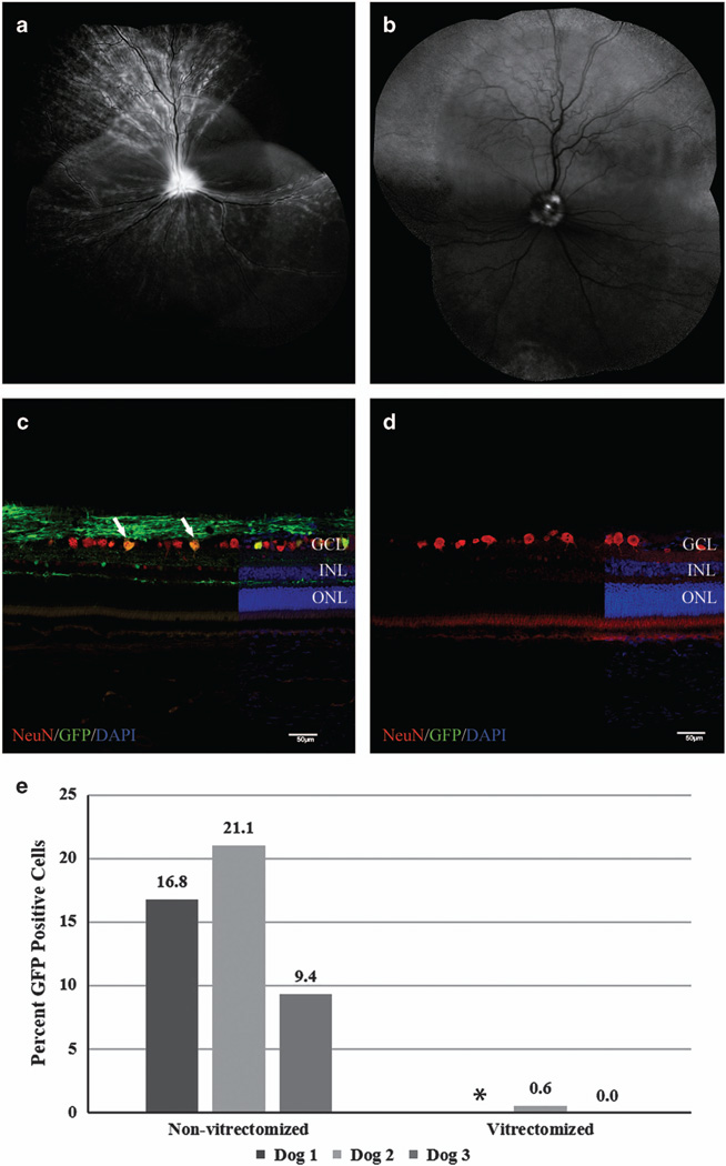
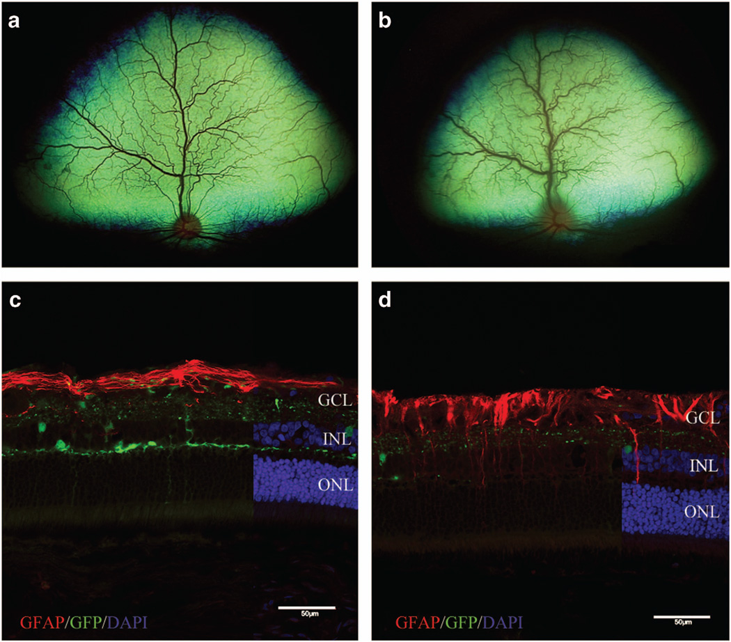
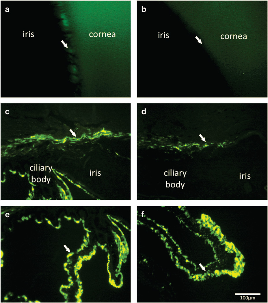
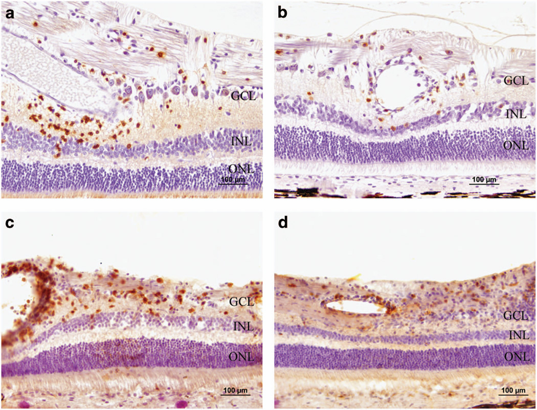
Adeno-associated virus (AAV) vector-based gene therapy is a promising treatment strategy for delivery of neurotrophic transgenes to retinal ganglion cells (RGCs) in glaucoma patients. Retinal distribution of transgene expression following intravitreal injection (IVT) of AAV is variable in animal models and the vitreous humor may represent a barrier to initial vector penetration. The primary goal of our study was to investigate the effect of prior core vitrectomy with posterior hyaloid membrane peeling on pattern and efficiency of transduction of a capsid amino acid substituted AAV2 vector, carrying the green fluorescent protein (GFP) reporter transgene following IVT in dogs. When progressive intraocular inflammation developed starting 4 weeks post IVT, the study plan was modified to allow detailed characterization of the etiology as a secondary goal. Unexpectedly, surgical vitrectomy was found to significantly limit transduction, whereas in non-vitrectomized eyes transduction efficiency reached upwards to 37.3% of RGC layer cells. The developing retinitis was characterized by mononuclear cell infiltrates resulting from a delayed-type hypersensitivity reaction, which we suspect was directed at the GFP transgene. Our results, in a canine large animal model, support caution when considering surgical vitrectomy before IVT for retinal gene therapy in patients, as prior vitrectomy appears to significantly reduce transduction efficiency and may predispose the patient to development of vector-induced immune reactions.


Conflict of interest statement

CONFLICT OF INTEREST

WWH and the University of Florida have a financial interest in the use of AAV therapies and own equity in a company (AGTC Inc.) that might, in the future, commercialize some aspects of this work. The remaining authors declare no conflict of interest.

Figures

Figure 1
Figure 1
Comparison of ganglion cell layer transduction efficiency of non-vitrectomized and vitrectomized eyes following IVT of AAV2 (triple Y-F+T-V). Representative confocal scanning laser ophthalmoscopy images (a, b) obtained at 5 weeks post injection demonstrate widespread fluorescence in non-vitrectomized eye (a) versus fluorescence limited to the optic nerve head of the vitrectomized eye. (b) The low level of background autofluorescence in the superior retina of image (b) is the expected appearance of the canine tapetum. Immunohistochemical labeling of retinal cryosections with NeuN antibody (c, d) demonstrating a higher number of cells colabeling with GFP in non-vitrectomized eye (c, white arrows) compared with absent transduction in vitrectomized eye (d). (e) Overall ganglion cell layer transduction efficiency for non-vitrectomized eyes and vitrectomized eyes 6 weeks post IVT. Non-vitrectomized eyes had significantly higher levels of ganglion cell layer transduction (P=0.04; unpaired t-test). DAPI, 4',6-diamidino-2-phenylindole; GCL, ganglion cell layer; GFP, green fluorescent protein; INL, inner nuclear layer; IVT, intravitreal injection; ONL, outer nuclear layer. Scale bar, 50 µm. *There is no transduction efficiency calculation for the vitrectomized eye of Dog 1 due to loss of sample for histopathological analysis.
Figure 2
Figure 2
White-light fundoscopic imaging showing the normal fundus of the non-vitrectomized eye of Dog 1 at 4 weeks post IVT (a) and acute retinitis that developed during week 6 (b). Fluorescent microscopic images of retinas from non-vitrectomized eyes labeled with GFAP shown in c and d to demonstrate glial activation of Müller cells as a result of inflammation. (c) Retina from Dog 2 without gross evidence of inflammation, showing expected strong GFAP labeling limited predominantly to the nerve fiber layer. (d) Retina from Dog 3 with chronic inflammation, showing GFAP labeling extending from the nerve fiber layer into the inner and outer retinal layers. DAPI, 4',6-diamidino-2-phenylindole; GCL, ganglion cell layer; GFP, green fluorescent protein; INL, inner nuclear layer; IVT, intravitreal injection; ONL, outer nuclear layer. Scale bar, 50 µm.
Figure 3
Figure 3
Fluorescent gonioscopic imaging showing fluorescence within the ciliary cleft of vitrectomized eye of Dog 2 at 6 weeks post IVT (a, arrow). Note the pectinate ligament fibrils demonstrated by the dark bands crossing the region of fluorescence. Non-vitrectomized eye of Dog 2 shows no appreciable fluorescence within the cliary cleft (b, arrow). Immunohistochemical labeling of cryosections of the anterior segment of vitrectomized eye (c, e) showed GFP expression within the trabecular meshwork outflow pathway (c, arrow), and expression within the ciliary body epithelium (e, arrow). The anterior segment sections from non-vitrectomized eye (d, f) showed less prominent GFP expression within the trabecular meshwork outflow pathway (d, arrow), but comparable expression within the ciliary body epithelium (f, arrow). GFP, green fluorescent protein; IVT, intravitreal injection. Scale bar, 100 µm.
Figure 4
Figure 4
Photomicrographs characterizing the cellular component of the retinal inflammatory response from vitrectomized eye of Dog 1 (a, b) and non-vitrectomized eye of Dog 3 (c, d) through the use of CD labeling. Positive immunolabeling of mononuclear inflammatory cells with CD20 (a) and CD3 (b) demonstrates a mixed response involving both B cells and T cells, respectively. Positive immunolabeling of inflammatory cells with CD4 (c) and CD8 (d) demonstrates that both T-helper and cytotoxic T cells, respectively, were present. GCL, ganglion cell layer; INL, inner nuclear layer; ONL, outer nuclear layer. Scale bar, 100 µm.

Comment on

References

    1. Chader GJ. Advances in glaucoma treatment and management: neurotrophic agents. Invest Ophthalmol Vis Sci. 2012;53:2501–2505. - PubMed
    1. Hellstrom M, Pollett MA, Harvey AR. Post-injury delivery of rAAV2-CNTF combined with short-term pharmacotherapy is neuroprotective and promotes extensive axonal regeneration after optic nerve trauma. J Neurotrauma. 2011;28:2475–2483. - PubMed
    1. van Adel BA, Kostic C, Deglon N, Ball AK, Arsenijevic Y. Delivery of ciliary neurotrophic factor via lentiviral-mediated transfer protects axotomized retinal ganglion cells for an extended period of time. Hum Gene Ther. 2003;14:103–115. - PubMed
    1. Hellstrom M, Harvey AR. Retinal ganglion cell gene therapy and visual system repair. Curr Gene Ther. 2011;11:116–131. - PubMed
    1. Harvey AR, Hellström M, Rodger J. Gene therapy and transplantation in the retinofugal pathway. Prog Brain Res. 2009;175:151–161. - PubMed

Publication types

Substances