. 2016 May 27;291(22):11776-86.
doi: 10.1074/jbc.M116.719351. Epub 2016 Apr 6.

Molecular Insights into the Impact of Oxidative Stress on the Quorum-Sensing Regulator Protein LasR

Affiliations

Molecular Insights into the Impact of Oxidative Stress on the Quorum-Sensing Regulator Protein LasR

Prapti Kafle et al. J Biol Chem. .

Abstract

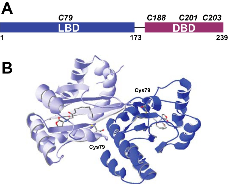
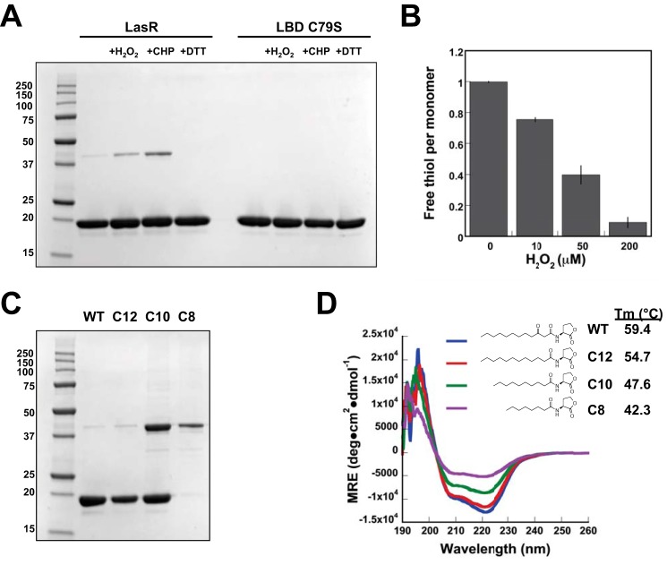
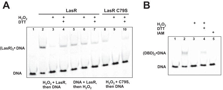
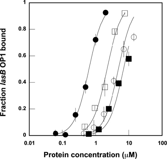
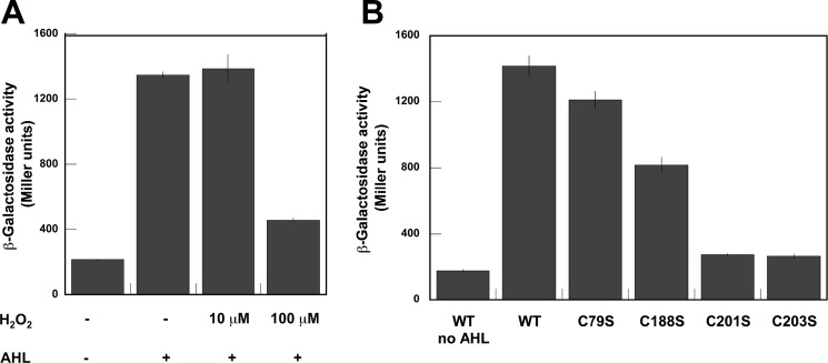
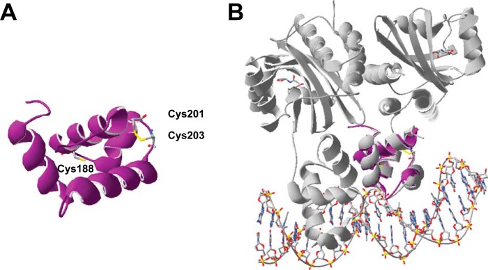
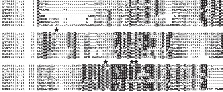
The LasR regulator protein functions at the top of the Pseudomonas aeruginosa quorum-sensing hierarchy and is implicated in promoting bacterial virulence. Of note is recent evidence that this transcription factor may also respond to oxidative stress. Here, all cysteines in LasR were inspected to deduce their redox sensitivity and to probe the connection between stress response and LasR activity using purified LasR and individual LasR domains. Cys(79) in the ligand binding domain of LasR appears to be important for ligand recognition and folding of this domain to potentiate DNA binding but does not seem to be sensitive to oxidative stress when bound to its native ligand. Two cysteines in the DNA binding domain of LasR do form a disulfide bond when treated with hydrogen peroxide, and formation of this Cys(201)-Cys(203) disulfide bond appears to disrupt the DNA binding activity of the transcription factor. Mutagenesis of either of these cysteines leads to expression of a protein that no longer binds DNA. A cell-based reporter assay linking LasR function with β-galactosidase activity gave results consistent with those obtained with purified LasR. This work provides a possible mechanism for oxidative stress response by LasR and indicates that multiple cysteines within the protein may prove to be useful targets for disabling its activity.

Keywords: DNA binding protein; Pseudomonas aeruginosa; antibiotic resistance; bacterial pathogenesis; bacterial transcription; cysteine; ligand-binding protein; quorum sensing.


Figures

FIGURE 1.
FIGURE 1.
Cysteine composition of LasR. A, domain structure of LasR with the relative positions of all LasR cysteines shown. B, dimeric crystal structure (22) of the LBD with 3O-C12-HSL bound by each monomer and both Cys79 residues shown. Within each monomer, the terminal carbon of 3O-C12-HSL is proximal to Cys79 (4.3 Å), whereas the two cysteines in the dimeric complex are 21.6 Å apart.
FIGURE 2.
FIGURE 2.
Impact of oxidative stress and unnatural ligands on LasR LBD. A, SDS-PAGE of 25 μm LBD under non-reducing conditions showing both monomer and dimer bands. Protein bands were excised from the third lane of a duplicate gel and subjected to LC-MS to confirm the predicted molecular weight of monomer (20,453.0 Da observed and 20,451.1 Da calculated) and dimer (40,906.5 Da observed and 40,900.2 Da calculated) LBD. Dimer formation is enhanced in the presence of 2.5 mm H2O2 and cumene hydroperoxide (CHP), whereas only monomer LBD is observed in the presence of 100 mm DTT or with the LBD C79S mutant. B, DTNB quantification of free thiols under denaturing conditions illustrates the impact of 10–200 μm H2O2 on 25 μm LBD. Error bars denote standard deviation for triplicate data. C, non-reducing SDS-PAGE of LBD expressed in the presence of 3O-C12-HSL (WT) and the non-native ligands C12-HSL, C10-HSL, and C8-HSL. D, wavelength-dependent CD spectra comparing relative folding of 15 μm LBD expressed in the presence of varied AHLs. The thermal stability (Tm) of each was also determined.
FIGURE 3.
FIGURE 3.
Oxidative stress impacts LasR-DNA binding. A, representative EMSA with 0.5 μm LasR and 5.0 μm LasR C79S. Target lasB OP1 is shown alone (lane 1). Both proteins shifted DNA in the absence of oxidant (lanes 2, 5, and 8), and the addition of 7.5 mm H2O2 significantly reduced DNA binding observed (lanes 3 and 9). The concentration of H2O2 used in these assays is similar to that reported in prior EMSA studies to evaluate the impact of oxidant on transcription factor-DNA binding (18–20, 28–30). The incubation of LasR with DNA for 30 min prior to the addition of oxidant appears to protect LasR from oxidation, preserving DNA binding (compare lanes 3 and 6). DNA binding for both proteins is restored with the addition of 25 mm DTT following incubation with H2O2 (compare lanes 3 and 4 for LasR and lanes 9 and 10 for LasR C79S). B, representative EMSA with 14 μm LasR DBD indicates binding of this domain to lasB OP1 (lane 2) that is disrupted in the presence of 7.5 mm H2O2 (lane 3) but restored with the addition of 25 mm DTT (lane 4). DBD reacted with iodoacetamide (IAM) prior to the binding assay did not show DNA binding (lane 5).
FIGURE 4.
FIGURE 4.
Quantitative EMSA comparing equilibrium binding affinity for wild-type LasR (), LasR C79S (), LasR C79A (), and LasR DBD (). The apparent Kd for each was determined by fitting data to the Langmuir equation as described in Ref. . Error bars denote standard deviation for triplicate data.
FIGURE 5.
FIGURE 5.
Mass spectrometric mapping of the Cys201-Cys203 disulfide bond in LasR. A, graphical fragment map correlating the relevant peptide sequence with the observed fragmentation ions. B, representative MS/MS fragmentation of the ion at m/z 965.40 corresponding to the disulfide-linked Glu-C peptide fragment 197–205.
FIGURE 6.
FIGURE 6.
Miller assay to detect the activity of β-galactosidase produced by the E. coli lasR lasB::lacZ reporter gene system. A, impact of adding H2O2 and 20 μm AHL (3O-C12-HSL) to cell cultures. Error bars denote standard deviation for triplicate data. B, impact of the indicated lasR gene mutations reflected in the reporter assay. Cultures were grown in the presence of 20 μm 3O-C12-HSL except where noted. Error bars denote standard deviation for triplicate data.
FIGURE 7.
FIGURE 7.
Modeling the LasR DBD. A, homology model of DBD generated using the Phyre2 web server (40). The three DBD cysteines are shown, including the Cys201-Cys203 disulfide bond. B, overlay of the DBD homology model (purple) with the TraR DBD (right monomer, shown in gray) within the crystal structure (41) of TraR bound to its autoinducer and target DNA.
FIGURE 8.
FIGURE 8.
Multiple sequence alignment of LasR and its homologs. Protein sequences shown include P. aeruginosa LasR (UniProt P25084), Vibrio fischeri LuxR (UniProt P12746), Burkholderia kururiensis BraR (UniProt B1VD86), Pseudomonas putida PpuR (UniProt Q348H6), Pseudomonas fluorescens MupR (UniProt Q9AH79), A. tumefaciens TraR (UniProt A5WYC9), E. coli SdiA (UniProt P07026), P. aeruginosa RhlR (UniProt B6E4Z5), and Chromobacterium violaceum CviR (UniProt D3W065). The positions of the four LasR cysteines are indicated (stars). Cys79 is shared by BraR (47), PpuR (48), and MupR (49), all believed to recognize 3O-C12-HSL. This is not the autoinducer for the other proteins shown. With LuxR, these proteins also share Cys203. Interestingly, there is no sequence homology to Cys201 even in proteins that share Cys203, suggesting that the potential for a disulfide bond at that position is unique to LasR among known regulator sequences. It is possible that Cys201 in its reduced state may be an important hydrophobic amino acid, based on the conserved leucine at this position in most homologous sequences and the known hydrophobic nature of free cysteines (50).

References

    1. Lyczak J. B., Cannon C. L., and Pier G. B. (2000) Establishment of Pseudomonas aeruginosa infection: lessons from a versatile opportunist. Microbes Infect. 2, 1051–1060 - PubMed
    1. Sadikot R. T., Blackwell T. S., Christman J. W., and Prince A. S. (2005) Pathogen-host interactions in Pseudomonas aeruginosa pneumonia. Am. J. Respir. Crit. Care Med. 171, 1209–1223 - PMC - PubMed
    1. Bjarnsholt T., Jensen P. Ø., Jakobsen T. H., Phipps R., Nielsen A. K., Rybtke M. T., Tolker-Nielsen T., Givskov M., Høiby N., Ciofu O., and Scandinavian Cystic Fibrosis Study Consortium (2010) Quorum sensing and virulence of Pseudomonas aeruginosa during lung infection of cystic fibrosis patients. PLoS ONE 5, e10115. - PMC - PubMed
    1. Waters C. M., and Bassler B. L. (2005) Quorum sensing: cell-to-cell communication in bacteria. Annu. Rev. Cell Dev. Biol. 21, 319–346 - PubMed
    1. Schuster M., Sexton D. J., Diggle S. P., and Greenberg E. P. (2013) Acyl-homoserine lactone quorum sensing: from evolution to application. Annu. Rev. Microbiol. 67, 43–63 - PubMed

Publication types

MeSH terms

LinkOut - more resources