. 2016 May;13(5):4135-42.
doi: 10.3892/mmr.2016.5090. Epub 2016 Apr 5.

Effects of NOX1 on fibroblastic changes of endothelial cells in radiation‑induced pulmonary fibrosis

Affiliations

Effects of NOX1 on fibroblastic changes of endothelial cells in radiation‑induced pulmonary fibrosis

Seo-Hyun Choi et al. Mol Med Rep. 2016 May.

Abstract

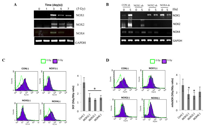
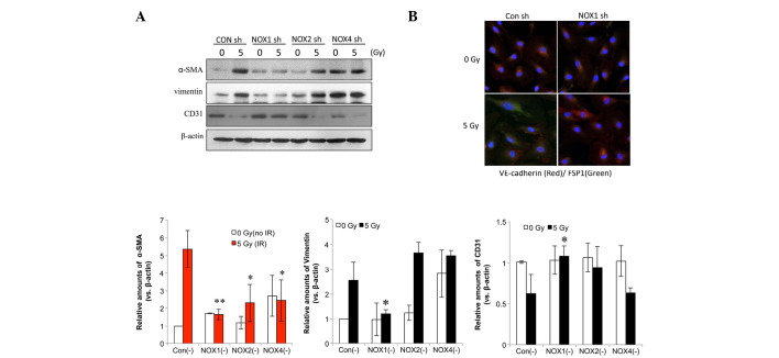
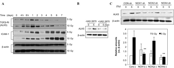
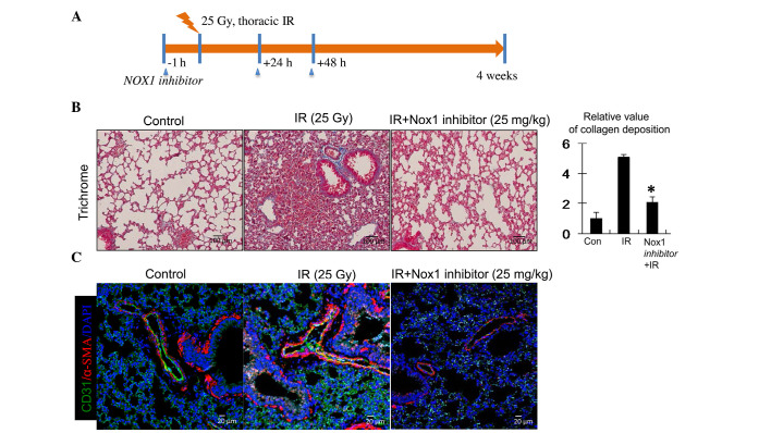
Lung fibrosis is a major complication in radiation‑induced lung damage following thoracic radiotherapy, while the underlying mechanism has remained to be elucidated. The present study performed immunofluorescence and immunoblot assays on irradiated human pulmonary artery endothelial cells (HPAECs) with or without pre‑treatment with VAS2870, a novel NADPH oxidase (NOX) inhibitor, or small hairpin (sh)RNA against NOX1, ‑2 or ‑4. VAS2870 reduced the cellular reactive oxygen species content induced by 5 Gy radiation in HPAECs and inhibited phenotypic changes in fibrotic cells, including increased alpha smooth muscle actin and vimentin, and decreased CD31 and vascular endothelial cadherin expression. These fibrotic changes were significantly inhibited by treatment with NOX1 shRNA, but not by NOX2 or NOX4 shRNA. Next, the role of NOX1 in pulmonary fibrosis development was assessed in the lung tissues of C57BL/6J mice following thoracic irradiation using trichrome staining. Administration of an NOX1‑specific inhibitor suppressed radiation‑induced collagen deposition and fibroblastic changes in the endothelial cells (ECs) of these mice. The results suggested that radiation‑induced pulmonary fibrosis may be efficiently reduced by specific inhibition of NOX1, an effect mediated by reduction of fibrotic changes of ECs.


Figures

Figure 1
Figure 1
VAS2870 inhibits radiation-induced fibroblastic changes in ECs. (A) HPAECs were irradiated with 5 Gy and incubated for 72 h. VAS2870 (1 µm) was added to cells 1 h prior to irradiation. To measure ROS, cells were incubated for 30 min with 1 µm 2′,7′-dichlorodihydrofluorescein diacetate and analyzed by flow cytometry (*P<0.05 vs. no VAS2870). (B) HPAECs were irradiated with 5 Gy and incubated for 72 h. VAS2870 (1 µm) was added to cells 1 h prior to irradiation and analysis by immunofluorescence with Alexa 488-conjugated anti-α-SMA and Alexa 594-conjugated anti-CD31 antibodies (green and red, respectively). Nuclei were counterstained with 4′,6-diamidino-2-phenylindole (blue) (scale bars, 20 µm). (C) Samples were subjected to western blot analysis of α-SMA, vimentin, CD31 and VE-cadherin. β-actin served as the loading control. Protein expression was quantified by densitometric analysis. Values are expressed as the mean ± standard deviation (n=3). *P<0.05 and **P<0.01 vs. VAS2870-untreated. HPAEC, human pulmonary artery endothelial cell; SMA, smooth-muscle actin; VAS, nicotinamide adenine dinucleotide phosphate oxidase inhibitor VAS2870; ROS, reactive oxygen species; VE, vascular endothelial.
Figure 2
Figure 2
NOX1, 2 and 4 shRNAs decrease radiation-induced ROS in HPAECs. (A) Following irradiation, NOX1, 2 and 4 expression was evaluated by RT-qPCR. HPAECs were cultured for the indicated number of days after receiving 5 Gy irradiation. (B) Each lentiviral vector contained a NOX1-, 2- or 4-targeted shRNA, which was then transfected into cultured HPAECs. A lentiviral vector containing a scrambled sequence served as the control. To confirm lentiviral-mediated gene knockdown, NOX1, 2 and 4 expression was analyzed by RT-qPCR. (C) Assessment of ROS and (D) determination of mitochondrial superoxide. Infected cells were irradiated with 5 Gy, followed by incubation with 1 µm H2DCFDA or 2.5 µm mitoSOX™, respectively, for 30 min and flow cytometric analysis. Values are expressed as the mean ± standard deviation. *P<0.05 (n=6) in C; *P=0.05 (n=3) in D vs. control shRNA. HPAEC, human pulmonary artery endothelial cell; NOX, nicotinamide adenine dinucleotide phosphate oxidase; ROS, reactive oxygen species; GAPDH, glyceraldehyde 3-phosphate dehydrogenase; shRNA, small hairpin RNA; CON, control; RT-qPCR, reverse-transcription quantitative polymerase chain reaction; H2DCFDA, 2′,7′-dichlorodihydrofluorescein diacetate.
Figure 3
Figure 3
NOX1 shRNA decreases radiation-induced fibrotic changes in HPAECs. (A) Cells transfected with NOX1-, 2- or 4-targeted shRNA were irradiated and incubated for 72 h, followed by western blot analysis of α-SMA, vimentin and CD31. Protein levels were quantified by densitometric analysis of the blots. Values are expressed as the mean ± standard deviation (n=3). **P<0.005; *P<0.05, α-SMA vs. Con (-). (B) HPAECs transfected with NOX1 shRNA were irradiated with 5 Gy and incubated for 72 h. Alexa 488-conjugated anti-FSP1 and Alexa 594-conjugated anti-VE-cadherin antibodies (green and red, respectively) were used to stain cells and nuclei were counterstained with 4′,6-diamidino-2-phenylindole (blue). HPAEC, human pulmonary artery endothelial cell; SMA, smooth-muscle actin; VE, vascular endothelial; NOX, nicotinamide adenine dinucleotide phosphate oxidase; CON, control; shRNA, small hairpin RNA; IR, irradiation; FSP fibroblast-specific protein.
Figure 4
Figure 4
NOX1 shRNA reverses the radiation-induced increase in ALK5. (A) human pulmonary artery endothelial cells were irradiated with 5 or 10 Gy and incubated for 7 days. Cell lysates were used for western blot analysis of ALK5, ICAM-1 and β-actin. Cells treated with (B) VAS2870 or (C) transfected with NOX1-, 2- or 4-targeted shRNA were subjected to western blot analysis of ALK5 and β-actin. Protein levels were quantified by densitometric analysis of the blots. Values are expressed as the mean ± standard deviation (n=3). **P<0.01 ALK5 vs. Con (−). NOX, nicotinamide adenine dinucleotide phosphate oxidase; CON, control; shRNA, small hairpin RNA; TGF, transforming growth factor; ICAM, intercellular adhesion molecule; ALK5, TGF-β type I receptor kinase.
Figure 5
Figure 5
Collagen deposition and fibrotic changes in endothelial-cells of irradiated lung tissue. (A) Schematic illustrating the experimental design. C57BL/6 mice were subjected to thoracic irradiation with 25 Gy. Lung samples (n=4–5 per condition) were obtained from mice prior to and 4 weeks after irradiation. (B) Representative trichrome-stained images of collagen deposition. Collagen is stained blue, nuclei are purple and cytoplasm is red/pink (scale bar, 50 µm). The graph shows the relative area of collagen deposition in irradiated lung tissue, determined from determined from randomly selected microscopic field with Image J software (five field per mouse at ×200 magnification). The relative levels of collagen deposition per 200x field are expressed as the mean ± standard deviation (n=3); *P<0.05 vs. Nox1 inhibitor-untreated. (C) Fibroblastic changes in ECs in the irradiated samples were analyzed by immunofluorescence using Alexa 594-conjugated anti-α-SMA and Alexa 488-conjugated anti-CD31 antibodies (red and green, respectively) with nuclei counterstained with DAPI. NOX, nicotinamide adenine dinucleotide phosphate oxidase; IR, irradiation; CON, control; DAPI, 4′,6-diamidino-2-phenylindole.

References

    1. Mehta V. Radiation pneumonitis and pulmonary fibrosis in non-small-cell lung cancer: Pulmonary function, prediction and prevention. Int J Radiat Oncol Biol Phys. 2005;63:5–24. doi: 10.1016/j.ijrobp.2005.03.047. - DOI - PubMed
    1. Fleckenstein K, Gauter-Fleckenstein B, Jackson IL, Rabbani Z, Anscher M, Vujaskovic Z. Using biological markers to predict risk of radiation injury. Semin Radiat Oncol. 2007;17:89–98. doi: 10.1016/j.semradonc.2006.11.004. - DOI - PubMed
    1. Ueno M, Maeno T, Nomura M, Aoyagi-Ikeda K, Matsui H, Hara K, Tanaka T, Iso T, Suga T, Kurabayashi M. Hypoxia-inducible factor-1α mediates TGF-β-induced PAI-1 production in alveolar macrophages in pulmonary fibrosis. Am J Physiol Lung Cell Mol Physiol. 2011;300:L740–L752. doi: 10.1152/ajplung.00146.2010. - DOI - PubMed
    1. Rodemann HP, Bamberg M. Cellular basis of radiation-induced fibrosis. Radiother Oncol. 1995;35:83–90. doi: 10.1016/0167-8140(95)01540-W. - DOI - PubMed
    1. Molteni A, Moulder JE, Cohen EF, Ward WF, Fish BL, Taylor JM, Wolfe LF, Brizio-Molteni L, Veno P. Control of radiation-induced pneumopathy and lung fibrosis by angiotensin-converting enzyme inhibitors and an angiotensin II type 1 receptor blocker. Int J Radiat Biol. 2000;76:523–532. doi: 10.1080/095530000138538. - DOI - PubMed

MeSH terms