. 2016 Apr 7:6:24248.
doi: 10.1038/srep24248.

Deficiency of the TLR4 analogue RP105 aggravates vein graft disease by inducing a pro-inflammatory response

Affiliations

Deficiency of the TLR4 analogue RP105 aggravates vein graft disease by inducing a pro-inflammatory response

Anouk Wezel et al. Sci Rep. .

Abstract

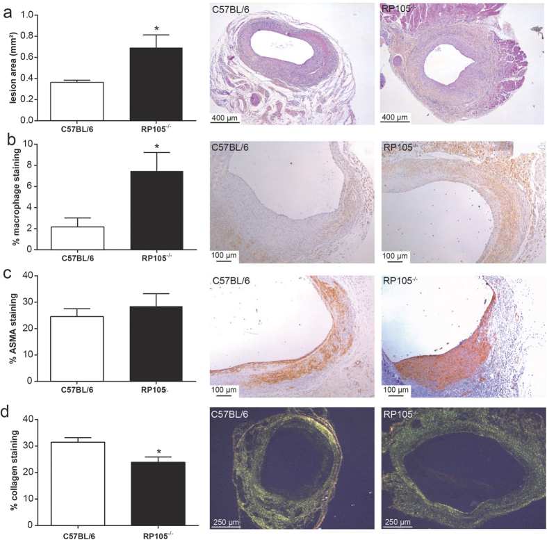
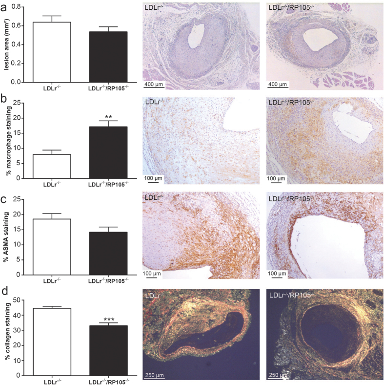
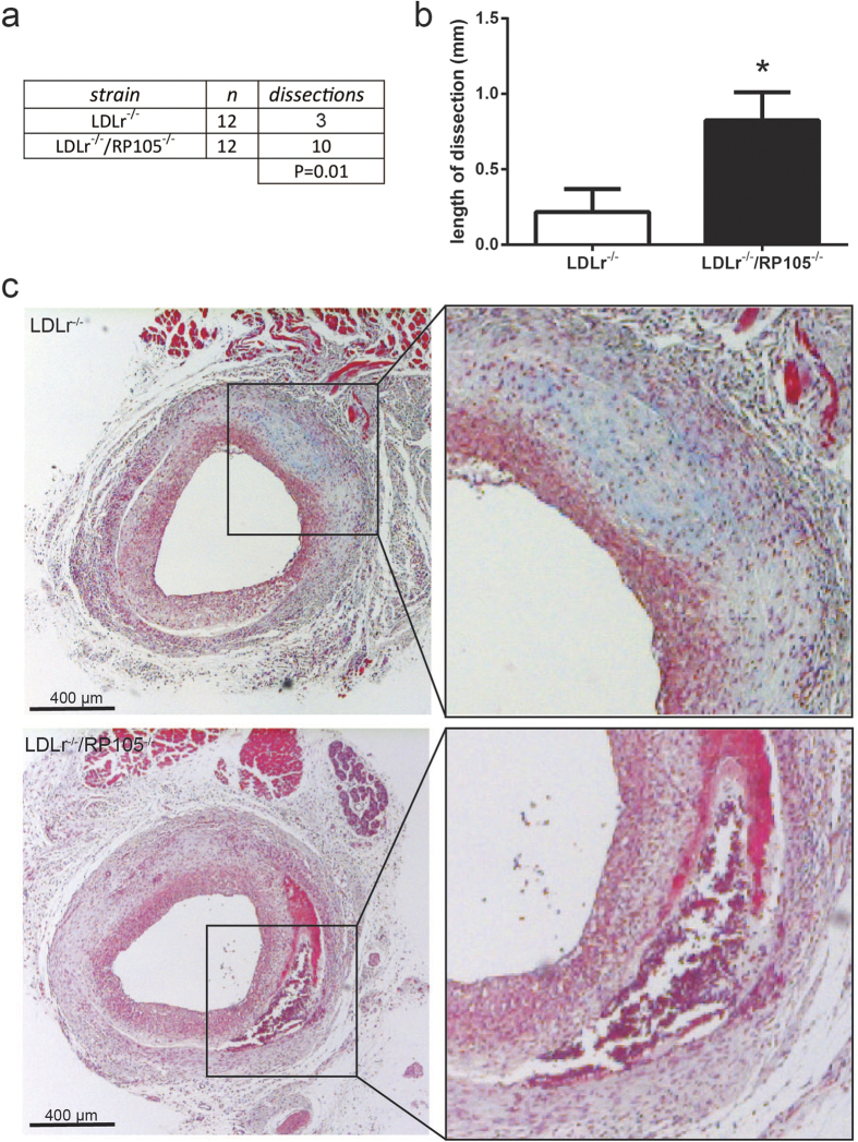
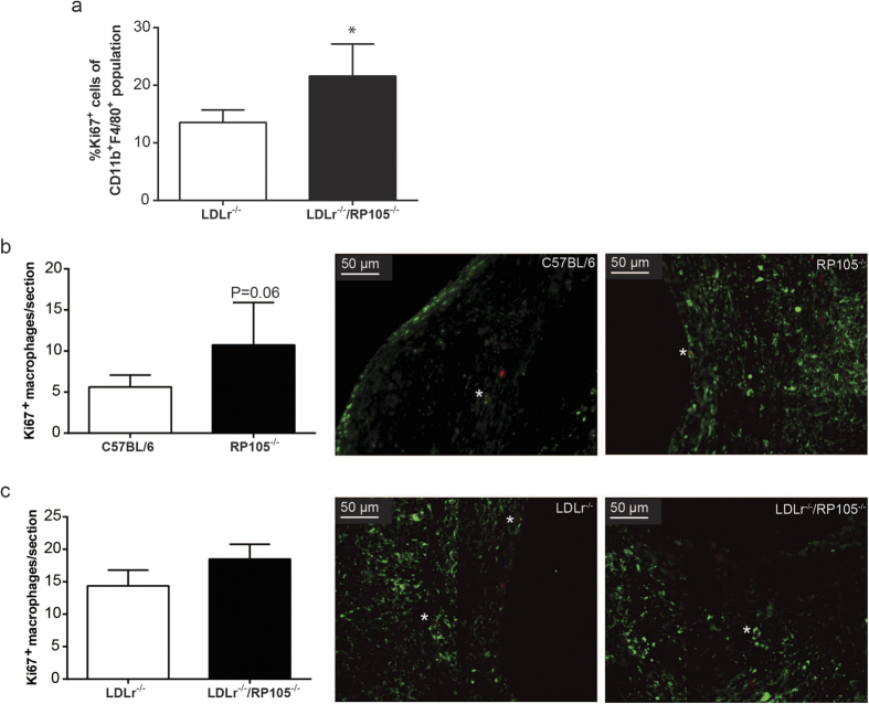
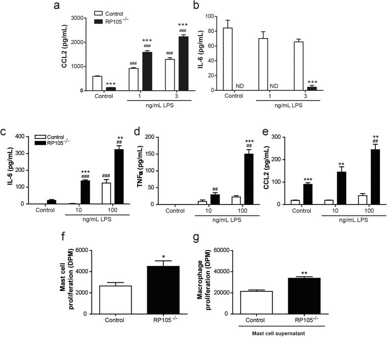
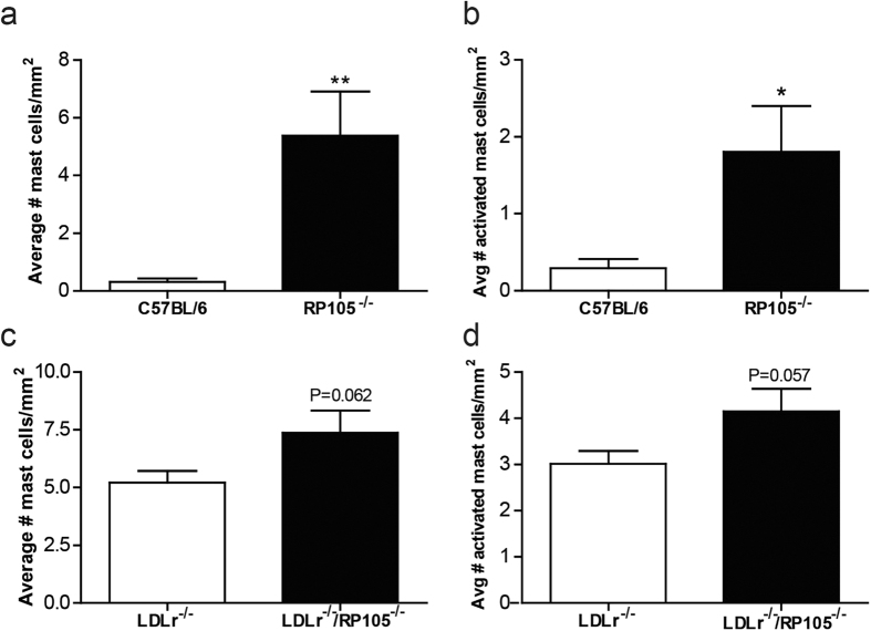
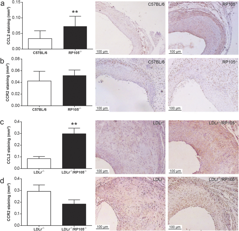
Venous grafts are often used to bypass occlusive atherosclerotic lesions; however, poor patency leads to vein graft disease. Deficiency of TLR4, an inflammatory regulator, reduces vein graft disease. Here, we investigate the effects of the accessory molecule and TLR4 analogue RadioProtective 105 (RP105) on vein graft disease. RP105 deficiency resulted in a 90% increase in vein graft lesion area compared to controls. In a hypercholesterolemic setting (LDLr(-/-)/RP105(-/-) versus LDLr(-/-) mice), which is of importance as vein graft disease is usually characterized by excessive atherosclerosis, total lesion area was not affected. However we did observe an increased number of unstable lesions and intraplaque hemorrhage upon RP105 deficiency. In both setups, lesional macrophage content, and lesional CCL2 was increased. In vitro, RP105(-/-) smooth muscle cells and mast cells secreted higher levels of CCL2. In conclusion, aggravated vein graft disease caused by RP105 deficiency results from an increased local inflammatory response.


Conflict of interest statement

The authors declare no competing financial interests.

Figures

Figure 1
Figure 1. RP105 deficiency aggravates vein graft disease.
Vein graft lesion area was significantly increased in RP105−/− mice compared to control C57BL/6 mice (a). The macrophage content, expressed as the percentage of stained area in the intimal hyperplasia, was higher in RP105−/− mice compared to control (b), while no changes were found in the percentage of smooth muscle cells (ASMA, alpha-smooth muscle cell actin, (c). Collagen content was decreased in RP105−/− mice, indicative of less stable lesions (d). The micrographs show representative images of each group (50x). N = 11 C57BL/6 mice/group, N = 13 RP105−/− mice/group. *P < 0.05.
Figure 2
Figure 2. Lesion size and plaque morphology in LDLr−/−/RP105−/− mice on a western type diet.
No changes were observed in the vessel wall area of LDLr−/−/RP105−/− mice compared to control LDLr−/− mice (a). Similar to mice fed a chow diet, a higher percentage of macrophages was observed in LDLr−/−/RP105−/− (b). Smooth muscle cell content was unaltered between the two groups (ASMA, (c)) and again a decrease in lesional collagen was observed (d). N = 12 LDLr−/− mice/group. N = 12 LDLr−/−/RP105−/− mice/group. *P < 0.05, **P < 0.01.
Figure 3
Figure 3. Increased plaque dissections in LDLr−/−/RP105−/− mice.
The number of plaque dissections in LDLr−/−/RP105−/− mice was significantly higher compared to control LDLr−/− mice (a). Also, the total length of plaque dissections was increased in LDLr−/−/RP105−/− mice compared to control (b). The micrographs show representative images of each group 50x. N = 12 LDLr−/− mice/group. N = 12 LDLr−/−/RP105−/− mice/group. *P < 0.05.
Figure 4
Figure 4. Macrophage proliferation.
Peritoneal macrophages isolated from LDLr−/−/RP105−/− mice showed an increased expression of the cellular proliferation marker Ki67 compared to peritoneal macrophages isolated from LDLr−/− mice (a), *P < 0.05). Intraplaque macrophage proliferation tended to be increased in RP105−/− compared to C57Bl/6 mice (P = 0.06) as demonstrated by a MAC3/Ki67 double staining (b) (right micrographs, N = 11 C57BL/6 mice/group, N = 13 RP105−/− mice/group). No significant differences could be detected in lesional macrophage proliferation between LDLr−/− and LDLr−/−/RP105−/− mice (c). N = 12 LDLr−/− mice/group. N = 12 LDLr−/−/RP105−/− mice/group.
Figure 5
Figure 5. Effect of RP105 deficiency on smooth muscle cell and mast cell function.
After 24 hours stimulation with 1 and 3 ng/mL LPS, RP105−/− vascular smooth muscle cells (v SMCs) secrete dose-dependent increased levels of CCL2 (a), which was significantly higher compared to control v SMCs (N = 4). IL-6 secretion was significantly reduced in RP105−/− v SMCs (b). ***P < 0.001 compared to control v SMCs. ###P < 0.001 compared to unstimulated v SMCs, nd =  not detectable. After 4 hours stimulation with 10 ng/mL and 100 ng/mL LPS, bone marrow derived RP105−/− mast cells secreted dose-dependently increased levels of IL6 (c), TNFα (d) and CCL2 (e), which was significantly higher compared to control mast cells (N = 3, typical example of multiple experiments). The basal proliferation rate of RP105−/− mast cells was significantly higher compared to control mast cells (f) (N = 4). Addition of the supernatant from RP105−/− mast cells to macrophages resulted in increased proliferation compared to the addition of supernatant from control mast cells (g) (N = 4). *P < 0.05. ***P < 0.001 compared to control mast cells. #P < 0.05, ###P < 0.001 compared to unstimulated mast cells.
Figure 6
Figure 6. In vivo mast cell activation.
The amount of perivascular mast cells was markedly increased in the perivascular tissue from vein grafts of RP105 deficient mice compared to control C57BL/6 mice (a). Also, the number of activated mast cells was significantly higher in RP105−/− mice (b). Mast cell numbers, as well as the amount of activated mast cells, showed a trend towards an increase in LDLr−/−/RP105−/− mice fed a western type diet compared to LDLr−/− mice (c,d). N = 11 C57BL/6 mice/group, N = 13 RP105−/− mice/group, N = 12 LDLr−/− mice/group. N = 12 LDLr−/−/RP105−/− mice/group. (1000x). *P < 0.05. **P < 0.01.
Figure 7
Figure 7. In vivo CCL2 and CCR2 staining.
Immunohistochemical stainings revealed a significant increase in the CCL2 positive area in the lesions of RP105−/− mice compared to control C57BL/6 mice (a). Also, the lesional CCL2 area was profoundly increased in hypercholesterolemic LDLr−/−/RP105−/− mice compared to control LDLr−/− mice (c). No differences were observed in lesional CCR2 expression between both experimental groups (b,d). N = 11 C57BL/6 mice/group, N = 13 RP105−/− mice/group, N = 12 LDLr−/− mice/group. N = 12 LDLr−/−/RP105−/− mice/group. **P < 0.01.

Similar articles

Cited by

References

    1. Frostegård J. Immunity, atherosclerosis and cardiovascular disease. BMC Med 11, 117 (2013). - PMC - PubMed
    1. Une D., Kulik A., Voisine P., Le May M. & Ruel M. Correlates of saphenous vein graft hyperplasia and occlusion 1 year after coronary artery bypass grafting: analysis from the CASCADE randomized trial. Circulation 128, S213–8 (2013). - PubMed
    1. Donker J. M. W. et al.. Midterm results of autologous saphenous vein and ePTFE pre-cuffed bypass surgery in peripheral arterial occlusive disease. Vasc Endovascular Surg 45, 598–603 (2011). - PubMed
    1. Al-Sabti H. A. et al.. Saphenous vein graft vs. radial artery graft searching for the best second coronary artery bypass graft. J Saudi Heart Assoc 25, 247–54 (2013). - PMC - PubMed
    1. Fitzgibbon G. M. et al.. Coronary bypass graft fate and patient outcome: angiographic follow-up of 5,065 grafts related to survival and reoperation in 1,388 patients during 25 years. J Am Coll Cardiol 28, 616–26 (1996). - PubMed

Publication types

MeSH terms