. 2016 May 27;90(12):5693-5699.
doi: 10.1128/JVI.00075-16. Print 2016 Jun 15.

Life Cycle Characterization of Sulfolobus Monocaudavirus 1, an Extremophilic Spindle-Shaped Virus with Extracellular Tail Development

Affiliations

Life Cycle Characterization of Sulfolobus Monocaudavirus 1, an Extremophilic Spindle-Shaped Virus with Extracellular Tail Development

Kristine B Uldahl et al. J Virol. .

Abstract

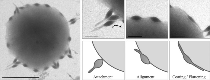
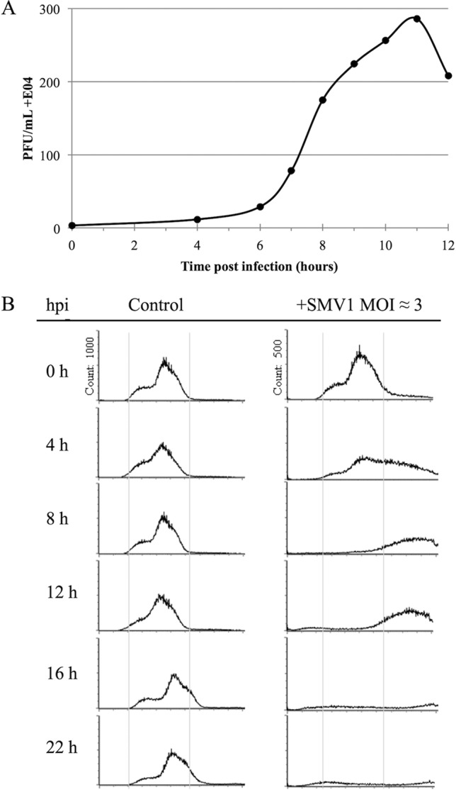
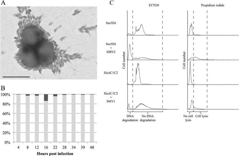
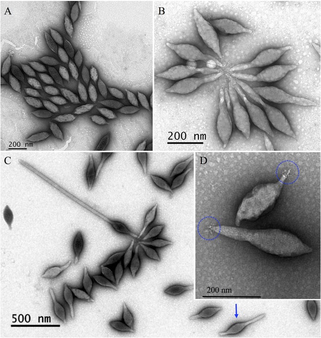
We provide here, for the first time, insights into the initial infection stages of a large spindle-shaped archaeal virus and explore the following life cycle events. Our observations suggest that Sulfolobus monocaudavirus 1 (SMV1) exhibits a high adsorption rate and that virions adsorb to the host cells via three distinct attachment modes: nosecone association, body association, and body/tail association. In the body/tail association mode, the entire virion, including the tail(s), aligns to the host cell surface and the main body is greatly flattened, suggesting a possible fusion entry mechanism. Upon infection, the intracellular replication cycle lasts about 8 h, at which point the virions are released as spindle-shaped tailless particles. Replication of the virus retarded host growth but did not cause lysis of the host cells. Once released from the host and at temperatures resembling that of its natural habitat, SMV1 starts developing one or two tails. This exceptional property of undergoing a major morphological development outside, and independently of, the host cell has been reported only once before for the related Acidianus two-tailed virus. Here, we show that SMV1 can develop tails of more than 900 nm in length, more than quadrupling the total virion length.

Importance: Very little is known about the initial life cycle stages of viruses infecting hosts of the third domain of life, Archaea This work describes the first example of an archaeal virus employing three distinct association modes. The virus under study, Sulfolobus monocaudavirus 1, is a representative of the large spindle-shaped viruses that are frequently found in acidic hot springs. The results described here will add valuable knowledge about Archaea, the least studied domain in the virology field.


Figures

FIG 1
FIG 1
(A and B) Susceptibility of different Sulfolobus strains to SMV1 infection. The OD600 was measured over a time period of 48 h in cultures with (open symbols) or without (filled symbols) the addition of SMV1 (MOI of 5). Cultures were incubated in triplicates at 78°C. (C) Growth inhibition of S. islandicus ΔC1C2 by SMV1 infection at different MOIs (0.01 to 5). Cultures were incubated in triplicates at 78°C.
FIG 2
FIG 2
Kinetics of SMV1 adsorption. Cells were infected with SMV1 using an MOI of 0.1 at 78°C. The number of unbound virus particles was determined at different time points postinfection as described in Materials and Methods.
FIG 3
FIG 3
Electron micrographs of SMV1 interaction with S. islandicus ΔC1C2 cells. Samples were collected at 10 min postinfection and negatively stained for TEM. Left scale bar, 1 μm; right scale bar, 200 nm. The three different virus-host association modes are indicated: attachment, alignment, and coating/flattening.
FIG 4
FIG 4
(A) One-step growth curve of SMV1 infection of S. islandicus ΔC1C2. SMV1 was added at an MOI of about 0.1. The PFU are plotted against time (hours). (B) Flow cytometry time course analysis of S. islandicus ΔC1C2 cells infected by SMV1. Left panel, DNA content distribution from an uninfected culture. Right panel, DNA distribution from a culture infected with SMV1 (MOI ≈ 3). The virus was added just before time point 0 h. The experiment was repeated twice.
FIG 5
FIG 5
(A) Electron micrograph of an S. islandicus ΔC1C2 culture infected with SMV1 (high MOI) at 22 hpi. Scale bar, 1 μm. (B) Time course analysis of the percentage of live (light gray) and dead (dark gray) cells in an S. islandicus ΔC1C2 culture infected with SMV1 (MOI ≈ 3) compared to an uninfected control. (C) Flow cytometry analysis of live/dead staining of S. islandicus ΔC1C2 before and after infection with SMV1 (MOI of 3) at 22 hpi. S. solfataricus 5E6 cells infected with SIRV2 (MOI of 5) were used as a positive control for DNA degradation and cell lysis.
FIG 6
FIG 6
Electron micrographs of different forms of SMV1. (A) Tailless SMV1 particles isolated immediately after release. (B and C) SMV1 develops 1 or 2 tails outside the host cell. The development occurs at high temperatures (>75°C). Often one longer and one shorter tail are observed (blue arrow). (D) Only one pole appears to have short tail fibers, which can attach to tail fibers of other virions to form characteristic rosettes (B). All preparations were negatively stained with 2% uranyl acetate.

References

    1. Menzel P, Gudbergsdottir SR, Rike AG, Lin L, Zhang Q, Contursi P, Moracci M, Kristjansson JK, Bolduc B, Gavrilov S, Ravin N, Mardanov A, Bonch-Osmolovskaya E, Young M, Krogh A, Peng X. 2015. Comparative metagenomics of eight geographically remote terrestrial hot springs. Microb Ecol 70:411–424. doi:10.1007/s00248-015-0576-9. - DOI - PubMed
    1. Narasingarao P, Podell S, Ugalde JA, Brochier-Armanet C, Emerson JB, Brocks JJ, Heidelberg KB, Banfield JF, Allen EE. 2012. De novo metagenomic assembly reveals abundant novel major lineage of Archaea in hypersaline microbial communities. ISME J 6:81–93. doi:10.1038/ismej.2011.78. - DOI - PMC - PubMed
    1. Andersson AF, Banfield JF. 2008. Virus population dynamics and acquired virus resistance in natural microbial communities. Science 320:1047–1050. doi:10.1126/science.1157358. - DOI - PubMed
    1. Mojica KD, Brussaard CP. 2014. Factors affecting virus dynamics and microbial host-virus interactions in marine environments. FEMS Microbiol Ecol 89:495–515. doi:10.1111/1574-6941.12343. - DOI - PubMed
    1. Atanasova NS, Roine E, Oren A, Bamford DH, Oksanen HM. 2012. Global network of specific virus-host interactions in hypersaline environments. Environ Microbiol 14:426–440. doi:10.1111/j.1462-2920.2011.02603.x. - DOI - PubMed

LinkOut - more resources