Review
. 2016 Jun 1;310(11):R1193-211.
doi: 10.1152/ajpregu.00250.2015. Epub 2016 Apr 6.

Mechanisms of animal diapause: recent developments from nematodes, crustaceans, insects, and fish

Affiliations
Review

Mechanisms of animal diapause: recent developments from nematodes, crustaceans, insects, and fish

Steven C Hand et al. Am J Physiol Regul Integr Comp Physiol. .

Abstract

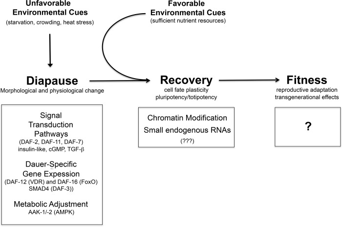
Life cycle delays are beneficial for opportunistic species encountering suboptimal environments. Many animals display a programmed arrest of development (diapause) at some stage(s) of their development, and the diapause state may or may not be associated with some degree of metabolic depression. In this review, we will evaluate current advancements in our understanding of the mechanisms responsible for the remarkable phenotype, as well as environmental cues that signal entry and termination of the state. The developmental stage at which diapause occurs dictates and constrains the mechanisms governing diapause. Considerable progress has been made in clarifying proximal mechanisms of metabolic arrest and the signaling pathways like insulin/Foxo that control gene expression patterns. Overlapping themes are also seen in mechanisms that control cell cycle arrest. Evidence is emerging for epigenetic contributions to diapause regulation via small RNAs in nematodes, crustaceans, insects, and fish. Knockdown of circadian clock genes in selected insect species supports the importance of clock genes in the photoperiodic response that cues diapause. A large suite of chaperone-like proteins, expressed during diapause, protects biological structures during long periods of energy-limited stasis. More information is needed to paint a complete picture of how environmental cues are coupled to the signal transduction that initiates the complex diapause phenotype, as well as molecular explanations for how the state is terminated. Excellent examples of molecular memory in post-dauer animals have been documented in Caenorhabditis elegans It is clear that a single suite of mechanisms does not regulate diapause across all species and developmental stages.

Keywords: cell cycle; development; diapause; dormancy; metabolism.


Figures

Fig. 1.
Fig. 1.
A molecular summary of dauer and its consequences in Caenorhabditis elegans. The diapause-like dauer stage is regulated by signal transduction pathways that control the decision between continuous or dauer development. Key effectors of each of these pathways are indicated: DAF-2, an insulin-like receptor; DAF-11,-a guanylyl cyclase transmembrane receptor required for chemosensory signaling and the cGMP regulatory branch of dauer formation; and DAF-7, a TGF-β-like molecule. Execution of dauer development results in a change in gene expression that is mediated by transcription factors that adjust gene expression to mediate developmental and physiological modification. Upon recovery, the dauer genome is altered compared with nondauer larvae, which is reflected in the modified distribution of chromatin marks and the small RNA population. These changes likely change the genomic ground state, allowing for the correction of specific mutant phenotypes. Precisely how these effects on gene expression and metabolism impinge on brood size and lifespan remain to be elucidated.
Fig. 2.
Fig. 2.
A: aerial view of diapausing embryos of Artemia franciscana floating in wind-blown “streaks” on the Great Salt Lake, Utah, in autumn. The streaks of embryos typically are kilometers long. B: kilogram quantities of diapausing embryos are easily collected from these streaks on the lake surface.
Fig. 3.
Fig. 3.
Product-to-substrate ratios for diapausing embryos (open bars) of Artemia franciscana compared with postdiapause embryos (shaded bars). The significantly lower values seen in diapause indicate inhibition at the trehalase (A), hexokinase (B), pyruvate kinase (C), and pyruvate dehydrogenase (D) reactions in the pathway for trehalose catabolism. Values are expressed as means ± SE for n = 4 samples in A–C and n = 6 for D. *Statistical significance with P < 0.0001. [Modified from Patil Y, Marden B, Brand MD, SC. “Metabolic downregulation and inhibition of carbohydrate catabolism.” Physiol Biochem Zool 86: 111–113, 2013, published by the University of Chicago; Ref. 142].
Fig. 4.
Fig. 4.
Aspects of mitochondrial energetics during active metabolism (A) vs. the diapause state (B), as predicted for embryos of Artemia franciscana. A: the TCA cycle and electron transport system (ETS) are active with high oxygen consumption measurable. Under this condition, a large ΔpH and ΔΨ would be predicted with an alkaline pH present in the matrix. The F1Fo ATP synthase (ComplexV) operates in the direction of ATP production, and the adenine nucleotide translocator (ANT) and phosphate carrier would run in their typical directions. Alkaline conditions in the matrix could potentially foster the tetrameric (inactive) state of the inhibitor protein IF1. B: during diapause, carbon delivery to the mitochondrion is arrested and the TCA cycle, ETS, and respiration are depressed, which would be predicted to result in loss of membrane potential and proton gradient, because proton conductance is known not to change during diapause compared with the active state in mitochondria from A. francicana. Consequently, the F1Fo ATP synthase and ANT could potentially reverse, leading to consumption of cellular ATP stores. Proton leak would promptly cease due to loss of ΔpH. However, under the more acidic conditions in the matrix, one might hypothesize that the IF1 protein might depolymerize to active dimers and bind to the F1Fo ATP synthase, thereby preventing reversal of Complex V.
Fig. 5.
Fig. 5.
Knock-down experiments targeting the clock genes shown in this figure suggest that suppression of period (per), timeless (tim), or cryptochrome2 (cry2) leads to elevation of pigment dispersing factor (pdf) and, consequently, the nondiapause phenotype in the mosquito Culex pipiens. By contrast, knocking down pdf in females reared under long daylengths generates the diapause phenotype. These observations suggest that per, tim, and cry2 transcripts are elevated under short-day, diapause-inducing conditions and that pdf is elevated under long day, diapause-averting conditions. Two additional clock elements shown here, clock (clk) and cycle (cyc), remain to be tested. [Based on results discussed in Refs. and 130].
Fig. 6.
Fig. 6.
Model depicting a role for the insulin/FoxO signaling pathway in diapause regulation of the mosquito Culex pipiens. A: in response to the long daylengths of early summer, insulin signaling leads to juvenile hormone (JH) synthesis and ovarian development and the concurrent suppression of the transcription factor Foxo. B: in response to short daylengths of autumn, insulin signaling is shut down, allowing the activation of FoxO, which, in turn, is hypothesized to generate many features of the diapause phenotype. [Based on results discussed in Refs. and 172].
Fig. 7.
Fig. 7.
A proposed model for the environmental alternation of maternally programmed diapause in embryos of the annual killifish Austrofundulus limnaeus. In this model, embryos are programmed to enter into diapause II through the provisioning of specific maternal mRNA transcripts during oogenesis. In response to the correct environmental signals, such as increases in incubation temperature or exposure to long-day photoperiods, the developing embryo expresses specific miR-430 microRNAs that clear the maternal mRNAs and favor direct development. Preliminary data suggest overexpression of miR-430 RNAs in embryos that bypass diapause II due to increased incubation temperature (Romney and Podrabsky J, personal observation).
Fig. 8.
Fig. 8.
Initial survival is high in diapause II embryos of Austrofundulus limnaeus exposed to UV-C light at 254 nm and allowed to recover in the dark. However, after several weeks, embryos begin to die, and even more embryos die during postdiapause II development after diapause is experimentally broken. Symbols are means ± SE (n = 3) *Significantly different from control levels (P < 005). Wk = weeks of recovery in the dark. Final = final survival of embryos that developed to diapause III 31 days after diapause II was experimentally broken. [Modified from Wagner JT, Podrabsky JE. “Extreme tolerance and developmental buffering of UV-C-induced DNA damage in embryos of the annual killifish Austrofundulus limnaeus.”J Exp Zool A Ecol Genet Physiol 323A: 10–30, 2015; Published by John Wiley and Sons; Ref. 188].
Fig. 9.
Fig. 9.
Survival (top) and proportion of abnormal embryos (bottom) of Austrofundulus limnaeus following exposure to UV-C radiation at 254 nm. Embryos were exposed during the dispersed cell phase (4 dpf) or after formation of the embryonic axis (10 dpf) and allowed to recover in the light or dark. Note the lack of abnormal embryos at 4 dpf, even at levels of irradiation that cause significant teratogenic effects in 10 dpf embryos. [Modified from Wagner JT, Podrabsky JE. “Extreme tolerance and developmental buffering of UV-C-induced DNA damage in embryos of the annual killifish Austrofundulus limnaeus.”J Exp Zool A Ecol Genet Physiol 323A: 10–30, 2015; Published by John Wiley and Sons; Ref. 188].

References

    1. Adam S, Polo SE. Blurring the line between the DNA damage response and transcription: The importance of chromatin dynamics. Exp Cell Res 329: 148–153, 2014. - PMC - PubMed
    1. Altiero T, Guidetti R, Caselli V, Cesari M, Rebecchi L. Ultraviolet radiation tolerance in hydrated and desiccated eutardigrades. J Zool System Evol Res 49: 104–110, 2011.
    1. Anchordoguy TJ, Hand SC. Acute blockage of the ubiquitin-mediated proteolytic pathway during invertebrate quiescence. Am J Physiol Regul Integr Comp Physiol 267: R895–R900, 1994. - PubMed
    1. Anchordoguy TJ, Hand SC. Reactivation of ubiquitination in Artemia franciscana embryos during recovery from anoxia-induced quiescence. J Exp Biol 198: 1299–1305, 1995. - PubMed
    1. Anchordoguy TJ, Hofmann G, Hand SC. Extension of enzyme half-life during quiescence in Artemia embryos. Am J Physiol Regul Integr Comp Physiol 264: R85–R89, 1993. - PubMed

Publication types

LinkOut - more resources