. 2016 Jun 1;25(11):2182-2193.
doi: 10.1093/hmg/ddw085. Epub 2016 Apr 5.

A mouse model of a human congenital disorder of glycosylation caused by loss of PMM2

Affiliations

A mouse model of a human congenital disorder of glycosylation caused by loss of PMM2

Barden Chan et al. Hum Mol Genet. .

Abstract

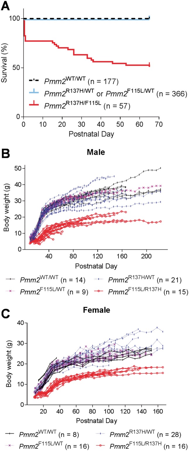
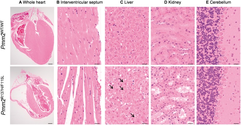
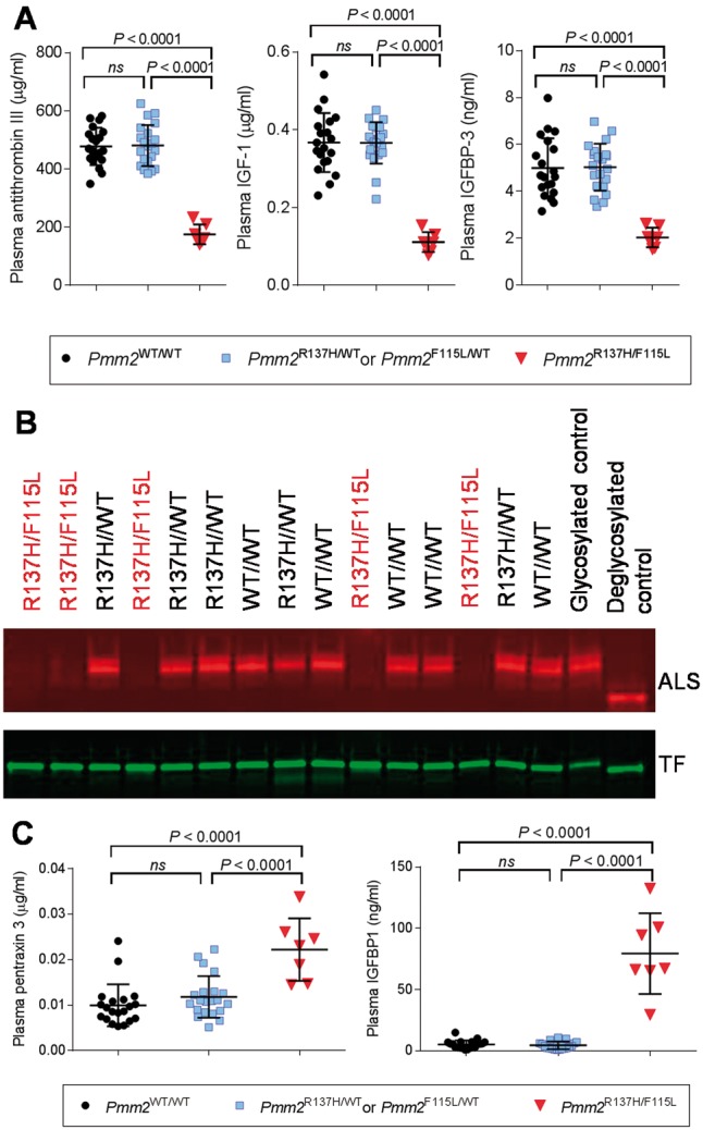
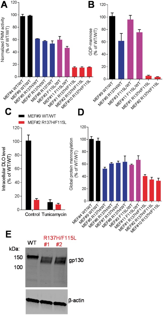
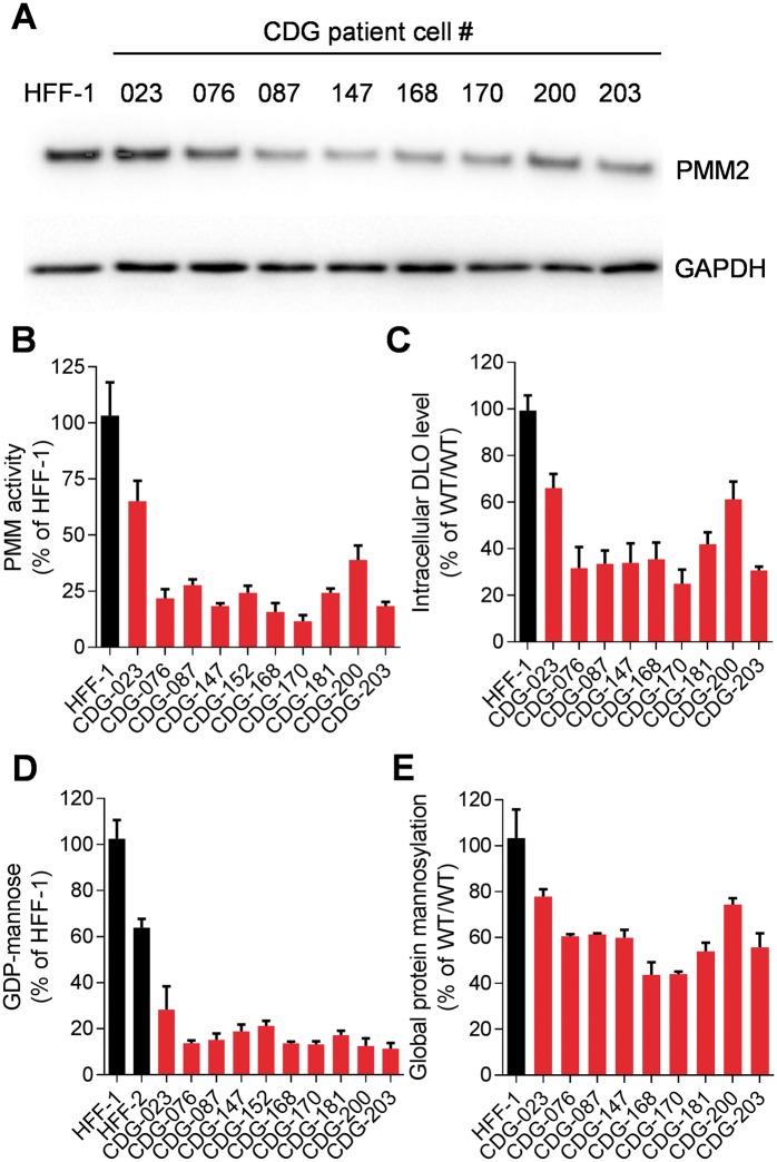
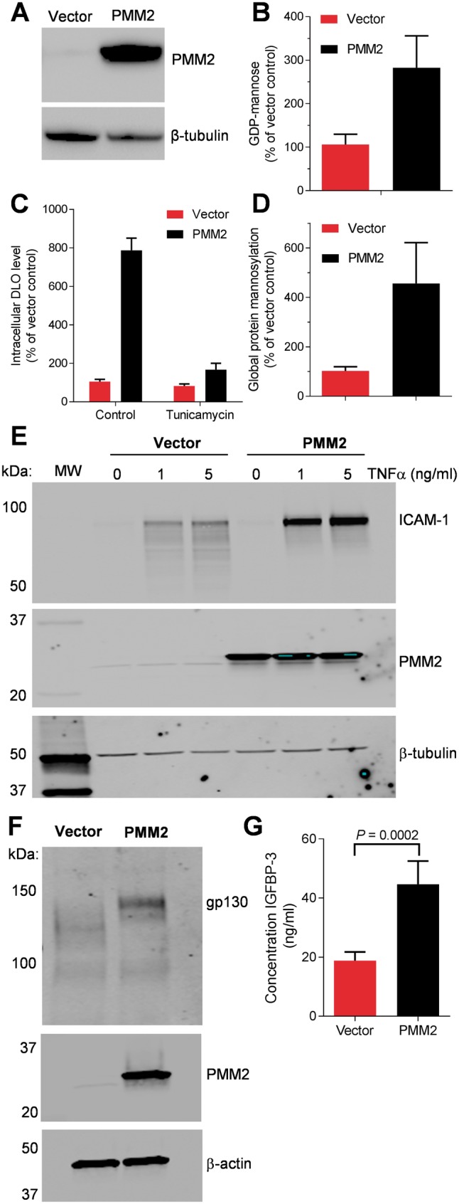
The most common congenital disorder of glycosylation (CDG), phosphomannomutase 2 (PMM2)-CDG, is caused by mutations in PMM2 that limit availability of mannose precursors required for protein N-glycosylation. The disorder has no therapy and there are no models to test new treatments. We generated compound heterozygous mice with the R137H and F115L mutations in Pmm2 that correspond to the most prevalent alleles found in patients with PMM2-CDG. Many Pmm2R137H/F115L mice died prenatally, while survivors had significantly stunted growth. These animals and cells derived from them showed protein glycosylation deficiencies similar to those found in patients with PMM2-CDG. Growth-related glycoproteins insulin-like growth factor (IGF) 1, IGF binding protein-3 and acid-labile subunit, along with antithrombin III, were all deficient in Pmm2R137H/F115L mice, but their levels in heterozygous mice were comparable to wild-type (WT) littermates. These imbalances, resulting from defective glycosylation, are likely the cause of the stunted growth seen both in our model and in PMM2-CDG patients. Both Pmm2R137H/F115L mouse and PMM2-CDG patient-derived fibroblasts displayed reductions in PMM activity, guanosine diphosphate mannose, lipid-linked oligosaccharide precursor and total cellular protein glycosylation, along with hypoglycosylation of a new endogenous biomarker, glycoprotein 130 (gp130). Over-expression of WT-PMM2 in patient-derived fibroblasts rescued all these defects, showing that restoration of mutant PMM2 activity is a viable therapeutic strategy. This functional mouse model of PMM2-CDG, in vitro assays and identification of the novel gp130 biomarker all shed light on the human disease, and moreover, provide the essential tools to test potential therapeutics for this untreatable disease.


Figures

Figure 1.
Figure 1.
Survival and growth of Pmm2R137H/F115L mice was reduced. (A) 600 pups from 80 matings of Pmm2R137H/WT and Pmm2F115L/WT were monitored for postnatal lethality. The Pmm2R137H/F115L genotype induced postnatal death. The four deaths in the heterozygotes all occurred on postnatal Day 1 due to abandonment of pups by young mothers because the mother was disturbed. Pmm2R137H/F115L mice demonstrated lower body weight in males (B) and females (C) throughout the period monitored. Only litters with surviving Pmm2R137H/F115L mice were used to generate B and C.
Figure 2
Figure 2
Surviving Pmm2R137H/F115L mice showed additional postnatal defects in multiple organ systems. Three pairs of sex-matched 4-week-old Pmm2WT/WT and Pmm2R137H/F115L mice were examined. Representative H&E staining of sections from (A) the whole heart (bar = 500 μm), (B) interventricular septum, (C) liver, (D) kidney and (E) cerebellum shown (bar = 20 μm for B, C, D and E). A and B demonstrate the decreased wall thickness of both ventricles and the interventricular septum, and decreased diameter (amount of cytoplasm) of individual myofibers in Pmm2R137H/F115L compared with Pmm2WT/WT mice. C demonstrates the accumulation of cytoplasmic hyaline bodies in the liver of the mutant mice, indicated by arrows. D demonstrates the tubular dilation observed in the kidneys of mutant mice. E illustrates the lack of an effect on the Purkinje cell density in the cerebellum of mutant mice.
Figure 3
Figure 3
Pmm2R137H/F115L mice have significantly altered levels of disease-relevant plasma biomarkers. (A) Mean (±SD) levels of ATIII, IGF-1 and IGFBP-3 measured by ELISA in plasma of Pmm2WT/WT (n = 21), Pmm2R137H/WT or Pmm2F115L/WT (n = 20) and Pmm2R137H/F115L (n = 7) mice. P-values were generated using one-way ANOVA test followed by Tukey post-hoc tests for multiple comparisons. (B) Glycosylation and expression level of ALS and transferrin in mouse plasma (genotype indicated above, hypomorphic mutants labeled in red). The two lanes on the far right are controls. Glycosylated control was a sample of normal B6 mouse plasma. Deglycosylated control was a sample of normal B6 mouse plasma de-glycosylated in vitro by N-glycanase. Note an increase in migration rate for both ALS and transferrin after N-glycanase treatment. (C) Mean (±SD) levels of PTX3 and IGFBP-1 measured by ELISA in plasma of Pmm2WT/WT (n = 21), Pmm2R137H/WT or Pmm2F115L/WT (n = 20) and Pmm2R137H/F115L (n = 7) mice. P-values were generated using one-way ANOVA test followed by Tukey post-hoc tests for multiple comparisons.
Figure 4
Figure 4
Mutant Pmm2 knock-in mice have reduced PMM activity. (A) Total PMM enzymatic activity was reduced by Pmm2 R137H and F115L mutations. MEFs isolated from individual mice were numerically labeled. (B) GDP-mannose level was significantly reduced in Pmm2R137H/F115L MEFs when incubated for 5 h without glucose. This difference was rescued in vitro by high glucose concentration (data not shown) as reported previously (26). (C) DLO levels in Pmm2R137H/F115L MEFs were significantly lower than that in Pmm2WT/WT. Levels of endogenous DLO were completely suppressed by addition of tunicamycin at 10 μg/ml overnight prior to harvesting. (D) Global mannosylation was reduced in Pmm2-mutant MEFs as measured by incorporation of 3H mannose into cellular protein fractions. (E) Hypoglycosylation of gp130 was detected in Pmm2R137H/F115L MEFs as judged by SDS-PAGE migration detected by western blotting. Each experiment was repeated at least twice. In AD, representative data from one experiment are shown as mean ± SD from triplicate.
Figure 5
Figure 5
PMM2 pathway activity characterization of PMM2-CDG patient fibroblasts. (A) Western blot showing lower expression level of PMM2 protein in a panel of PMM2-CDG patient fibroblasts (B) PMM activity was diminished in multiple PMM2-CDG patient fibroblast cell lines with different genotypes compared with normal fibroblasts. Data represent the average of three assays run in quadruplicate and error bars represent SD. (C) DLO levels were reduced in patient fibroblasts. (D) Intracellular levels of GDP-mannose were diminished in multiple PMM2-CDG patient fibroblast cell lines with different genotypes compared with normal fibroblasts, under glucose-free conditions. Data represent the average of three experiments and error bars represent SD. This difference was rescued in vitro by high glucose concentration (data not shown) as reported previously (26). (E) Protein mannosylation levels were also reduced in patient fibroblasts. Each experiment was repeated at least twice. Representative data from one experiment are shown as mean±SD from triplicates.
Figure 6
Figure 6
WT-PMM2 over-expression restored PMM2 pathway activity and rescued expression of three endogenous glycoproteins in CDG-168 patient fibroblasts. (A) Western blot showing over-expression of human WT-PMM2 in CDG-168 cells. (B) GDP-mannose, (C) DLO and (D) global mannosylation levels were all elevated by WT-PMM2 over-expression (mean ± SD from four independent experiments). For B and C, each experiment was repeated at least twice. Representative data from one experiment are shown as mean±SD from triplicate. (E) CDG-168 cells with and without WT-PMM2 over-expression were treated with TNFα at the indicated concentrations for 18 h, cell lysates were prepared and equal volumes of sample were probed for ICAM-1, PMM2 and β-tubulin (as a loading control) by western blotting. (F) CDG-168 cells with and without WT-PMM2 over-expression were probed for gp130, PMM2 and β-actin levels. (G) Human IGFBP-3 levels in cell culture supernatants were analyzed by ELISA, mean ± SD, statistical significance tested with Student t-test. Representative data from three independent experiments are shown.

Similar articles

Cited by

References

    1. Freeze H.H, Schachter H. (2009) Genetic disorders of glycosylation In Varki A., Cummings R.D., Esko J.D., Freeze H.H., Stanley P., Bertozzi C.R., Hart G.W., Etzler M.E. (eds), Essentials of Glycobiology. Cold Spring Harbor Laboratory Press, Cold Spring Harbor, NY, Chapter 42. - PubMed
    1. Cylwi B., Naklicki M., Chrostek L., Gruszewska E. (2013) Congenital disorders of glycosylation. Part I. Defects of protein N-glycosylation. Acta Biochim. Pol, 20, 151–161. - PubMed
    1. Freeze H.H. (2009) Towards a therapy for phosphomannomutase 2 deficiency, the defect in CDG-Ia patients. Biochim. Biophys. Acta, 1792, 835–840. - PMC - PubMed
    1. Jaeken J., van Eijk H.G., van der Heul C., Corbeel L., Eeckels R., Eggermont E. (1984) Sialic acid-deficient serum and cerebrospinal fluid transferrin in a newly recognized genetic syndrome. Clin. Chim. Acta, 144, 245–247. - PubMed
    1. Sharma V., Ichikawa M., Freeze H.H. (2014) Mannose metabolism: More than meets the eye. Biochem. Biophys. Res. Commun., 453, 220–228. - PMC - PubMed

Publication types