

Proposal of a computed tomography classification for hepatic alveolar echinococcosis
- PMID: 27053854
- PMCID: PMC4814648
- DOI: 10.3748/wjg.v22.i13.3621
Proposal of a computed tomography classification for hepatic alveolar echinococcosis
Abstract
Aim: To establish a computed tomography (CT)-morphological classification for hepatic alveolar echinococcosis was the aim of the study.
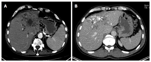
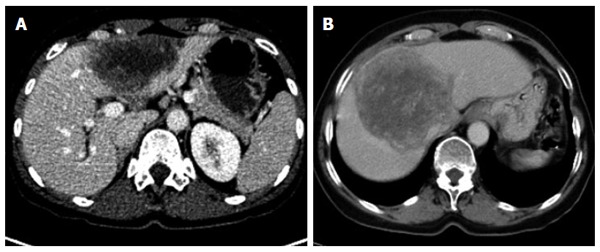
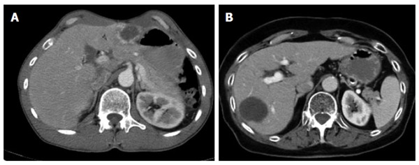
Methods: The CT morphology of hepatic lesions in 228 patients with confirmed alveolar echinococcosis (AE) drawn from the Echinococcus Databank of the University Hospital of Ulm was reviewed retrospectively. For this reason, CT datasets of combined positron emission tomography (PET)-CT examinations were evaluated. The diagnosis of AE was made in patients with unequivocal seropositivity; positive histological findings following diagnostic puncture or partial resection of the liver; and/or findings typical for AE at either ultrasonography, CT, magnetic resonance imaging or PET-CT. The CT-morphological findings were grouped into the new classification scheme.
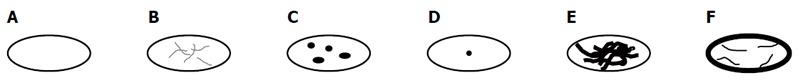
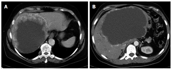
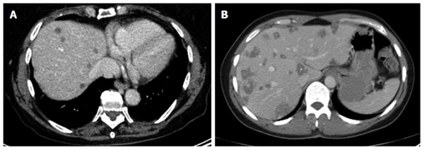
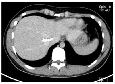
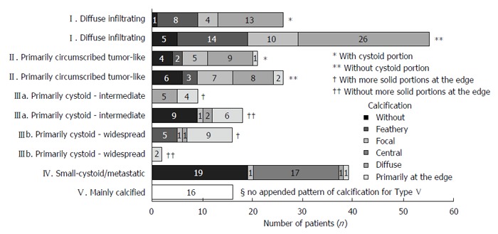
Results: Within the classification a lesion was dedicated to one out of five "primary morphologies" as well as to one out of six "patterns of calcification". "primary morphology" and "pattern of calcification" are primarily focussed on separately from each other and combined, whereas the "primary morphology" V is not further characterized by a "pattern of calcification". Based on the five primary morphologies, further descriptive sub-criteria were appended to types I-III. An analysis of the calcification pattern in relation to the primary morphology revealed the exclusive association of the central calcification with type IV primary morphology. Similarly, certain calcification patterns exhibited a clear predominance for other primary morphologies, which underscores the delimitation of the individual primary morphological types from each other. These relationships in terms of calcification patterns extend into the primary morphological sub-criteria, demonstrating the clear subordination of those criteria.
Conclusion: The proposed CT-morphological classification (EMUC-CT) is intended to facilitate the recognition and interpretation of lesions in hepatic alveolar echinococcosis. This could help to interpret different clinical courses better and shall assist in the context of scientific studies to improve the comparability of CT findings.
Keywords: Alveolar echinococcosis; Classification; Computed tomography; Diagnosis; Echinococcus multilocularis; Hepatic alveolar echinococcosis.
Figures
References
-
- Brunetti E, Kern P, Vuitton DA, Writing Panel for the WHO-IWGE. Expert consensus for the diagnosis and treatment of cystic and alveolar echinococcosis in humans. Acta Trop. 2010;114:1–16. - PubMed
-
- Stojkovic M, Junghanss T. Cystic and alveolar echinococcosis. Handb Clin Neurol. 2013;114:327–334. - PubMed
-
- Moro P, Schantz PM. Echinococcosis: a review. Int J Infect Dis. 2009;13:125–133. - PubMed
-
- Romig T, Dinkel A, Mackenstedt U. The present situation of echinococcosis in Europe. Parasitol Int. 2006;55 Suppl:S187–S191. - PubMed
MeSH terms
LinkOut - more resources
Full Text Sources
Other Literature Sources
