Review
. 2016 Apr 6:6:16.
doi: 10.1186/s13395-016-0086-6. eCollection 2016.

The beneficial role of proteolysis in skeletal muscle growth and stress adaptation

Affiliations
Review

The beneficial role of proteolysis in skeletal muscle growth and stress adaptation

Ryan A V Bell et al. Skelet Muscle. .

Erratum in

Abstract

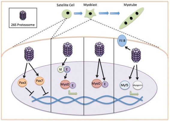
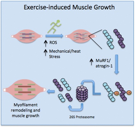
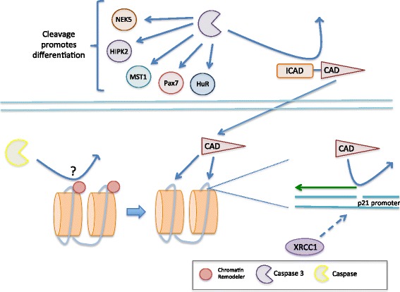
Muscle atrophy derived from excessive proteolysis is a hallmark of numerous disease conditions. Accordingly, the negative consequences of skeletal muscle protein breakdown often overshadow the critical nature of proteolytic systems in maintaining normal cellular function. Here, we discuss the major cellular proteolysis machinery-the ubiquitin/proteosome system, the autophagy/lysosomal system, and caspase-mediated protein cleavage-and the critical role of these protein machines in establishing and preserving muscle health. We examine how ordered degradation modifies (1) the spatiotemporal expression of myogenic regulatory factors during myoblast differentiation, (2) membrane fusion during myotube formation, (3) sarcomere remodeling and muscle growth following physical stress, and (4) energy homeostasis during nutrient deprivation. Finally, we review the origin and etiology of a number of myopathies and how these devastating conditions arise from inborn errors in proteolysis.

Keywords: Autophagy; Caspase; Muscle cell differentiation; Muscle growth; Proteasome; Proteolysis.


Figures

Fig. 1
Fig. 1
The role of the UPP in skeletal muscle cell differentiation. The fidelity of muscle cell differentiation is dependent upon the spatiotemporal expression of particular myogenic proteins. Indeed, UPP involvement in satellite cell differentiation begins with its role in the removal of Pax3 and Pax7, which maintain satellite cells in their stem cell niche. Further, the 26S proteasome appears critical for the early activation of a key myogenic factor, MyoD, through the removal of an endogenous MyoD inhibitor, Id. The continuation of the myogenic program relies on UPP-dependent degradation of MyoD and its binding partner E2A (E), as well as Myf5, myogenin, and filamin B (Fil B) during later stages of differentiation
Fig. 2
Fig. 2
The role of the UPP in skeletal muscle growth. Exercise-induced protein damage via increased ROS/mechanical and heat stress necessitates an increase in proteasome-mediated proteolysis to rid the cells of non-functional myofibrillar proteins. This is typically dependent on a prerequisite increase in key muscle-specific ubiquitin ligases, MuRF1 and atrogin-1 (MAFbx), which ubiquitinate and target damaged proteins for degradation by the 26S proteasome. Efficient removal of damaged proteins is critical to skeletal muscle growth and remodeling following exercise
Fig. 3
Fig. 3
Exercise- and starvation-induced autophagy pathways and their beneficial role in muscle stress adaptation. Nutrient deprivation decreases signaling through insulin/growth factor receptors, which decreases Akt activation and allows for the AMPK-dependent phosphorylation of FoxO3. FoxO3 is then able to translocate into the nucleus and initiate the transcription of autophagy-related genes. Activated AMPK also phosphorylates mTORC1 (preventing its action on ULK1, a key autophagy-related kinase) and ULK1 to allow for efficient autophagosome formation and clearance of encapsulated material. Moreover, the lack of intake of essential amino acids further prevents mTORC1 activation and promotes autophagy induction. Taken together, these processes recycle nutrients for muscle cells and the body as a whole during lean periods. While starvation-induced autophagy is undoubtedly a part of muscle biochemistry during exercise, physical activity also activates beclin-1 through its phosphorylation-dependent release from the BCL2-beclin-1 complex. Beclin-1 is critical to autophagosome formation and the efficient clearance of damaged organelles and proteins that arise from physical stress
Fig. 4
Fig. 4
The role of caspases in skeletal myoblast differentiation. Caspase 3 has a multifaceted role in regulating myogenesis. It is responsible for the proteolytic cleavage of the transcription factor Pax7, which maintains satellite cells in their stem cell niche and prevents myoblast differentiation. Moreover, caspase 3 cleaves the promyogenic kinases MST1, HIPK2, and NEK5 to promote myogenesis. The posttranscriptional regulator, HuR, is also cleaved by caspase 3 and is necessary for muscle fiber formation. Additionally, preliminary evidence (unpublished) suggests that the myogenic differentiation program appears to rely on the caspase-mediated cleavage of chromatin remodeling proteins to increase DNA accessibility for CAD (activated by caspase 3 cleavage of ICAD), which produces DNA strand breaks that are critical to regulating myogenic gene expression. For instance, CAD cleavage of the p21 promoter stimulates p21 expression, which is essential for cell cycle arrest and terminal differentiation. The CAD-derived DNA strand breaks require rapid resolution, which is mediated by the base excision repair protein XRCC1. This mending of DNA strand breaks is necessary to stabilize the genome and ensure the fidelity of the myogenic differentiation program

References

    1. Abu Hatoum O, Gross-Mesilaty S, Breitschopf K, Hoffman A, Gonen H, Ciechanover A, et al. Degradation of myogenic transcription factor MyoD by the ubiquitin pathway in vivo and in vitro: regulation by specific DNA binding. Mol Cell Biol. 1998;18:5670–5677. doi: 10.1128/MCB.18.10.5670. - DOI - PMC - PubMed
    1. Al-Khalaf MH, Blake LE, Larsen BD, Bell RAV, Brunette S, Parks RJ, et al. Temporal activation of XRCC1-mediated DNA repair is essential for muscle differentiation. Cell Disc. 2016. doi:10.1038/celldisc.2015.41. - PMC - PubMed
    1. Augusto V, Padovani CR, Campos GER. Skeletal muscle fibre types in C57BL6J mice. Braz J Morphol Sci. 2004;21:89–94.
    1. Angelin A, Tiepolo T, Sabatelli P, Grumati P, Bergamin N, Golfieri C, et al. Mitochondrial dysfunction in the pathogenesis of Ullrich congenital muscular dystrophy and prospective therapy with cyclosporins. Proc Natl Acad Sci U S A. 2007;104:991–6. - PMC - PubMed
    1. Askanas V, Engel WK, Nogalaska A. Sporadic inclusion-body myositis: a degenerative muscle disease associated with aging, impaired muscle protein homeostasis and abnormal mitophagy. Biochim Biophys Acta. 2015;1852:633–643. doi: 10.1016/j.bbadis.2014.09.005. - DOI - PubMed

Publication types

MeSH terms

LinkOut - more resources