. 2016 Apr 7;12(4):e1005932.
doi: 10.1371/journal.pgen.1005932. eCollection 2016 Apr.

Mutational History of a Human Cell Lineage from Somatic to Induced Pluripotent Stem Cells

Affiliations

Mutational History of a Human Cell Lineage from Somatic to Induced Pluripotent Stem Cells

Foad J Rouhani et al. PLoS Genet. .

Abstract

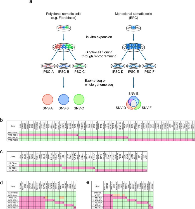
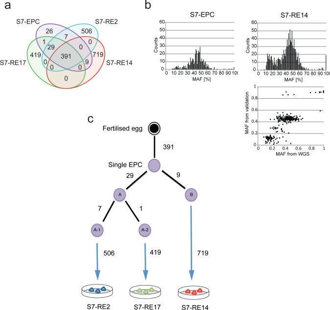
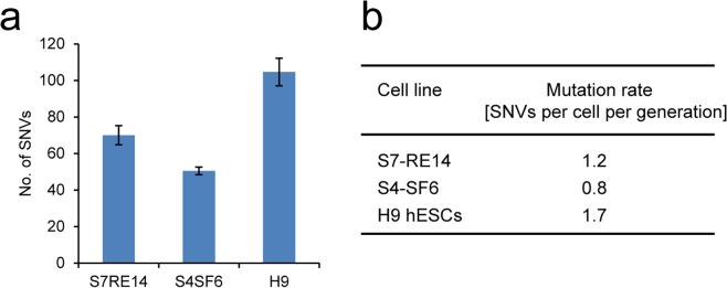
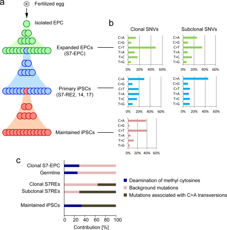
The accuracy of replicating the genetic code is fundamental. DNA repair mechanisms protect the fidelity of the genome ensuring a low error rate between generations. This sustains the similarity of individuals whilst providing a repertoire of variants for evolution. The mutation rate in the human genome has recently been measured to be 50-70 de novo single nucleotide variants (SNVs) between generations. During development mutations accumulate in somatic cells so that an organism is a mosaic. However, variation within a tissue and between tissues has not been analysed. By reprogramming somatic cells into induced pluripotent stem cells (iPSCs), their genomes and the associated mutational history are captured. By sequencing the genomes of polyclonal and monoclonal somatic cells and derived iPSCs we have determined the mutation rates and show how the patterns change from a somatic lineage in vivo through to iPSCs. Somatic cells have a mutation rate of 14 SNVs per cell per generation while iPSCs exhibited a ten-fold lower rate. Analyses of mutational signatures suggested that deamination of methylated cytosine may be the major mutagenic source in vivo, whilst oxidative DNA damage becomes dominant in vitro. Our results provide insights for better understanding of mutational processes and lineage relationships between human somatic cells. Furthermore it provides a foundation for interpretation of elevated mutation rates and patterns in cancer.


Conflict of interest statement

The authors have declared that no competing interests exist.

Figures

Fig 1
Fig 1. Comparing acquired SNVs in iPSCs derived from a polyclonal or a monoclonal origin.
a. Schematic comparing the reprogramming of polyclonal and monoclonal cells. Polyclonal cells such as fibroblasts (left panel) give rise to iPSCs which do not share a majority of mutations since they are derived from different progenitors. In contrast, iPSCs derived from monoclonal cells (right panel) such as EPCs share a proportion of their mutations and carry private mutations specific to each line. b, c. Exome sequencing of iPSCs generated using fibroblasts from two different individuals, a 65-year-old alpha-1 antitrypsin deficiency patient (AATD) (b) and a healthy subject, S2 (c). The data for iPSC-B were taken from our previous work [11]. Each column represents one SNV in the indicated gene. Duplicated genes indicate two adjacent SNVs. Green, mutation absent; pink, mutation present. See S2 and S3 Tables for the complete description. d, e. Exome sequencing of iPSC lines generated using monoclonal EPCs from the same AATD patient in b as well as a healthy subject, S7 (e). Orange, mutation detected by amplicon resequencing. See S4 and S5 Tables for the complete description.
Fig 2
Fig 2. Whole genome sequencing and detailed analysis of iPSCs derived from a monoclonal somatic cell reveal lineage relationships in vitro.
a. Venn diagram showing the overlap of mutations found in each cell line. b. Histogram showing mutant allele frequencies (MAF) of SNVs found in EPCs and S7-RE14. Note the presence of sub-clonal SNVs (<30% MAF). MAFs observed from amplicon resequencing revealed 3 distinct sub-populations, which represent clonal SNVs, SNVs fixed during the first and second cell division (bottom panel). c. Cellular phylogenetic tree showing the relationship between the first and second cell divisions of the originating EPC and the subsequent iPSC line. The minimum number of mutations (taking into account limitations of the sensitivity of detecting sub-clonal mutations by whole genome sequence) accrued by each daughter cell is shown.
Fig 3
Fig 3. Mutation rate of human pluripotent cells in culture.
a. The mean numbers of SNVs accumulated during 60 cell divisions in 2 iPSC lines, S7-RE14 (n = 3) and S4-SF6 (n = 2) and a human ESC line H9 (n = 3). Data are shown as mean ± SD. b. Mutation rate per cell per division in each pluripotent cell line.
Fig 4
Fig 4. Mutational signatures in vivo, in vitro and through reprogramming.
a. Schematic showing the longitudinal progression from in vivo development, in vitro culture of somatic cells through reprogramming and finally through to the experimental set-up used to calculate the mutation rate in iPSC maintenance culture. b. Mutational spectrum of SNVs found in the EPCs (top), and primary (middle) and sub-cloned (bottom) S7-RE14 iPSC lines. Clonal (left) and sub-clonal (right) mutations were shown separately. c. Contribution of mutational processes identified by the NNMF analysis. The germ line mutations described in ref 18 were analysed [18]. The NNMF analysis was not performed for the mutations in sub-clonal S7-EPC due to the limited number of mutations available.

References

    1. Stratton MR, Campbell PJ, Futreal PA (2009) The cancer genome. Nature 458: 719–724. 10.1038/nature07943 - DOI - PMC - PubMed
    1. Pleasance ED, Cheetham RK, Stephens PJ, McBride DJ, Humphray SJ, et al. (2010) A comprehensive catalogue of somatic mutations from a human cancer genome. Nature 463: 191–196. 10.1038/nature08658 - DOI - PMC - PubMed
    1. Nik-Zainal S, Alexandrov LB, Wedge DC, Van Loo P, Greenman CD, et al. (2012) Mutational processes molding the genomes of 21 breast cancers. Cell 149: 979–993. 10.1016/j.cell.2012.04.024 - DOI - PMC - PubMed
    1. Alexandrov LB, Nik-Zainal S, Wedge DC, Aparicio SA, Behjati S, et al. (2013) Signatures of mutational processes in human cancer. Nature 500: 415–421. 10.1038/nature12477 - DOI - PMC - PubMed
    1. Navin N, Kendall J, Troge J, Andrews P, Rodgers L, et al. (2011) Tumour evolution inferred by single-cell sequencing. Nature 472: 90–94. 10.1038/nature09807 - DOI - PMC - PubMed

Publication types