. 2016 Apr 7;11(4):e0153290.
doi: 10.1371/journal.pone.0153290. eCollection 2016.

How Soluble GARP Enhances TGFβ Activation

Affiliations

How Soluble GARP Enhances TGFβ Activation

Sven Fridrich et al. PLoS One. .

Abstract

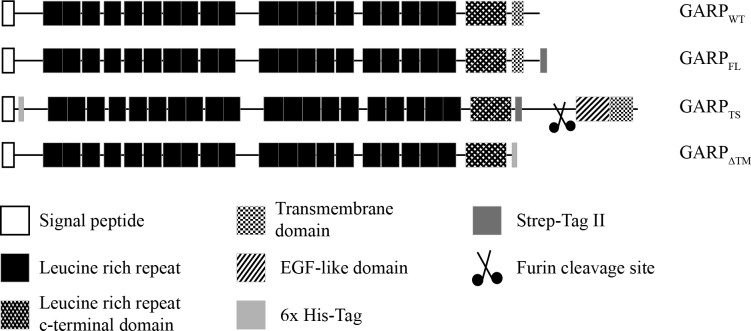
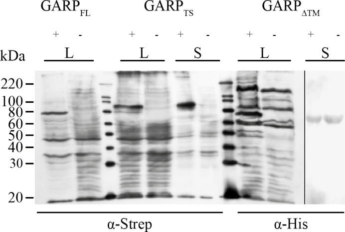
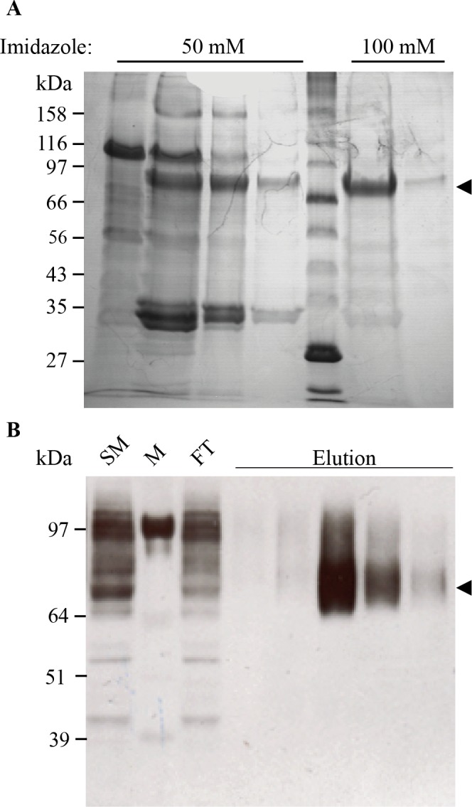
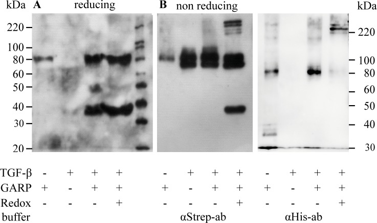
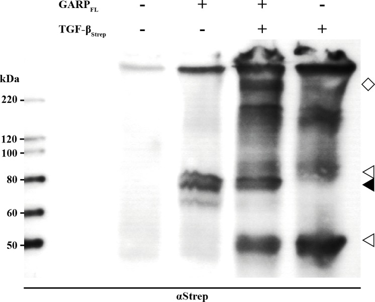
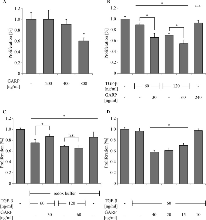
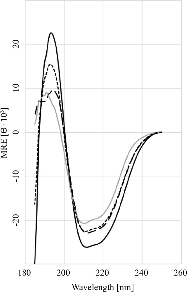
GARP (glycoprotein A repetitions predominant) is a cell surface receptor on regulatory T-lymphocytes, platelets, hepatic stellate cells and certain cancer cells. Its described function is the binding and accommodation of latent TGFβ (transforming growth factor), before the activation and release of the mature cytokine. For regulatory T cells it was shown that a knockdown of GARP or a treatment with blocking antibodies dramatically decreases their immune suppressive capacity. This confirms a fundamental role of GARP in the basic function of regulatory T cells. Prerequisites postulated for physiological GARP function include membrane anchorage of GARP, disulfide bridges between the propeptide of TGFβ and GARP and connection of this propeptide to αvβ6 or αvβ8 integrins of target cells during mechanical TGFβ release. Other studies indicate the existence of soluble GARP complexes and a functionality of soluble GARP alone. In order to clarify the underlying molecular mechanism, we expressed and purified recombinant TGFβ and a soluble variant of GARP. Surprisingly, soluble GARP and TGFβ formed stable non-covalent complexes in addition to disulfide-coupled complexes, depending on the redox conditions of the microenvironment. We also show that soluble GARP alone and the two variants of complexes mediate different levels of TGFβ activity. TGFβ activation is enhanced by the non-covalent GARP-TGFβ complex already at low (nanomolar) concentrations, at which GARP alone does not show any effect. This supports the idea of soluble GARP acting as immune modulator in vivo.


Conflict of interest statement

Competing Interests: The authors have declared that no competing interests exist.

Figures

Fig 1
Fig 1. Domain structure of GARP and its recombinant variants.
Schematic representation of human GARP and its recombinant variants used in this study. GARP consists of a signal peptide, 20 leucine rich repeats, a leucine rich repeat C-terminal flanking domain and a transmembrane region. For the construct GARPFL a Strep-tag was added at the intracellular C-terminus. Instead of the original GARP transmembrane region, the construct GARPTS possesses the transmembrane region of the protease meprin α and additionally its extracellular EGF-like and inserted domain. This construct was cleaved by furin in the trans-Golgi network and secreted into the extracellular space. For purification and detection a Strep-tag was inserted between the extracellular part of GARP and the meprin α part and a His-tag between the signal peptide and the mature chain. GARPΔTM lacks the complete transmembrane region of GARP, but contains a His-tag instead at the C-terminus of the extracellular part.
Fig 2
Fig 2. Transient Expression of the three recombinant GARP variants in HEK 293H cells.
HEK 293H cells were transfected with plasmids containing the cDNA of the constructs GARPFL, GARPTS and GARPΔTM, respectively. 48h after transfection, the culture medium was exchanged for FCS-free DMEM supplemented with NEAA. Supernatants (S) and cell lysates (L) were obtained after another 48h of incubation. 1 ml of supernatant was precipitated using 2% (w/v) Na-deoxycholate solution (1:100) and 100% TCA (1:10). Cell lysates were prepared using 200 μl RIPA buffer per 1x10⁶ cells. Samples were separated on a 10% PAA SDS-PAGE followed by western blotting on a PVDF membrane. For molecular size determination the magic mark XP marker (Invitrogen; Darmstadt, Germany) was used. For detection the blot was probed with α-Strep-tag and α-His-tag antibodies, respectively (Quiagen; Hilden, Germany). As secondary antibody a peroxidase coupled anti-mouse-IgG antibody (Dianova; Hamburg, Germany) was used.
Fig 3
Fig 3. Electrophoretic analysis of GARPTS fractions from the final Ni-NTA chromatography.
(A) Elution fractions of the Affigel Blue column were pooled and proteins bound on the Ni-NTA matrix overnight. Lane 1–4: Washing fractions, containing 50 mM imidazole; Lane 5: Protein Marker Broad Range (NEB; Ipswich, USA); Lane 6+7: Elution fractions containing 100 mM imidazole. The arrowhead marks the soluble form of GARPTS at a molecular weight of 75 kDa. Fractions were analyzed by 10% PAA SDS-PAGE und reducing conditions, followed by staining with coomassie brilliant blue. (B) The supernatant of TRP2TS transfected Expi293F cells (Invitrogen; Darmstadt, Germany) were concentrated and applied to a Strep-Tactin matrix. SM: Starting Material; M: Protein Marker SeeBlue Plus 2 (Invitrogen; Darmstadt, Germany); FT: Flowthrough; Elution: Eluted fractions with 2.5 mM D-Desthiobiotin. The arrowhead marks the soluble TRP2TS at a molecular weight of 75 kDa. Fractions were analyzed by 10% PAA SDS-PAGE und reducing conditions followed by western blotting on a PVDF membrane. For detection the blot was probed with anti-TRP2 antibody (Abcam; Cambridge, UK). As secondary antibody a peroxidase coupled anti-rabbit-IgG antibody (GE Healthcare; Solingen, Germany) was used.
Fig 4
Fig 4. In vitro coupling of GARPTS to recombinant TGFβ.
(A) 300 ng GARPTS and 600 ng recombinant latent TGFβ per lane were incubated overnight in the presence or absence of a redox-buffer containing 0.05 mM GSSH and 2 mM free cysteine at RT to allow the formation of GARP-LAP interactions. Then the samples were incubated for 4 hours at 4°C together with magnetic Ni-NTA beads to bind GARPTS and putative GARP-LAP complexes at the beads. Proteins bound to the beads were analyzed by western blot with anti-Strep-tag antibodies, which could be used to detect both GARPTS and recombinant TGFβ. (B) Equally treated controls were analyzed on a 10% PAA SDS-PAGE under non-reducing conditions but without the pull-down procedure. After gel electrophoresis, proteins were blotted onto a PVDF membrane and probed with anti-Strep-tag (left) or anti-His-tag antibodies (right), respectively.
Fig 5
Fig 5. In vivo coupling of GARPTS to recombinant TGFβ.
HEK 293H cells were transfected with plasmids containing the cDNA of the constructs GARPFL, TGFβStrep or both in combination. 48h after transfection, the culture medium was exchanged for FCS-free DMEM supplemented with NEAA. Cell lysates were prepared using 200 μl RIPA buffer per 1x10⁶ cells. Samples were separated on a 10% PAA SDS-PAGE followed by western blotting on a PVDF membrane. For molecular size determination the magic mark XP marker (Invitrogen; Darmstadt, Germany) was used. For detection the blot was probed with anti-Strep-tag and anti-His-tag antibodies, respectively (Quiagen; Hilden, Germany). As secondary antibody a peroxidase coupled anti-mouse-IgG antibody (Dianova; Hamburg, Germany) was used.
Fig 6
Fig 6. Inhibition of cell proliferation of Mv1Lu cells by GARP and TGFβ.
Bar diagrams show the proliferation rates (percentage of proliferation, n = 6) of treated versus untreated (100%) Mv1Lu cells. Cell numbers were determined using methylene blue staining of DNA [25]. (A) 2.5 x 10⁴ cells were grown for 48h and treated with 200 ng/ml to 800 ng/ml of recombinant GARPTS to determine its influence on cell proliferation alone. (B) Cells were grown for 48h in the presence of the indicated TGFβ concentrations pre-incubated with or without the indicated GARPTS concentration. (C) Cells were incubated the same way like in (B), except that the proteins were pre-incubated in redox buffer to provoke covalent bonding of TGFβ and GARP (D) 60 ng/ml TGFβ were pre-incubated with different amounts of GARPTS (40–10 ng/ml) to determine the minimal amount of GARPTS needed to achieve maximal enhancement of TGFβ activation. The asterisk (*) marks a statistical significance p < 0.01.
Fig 7
Fig 7. Far UV CD-Spectroscopy of GARP and TGFβ.
The far-UV spectra (25°C, 260–185 nm) for the following samples are as indicated: GARPTS alone (black solid line), latent TGFβ alone (grey solid line), summed spectrum of GARPTS and latent TGFβ (short-dashed line), measured spectra of the GARPTS and latent TGFβ complex (long dashed line). The decrease in the mean relative ellipticity below 200 nm between the calculated and measured spectra of the complex formation of GARPTS and TGFβ indicates a conformational change in GARP.

References

    1. Massagué J, Xi Q. TGF-β control of stem cell differentiation genes. FEBS Lett. 2012;586: 1953–1958. 10.1016/j.febslet.2012.03.023 - DOI - PMC - PubMed
    1. Kulkarni a B, Huh CG, Becker D, Geiser a, Lyght M, Flanders KC, et al. Transforming growth factor beta 1 null mutation in mice causes excessive inflammatory response and early death. Proc Natl Acad Sci U S A. 1993;90: 770–774. - PMC - PubMed
    1. Kehrl BYJH, Wakefield LM, Roberts AB, Jakowlew S, Alvarez-mon M, Derynck RIK, et al. Production of transforming growth factor beta by human T lymphocytes and its potential role in the regulation of T cell growth. J Exp Med. 1986;163: 1037–1050. - PMC - PubMed
    1. Robertson IB, Rifkin DB. Unchaining the beast; insights from structural and evolutionary studies on TGF beta secretion, sequestration, and activation. Cytokine and Growth Factor Reviews. 2013. pp. 355–372. 10.1016/j.cytogfr.2013.06.003 - DOI - PMC - PubMed
    1. Macaulay IC, Tijssen MR, Thijssen-timmer DC, Gusnanto A, Steward M, Burns P, et al. Comparative gene expression profiling of in vitro differentiated megakaryocytes and erythroblasts identifies novel activatory and inhibitory platelet membrane proteins. Blood. 2007; 3260–3269. 10.1182/blood-2006-07-036269 - DOI - PMC - PubMed

Publication types

MeSH terms