. 2016 Apr 7:22:1161-70.
doi: 10.12659/msm.897909.

MiR-206 Attenuates Denervation-Induced Skeletal Muscle Atrophy in Rats Through Regulation of Satellite Cell Differentiation via TGF-β1, Smad3, and HDAC4 Signaling

Affiliations

MiR-206 Attenuates Denervation-Induced Skeletal Muscle Atrophy in Rats Through Regulation of Satellite Cell Differentiation via TGF-β1, Smad3, and HDAC4 Signaling

Qiang Kai Huang et al. Med Sci Monit. .

Abstract

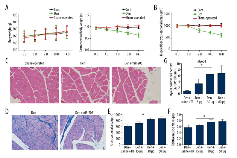
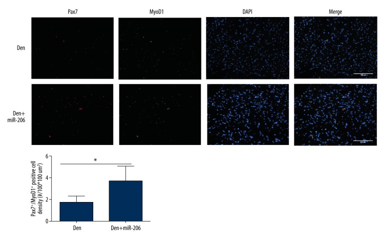
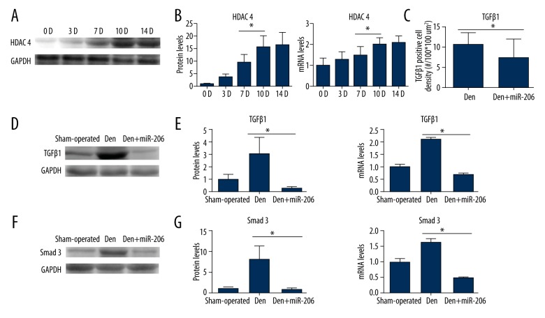
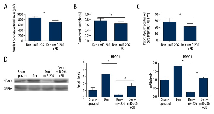
BACKGROUND Denervation-induced skeletal muscle atrophy results in significant biochemical and physiological changes potentially leading to devastating outcomes including increased mortality. Effective treatments for skeletal muscle diseases are currently not available. Muscle-specific miRNAs, such as miR-206, play an important role in the regulation of muscle regeneration. The aim of the present study was to examine the beneficial effects of miR-206 treatment during the early changes in skeletal muscle atrophy, and to study the underlying signaling pathways in a rat skeletal muscle atrophy model. MATERIAL AND METHODS The rat denervation-induced skeletal muscle atrophy model was established. miRNA-206 was overexpressed with or without TGF-β1 inhibitor in the rats. The mRNA and protein expression of HDAC4, TGF-β1, and Smad3 was determined by real-time PCR and western blot. The gastrocnemius muscle cross-sectional area and relative muscle mass were measured. MyoD1, TGF-β1, and Pax7 were determined by immunohistochemical staining. RESULTS After sciatic nerve surgical transection, basic muscle characteristics, such as relative muscle weight, deteriorated continuously during a 2-week period. Injection of miR-206 (30 μg/rat) attenuated morphological and physiological deterioration of muscle characteristics, prevented fibrosis effectively, and inhibited the expression of TGF-β1 and HDAC4 as assessed 2 weeks after denervation. Moreover, miR-206 treatment increased the number of differentiating (MyoD1+/Pax7+) satellite cells, thereby protecting denervated muscles from atrophy. Interestingly, the ability of miR-206 to govern HDAC4 expression and to attenuate muscle atrophy was weakened after pharmacological blockage of the TGF-b1/Smad3 axis. CONCLUSIONS TGF-β1/Smad3 signaling pathway is one of the crucial signaling pathways by which miR-206 counteracts skeletal muscle atrophy by affecting proliferation and differentiation of satellite cells. miR-206 may be a potential target for development of a new strategy for treatment of patients with early denervation-induced skeletal muscle atrophy.


Figures

Figure 1
Figure 1
Progression of denervation-induced muscle atrophy and improvement following miR-206 treatment. (A) Body weight and gastrocnemius relative muscle mass in the control, sham-operated, and Den groups 0 d, 3 d, 7 d, 10 d, and 14 d after surgery. (B) Changes of muscle fiber cross-sectional area in the control, sham-operated, and Den groups at 0 d, 3 d, 7 d, 10 d, and 14 d after sciatic nerve transection. (C) Histochemical staining (H&E) of GM for the sham-operated, Den, and Den+miR-206 groups. Normal muscle cell with round appearance and similar size are found in the sham-operated group. In the Den group, some cells show a polygonal shape, and a small number of hypertrophic and atrophic cells appear. In the Den+miR-206 group, the muscle cells show a less-rounded appearance. (D) Comparison of collagen fiber production between the Den and Den+miR-206 groups, as revealed by Masson’s trichrome staining. In the Den group muscle fibers were surrounded by collagen fibers, whereas in the Den+miR-206 group the production of collagen fiber was weakened. (E, F) Quantification of the cross-sectional area and gastrocnemius relative muscle mass (operated limb/normal limb) 14 d after surgery, as shown in Supplementary Figure 1. * P<0.05 in the miR-206 treatment group vs. Den+saline group. (G) Quantification of MyoD1 positive cell density, as shown in Supplementary Figure 2 in the Den+saline+RT (transfection reagent) and miR-206 treatment group (15 μg/rat, 30 μg/rat, and 60 μg/rat). * P<0.05.
Figure 2
Figure 2
Treatment with miR-206 promotes muscle cell differentiation following denervation-induced muscle atrophy. (A) Double immunofluorescence analysis of the GM in the Den and Den+miR-206 groups using the satellite cell marker Pax7 (red) and the muscle cell differentiation marker MyoD1 (green). DAPI (blue) was used as a nuclear stain. (B) Quantification of immunofluorescence staining showing the Pax7+/MyoD1+ positive cell density (#/100*100 um2). * P<0.05.
Figure 3
Figure 3
Expression of TGF-β1, Smad3, and HDAC4 in the GM following denervation-induced muscle atrophy. (A) Western blot analysis results showing the expression of HDAC4 protein levels 0 d, 3 d, 7 d, 10 d, and 14 d after sciatic nerve transection in comparison to GAPDH controls. (B) Quantification of Western blot and RT-PCR results showing protein and mRNA expression levels of HDAC4 at 0 d, 3 d, 7 d, 10 d, and 14 d after sciatic denervation surgery. (C) Quantification of immunofluorescence staining, as shown in Supplementary Figure 3, displaying TGF-β1 positive cell densities (#/100*100 um2). * P<0.05. (D–G) Representative Western blot for TGF-β1 and Smad3 expression in the sham-operated, Den, and Den+miR-206 groups. Quantification of Western blot and RT-qPCR results show relative protein and mRNA level of TGF-β1 and Smad3 expression in the sham-operated, Den, and Den+miR-206 groups. n=4, per group; * P<0.05.
Figure 4
Figure 4
The effect of the TGF-β1 inhibitor SB431542 combined with miR-206 treatment following denervation-induced muscle atrophy. (A, B) Measurement of GM cross-sectional area and relative muscle mass in the Den+miR-206 and Den+miR-206+SB groups. n=4, per group; * P<0.05. (C) Quantification of Pax7+/MyoD1+ positive cell density (#/100*100 um2) in the Den+miR-206 and Den+miR-206+SB groups, as shown in Supplementary Figure 4. n=4, per group; * P<0.05. (D) Representative Western blot showing relative HDAC4 protein level in the sham-operated, Den, Den+miR-206, and Den+miR1+SB groups. Quantification of Western blot and RT-qPCR results show relative protein and mRNA level of HDAC4 expression in the sham-operated, Den, Den+miR-206, and Den+miR-206+SB groups. n=4 per group, * P<0.05.

References

    1. Soldado F, Fontecha CG, Marotta M, et al. The role of muscle imbalance in the pathogenesis of shoulder contracture after neonatal brachial plexus palsy: A study in a rat model. J Shoulder Elbow Surg. 2014;23:1003–9. - PubMed
    1. Gwag T, Park K, Park J, et al. Celastrol overcomes HSP72 gene silencing-mediated muscle atrophy and induces myofiber preservation. J Physiol Pharmacol. 2015;66:273–83. - PubMed
    1. Bongers KS, Fox DK, Ebert SM, et al. Skeletal muscle denervation causes skeletal muscle atrophy through a pathway that involves both Gadd45a and HDAC4. Am J Physiol Endocrinol Metab. 2013;305:E907–15. - PMC - PubMed
    1. Li QT, Zhang PX, Yin XF, et al. Functional recovery of denervated skeletal muscle with sensory or mixed nerve protection: A pilot study. PLoS One. 2013;8:e79746. - PMC - PubMed
    1. Batt J, Bain J, Goncalves J, et al. Differential gene expression profiling of short and long term denervated muscle. FASEB J. 2006;20:115–17. - PubMed

Publication types

MeSH terms