Identification of biallelic LRRK1 mutations in osteosclerotic metaphyseal dysplasia and evidence for locus heterogeneity
- PMID: 27055475
- PMCID: PMC5769692
- DOI: 10.1136/jmedgenet-2016-103756
Identification of biallelic LRRK1 mutations in osteosclerotic metaphyseal dysplasia and evidence for locus heterogeneity
Abstract
Background: Osteosclerotic metaphyseal dysplasia (OSMD) is a unique form of osteopetrosis characterised by severe osteosclerosis localised to the bone ends. The mode of inheritance is autosomal recessive. Its genetic basis is not known.
Objective: To identify the disease gene for OSMD.
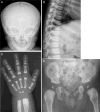
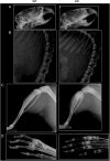
Methods and results: By whole exome sequencing in a boy with OSMD, we identified a homozygous 7 bp deletion (c.5938_5944delGAGTGGT) in the LRRK1 gene. His skeletal phenotype recapitulated that seen in the Lrrk1-deficient mouse. The shared skeletal hallmarks included severe sclerosis in the undermodelled metaphyses and epiphyseal margins of the tubular bones, costal ends, vertebral endplates and margins of the flat bones. The deletion is predicted to result in an elongated LRRK1 protein (p.E1980Afs*66) that lacks a part of its WD40 domains. In vitro functional studies using osteoclasts from Lrrk1-deficient mice showed that the deletion was a loss of function mutation. Genetic analysis of LRRK1 in two unrelated patients with OSMD suggested that OSMD is a genetically heterogeneous condition.
Conclusions: This is the first study to identify the causative gene of OSMD. Our study provides evidence that LRRK1 plays a critical role in the regulation of bone mass in humans.
Keywords: LRRK1 mutation; Lrrk1 deficient mouse; Osteosclerotic metaphyseal dysplasia; Whole exome sequencing.
Published by the BMJ Publishing Group Limited. For permission to use (where not already granted under a licence) please go to http://www.bmj.com/company/products-services/rights-and-licensing/
Conflict of interest statement
Figures


References
-
- Warman ML, Cormier-Daire V, Hall C, Krakow D, Lachman R, Lemerrer M, Mortier G, Mundlos S, Nishimura G, Rimoin DL, Robertson S, Savarirayan R, Sillence D, Spranger J, Unger S, Zabel B, Superti-Furga A. Nosology and classification of genetic skeletal disorders: 2010 revision. Am J Med Genet A. 2011;155A:943–68. - PMC - PubMed
-
- Nishimura G, Kozlowski K. Osteosclerotic metaphyseal dysplasia. Pediatr Radiol. 1993;23:450–2. - PubMed
-
- Mennel EA, John SD. Osteosclerotic metaphyseal dysplasia: a skeletal dysplasia that may mimic lead poisoning in a child with hypotonia and seizures. Pediatr Radiol. 2003;33:11–14. - PubMed
-
- Kasapkara CS, Kucukcongar A, Boyunağa O, Bedir T, Oncü F, Hasanoglu A, Tumer L. An extremely rare case: osteosclerotic metaphyseal dysplasia. Genet Couns. 2013;24:69–74. - PubMed
-
- Zheng H, Cai J, Wang L, He X. Osteosclerotic metaphyseal dysplasia with extensive interstitial pulmonary lesions: a case report and literature review. Skeletal Radiol. 2015;44:1529–33. - PubMed
MeSH terms
Substances
Supplementary concepts
Grants and funding
LinkOut - more resources
Full Text Sources
Other Literature Sources
Molecular Biology Databases
