Review
. 2016 Jul;30(7):901-16.
doi: 10.1038/eye.2016.47. Epub 2016 Apr 8.

Advances of optical coherence tomography in myopia and pathologic myopia

Affiliations
Review

Advances of optical coherence tomography in myopia and pathologic myopia

D S C Ng et al. Eye (Lond). 2016 Jul.

Abstract

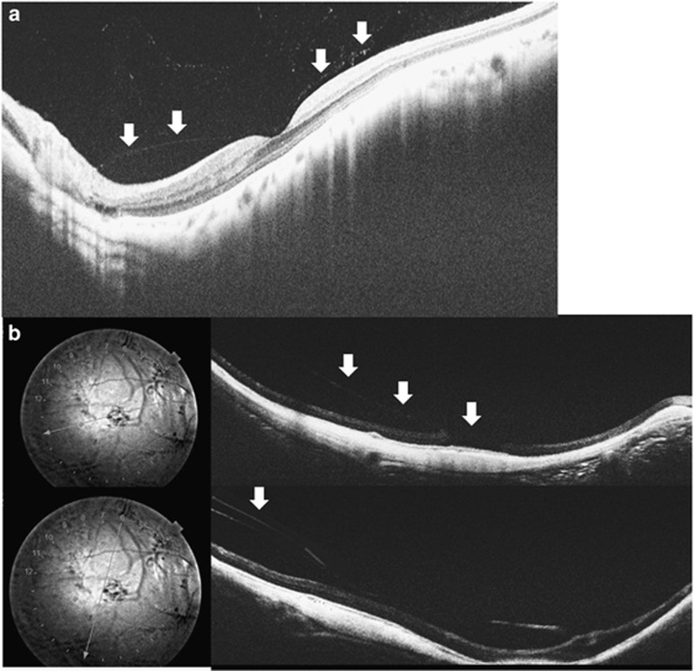
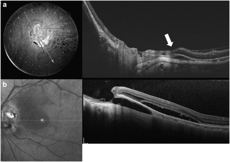
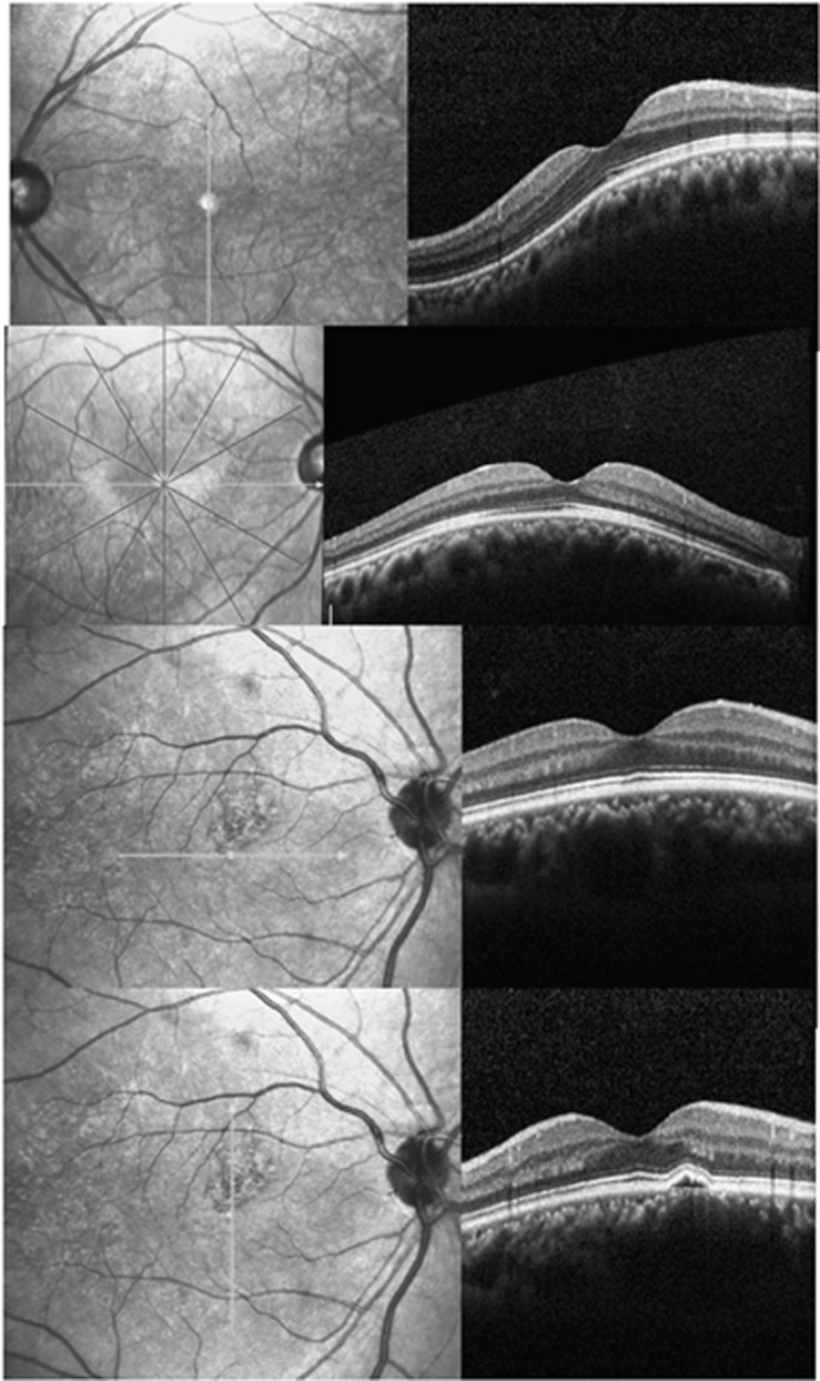
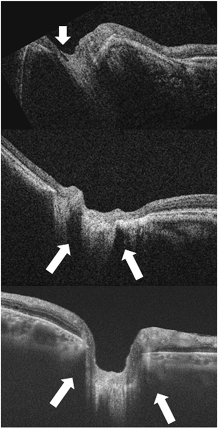
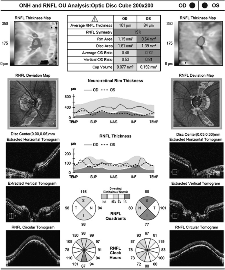
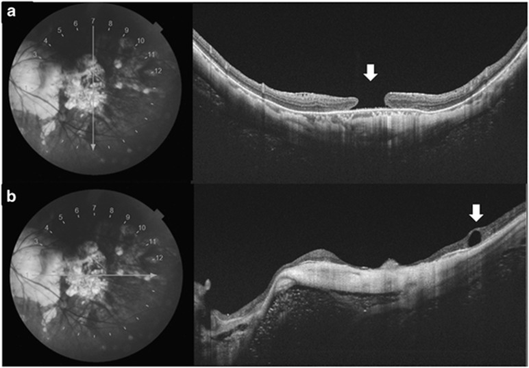
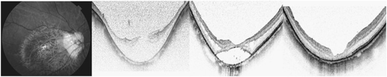
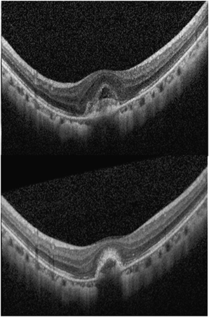
The natural course of high-axial myopia is variable and the development of pathologic myopia is not fully understood. Advancements in optical coherence tomography (OCT) technology have revealed peculiar intraocular structures in highly myopic eyes and unprecedented pathologies that cause visual impairment. New OCT findings include posterior precortical vitreous pocket and precursor stages of posterior vitreous detachment; peripapillary intrachoroidal cavitation; morphological patterns of scleral inner curvature and dome-shaped macula. Swept source OCT is capable of imaging deeper layers in the posterior pole for investigation of optic nerve pits, stretched and thinned lamina cribrosa, elongated dural attachment at posterior scleral canal, and enlargement of retrobulbar subarachnoid spaces. This has therefore enabled further evaluation of various visual field defects in high myopia and the pathogenesis of glaucomatous optic neuropathy. OCT has many potential clinical uses in managing visual impairing conditions in pathologic myopia. Understanding how retinal nerve fibers are redistributed in axial elongation will allow the development of auto-segmentation software for diagnosis and monitoring progression of glaucoma. OCT is indispensable in the diagnosis of various conditions associated with myopic traction maculopathy and monitoring of post-surgical outcomes. In addition, OCT is commonly used in the multimodal imaging assessment of myopic choroidal neovascularization. Biometry and topography of the retinal layers and choroid will soon be validated for the classification of myopic maculopathy for utilization in epidemiological studies as well as clinical trials.


Figures

Figure 1
Figure 1
Optical coherence tomography scans of abnormal vitreoretinal interface. (a) Swept source OCT (SS OCT) has high penetration that allows enhanced imaging of the posterior vitreous and vitreoretinal interface (outlined by white arrows) in cross sections. The posterior wall of the posterior precortical vitreous pocket is a thin vitreous cortex attached to the retina. The curvature of the inner sclera of this highly myopic eye is asymmetrical around the fovea. There is posterior vitreous detachment in the paramacular region just above the steepest point of the curvature and extending toward the perifovea. (b) SS OCT images of a highly myopic eye with the location of the cross sections indicated by the straight line lying on the infrared fundus photos on the left side. Top: a full-thickness macular hole with parafoveal vitreous traction (white arrows) in myopic traction maculopathy. Bottom: another cross-sectional OCT image of the same eye revealed vitreoschsis (white arrow).
Figure 2
Figure 2
Optical coherence tomography (OCT) images of intrachoroidal cavation (ICC). (a) Swept source OCT slice scanned along the line lying on the infrared fundus photo (left) shows ICC below the optic nerve. Hyporeflective space (white arrow) suggesting an existence of fluid is observed within the ICC. (b) Enhance depth imaging spectral domain OCT revealed a macular retinal detachment associated with ICC. During enlargement of the ICC in a highly myopic eye, the overlying retinal tissue develops a full-thickness defect, allowing the vitreous cavity to communicate directly with the cavity of the ICC. Macular retinal detachment occurs when the communication extends into the subretinal space.
Figure 3
Figure 3
Cross section enhanced depth imagining spectral domain OCT images with their orientation indicated by the thick straight lines lying on the left side fundi images. Top: vertically oriented dome-shaped maculopathy. Second from top: horizontally oriented dome-shaped maculopathy. Third from top and bottom: a bidirectional type dome-shaped maculopathy. A small juxtafoveal pigmented epithelial detachment is shown in the vertical orientation.
Figure 4
Figure 4
Enlargement of the optic nerve head in highly myopic eyes occurs due to stretching of the scleral canal and lamina cribrosa. The lamina is torn from the peripapillary sclera and eventually the overylying nerve fiber is disrupted, and this stage is observed as optic disc pits, especially at the superior and inferior poles of the optic disc. Top: optical coherence tomography (OCT) shows a hyporeflective gap indicating the acquired pit of the optic nerve (white arrow). Middle: OCT shows the subarachnoid spaces as hyporeflective triangular spaces along both the upper and lower borders of the optic nerve (white arrows). The elongated dural attachment at posterior scleral canal in highly myopic eye leads to widening of retrobulbar subarachnoid spaces comparing with that observed in an ametropic eye (Bottom).
Figure 5
Figure 5
Retinal nerve fiber layer (RNFL) analysis by Cirrus optical coherence tomography (OCT) shows abnormally reduced thickness in inferior sector of the right eye and superotemporal and inferotemporal sectors in the left eye of a high myopia patient. In glaucoma eyes, there is predilection of inferior and superior RNFL loss. However, the normative database of Cirrus OCT only comprises data collected from normal eyes with no or low myopia. The interpretation of RNFL thickness deviation may need to account for its altered topographical distribution in highly myopic eyes.
Figure 6
Figure 6
Optical coherence tomography (OCT) scans of myopic traction maculopathy (MTM). (a) A patient with MTM developed full-thickness macular hole (white arrow) in swept source OCT. (b) A horizontal OCT slice of the same eye revealed a paravascular retinal cyst (white arrow) associated with MTM.
Figure 7
Figure 7
Left: fundus photo of the posterior staphyloma and chorioretinal atrophy. Second from left: spectral domain optical coherence tomography (SD OCT) reveals retinoschisis with MH. Note that the sclera is strongly bowed posteriorly and the curve is symmetrical around the fovea. The patient underwent posterior vitrectomy with internal limiting-membrane peeling and gas tamponade. Third from left: SD OCT 3 months post operation shows that the macular gradually flattened. Right: SD OCT at 4 years after operation. The MH remains closed.
Figure 8
Figure 8
Optical coherence tomography (OCT) scans of myopic choroidal neovascularization (CNV). Top: SD OCT reveals intra- and subretinal fluid associated with an active myopic CNV. Bottom: after treatment with intravitreal anti-VEGF injection, there is resolution of both intra- and subretinal fluid. The lesion becomes more compact and the boundary between the lesion and retina becomes apparent.

Comment in

References

    1. Resnikoff S, Pascolini D, Mariotti SP, Pokharel GP. Global magnitude of visual impairment caused by uncorrected refractive errors in 2004. Bull World Health Organ 2008; 86(1): 63–70. - PMC - PubMed
    1. Saw SM, Katz J, Schein OD, Chew SJ, Chan TK. Epidemiology of myopia. Epidemiol Rev 1996; 18(2): 175–187. - PubMed
    1. Liu HH, Xu L, Wang YX, Wang S, You QS, Jonas JB. Prevalence and progression of myopic retinopathy in Chinese adults: the Beijing Eye Study. Ophthalmology 2010; 117(9): 1763–1768. - PubMed
    1. Jonas JB, Xu L. Histological changes of high axial myopia. Eye (Lond) 2014; 28(2): 113–117. - PMC - PubMed
    1. Wong TY, Ferreira A, Hughes R, Carter G, Mitchell P. Epidemiology and disease burden of pathologic myopia and myopic choroidal neovascularization: an evidence-based systematic review. Am J Ophthalmol 2014; 157(1): 9–25 e12. - PubMed

MeSH terms