. 2016 Apr 7;13(1):72.
doi: 10.1186/s12974-016-0538-2.

Complement activation at the motor end-plates in amyotrophic lateral sclerosis

Affiliations

Complement activation at the motor end-plates in amyotrophic lateral sclerosis

Nawal Bahia El Idrissi et al. J Neuroinflammation. .

Abstract

Background: Amyotrophic lateral sclerosis (ALS) is a fatal progressive neurodegenerative disease with no available therapy. Components of the innate immune system are activated in the spinal cord and central nervous system of ALS patients. Studies in the SOD1(G93A) mouse show deposition of C1q and C3/C3b at the motor end-plate before neurological symptoms are apparent, suggesting that complement activation precedes neurodegeneration in this model. To obtain a better understanding of the role of complement at the motor end-plates in human ALS pathology, we analyzed post-mortem tissue of ALS donors for complement activation and its regulators.

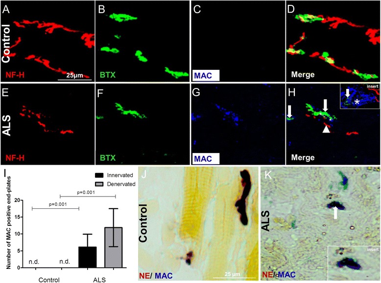
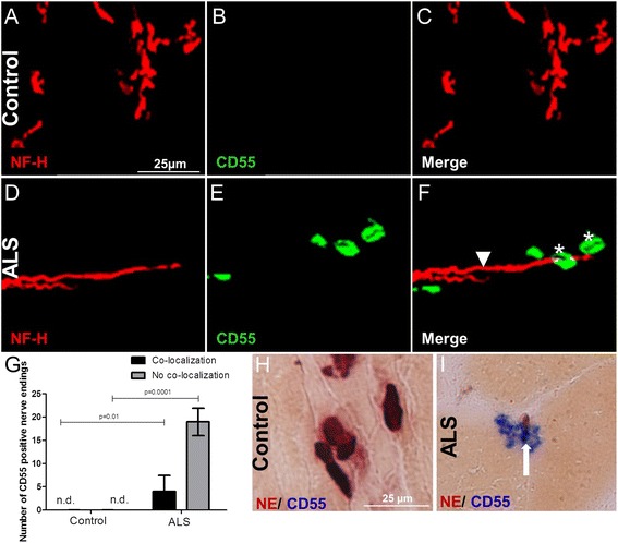
Methods: Post-mortem intercostal muscle biopsies were collected at autopsy from ALS (n = 11) and control (n = 6) donors. The samples were analyzed for C1q, membrane attack complex (MAC), CD55, and CD59 on the motor end-plates, using immunofluorescence or immunohistochemistry.

Results: Here, we show that complement activation products and regulators are deposited on the motor end-plates of ALS patients. C1q co-localized with neurofilament in the intercostal muscle of ALS donors and was absent in controls (P = 0.001). In addition, C1q was found deposited on the motor end-plates in the intercostal muscle. MAC was also found deposited on motor end-plates that were innervated by nerves in the intercostal muscle of ALS donors but not in controls (P = 0.001). High levels of the regulators CD55 and CD59 were detected at the motor end-plates of ALS donors but not in controls, suggesting an attempt to counteract complement activation and prevent MAC deposition on the end-plates before they are lost.

Conclusions: This study provides evidence that complement activation products are deposited on innervated motor end-plates in the intercostal muscle of ALS donors, indicating that complement activation may precede end-plate denervation in human ALS. This study adds to the understanding of ALS pathology in man and identifies complement as a potential modifier of the disease process.

Keywords: Amyotrophic lateral sclerosis; C1q; CD55; CD59; Complement; MAC; Motor end-plates.


Figures

Fig. 1
Fig. 1
Number and size of α-BTX-positive end-plates in intercostal muscle of ALS donors. Confocal microscopic images of motor end-plates from controls (a) and ALS donors (b, c) double-labeled with α-bungarotoxin (α-BTX, Alexa 488) and antibodies against neurofilament (NF-H, Cy3). All controls showed co-localization of NF-H with α-BTX (white arrows). In ALS, both innervated end-plates (panel b) and denervated end-plates (panel c) were detected. The number and size of α-BTX-positive end-plates in 20 non-overlapping Z-stacks in 40-μm thick intercostal muscle sections is shown in panels d and e. Both number and size of α-BTX-positive end-plates of ALS donors (n = 2) are reduced compared to controls (n = 2) (P = 0.0003 and P = <0.0001, respectively). Error bars represent standard deviation of the mean
Fig. 2
Fig. 2
Confocal microscopic images of intercostal muscle from controls (a, b, c) and ALS donors (d, e, f) double-labeled with antibodies against neurofilament (NF-H, Cy3) and antibodies against classical pathway component of the complement system C1q (C1q, FITC). C1q deposition was detected on the nerves as well as near the nerve endings (white asterisks in f) in muscle of ALS donors but not in controls. g Quantification showed C1q-positive staining co-localizing with nerves and in the vicinity of nerve endings (white arrowhead pointing to NF-H and asterisk on C1q in f) in the intercostal muscle of ALS donors but not in controls (P = 0.001 and P = 0.001, respectively). NE staining (dark brown) followed by an immune staining for C1q (blue) showed (i) C1q deposition on the end-plates of ALS donors (white arrow in i and enlargement of the area as insert) (h) by contrast, no C1q deposition was found deposited on the motor end-plates in the intercostal muscle of control donors. Numbers of C1q-positive nerve endings in 20 non-overlapping Z-stacks in 40-μm thick intercostal muscle sections is given on the y-axis. Error bars represent standard deviation of the mean. n.d. not detected
Fig. 3
Fig. 3
Representative confocal images of triple-immunofluorescence staining for neurofilament (NF-H, Cy3), motor end-plates with α-BTX (Alexa 488), and complement component C5b-9 with MAC (Cy5) in control (a, b, c, d) and ALS intercostal muscle (e, f, g, h), shows presence of MAC (white asterisks in h and enlarged in the insert) on end-plates (white arrows in h) and around nerves in ALS muscle (white arrowhead in h) but not in controls (c, d). Quantification showed a significantly higher percentage of MAC-positive innervated end-plates (P = 0.001) and denervated end-plates (P = 0.001) in ALS intercostal muscle compared to controls. Numbers of MAC-positive end-plates in 20 non-overlapping Z-stacks in 40-μm thick intercostal muscle sections is given on the y-axis. Error bars represent standard deviation of the mean (i). NE staining (dark brown) followed by an immune staining for MAC (blue) showed (k) MAC deposition deposited on the end-plates of ALS donors (white arrow in k enlarged in the insert), j but not on end-plates of control donors. n.d. not detected
Fig. 4
Fig. 4
Representative confocal double-immunofluorescence for neurofilament (NF-H, Cy3) and CD55 detected with anti-DAF (FITC) in control (a, b, c) and ALS (d, e, f) intercostal muscle shows CD55 deposition in ALS intercostal muscle on and around nerves (white asterisks on CD55 and arrowhead pointing to NF-H in f) but not in control tissue (c). Quantification showed CD55 deposition co-localizing with nerves or in the vicinity of nerves in the intercostal muscle of ALS donors but not in controls (P = 0.01 and P = 0.0001, respectively) (g). Numbers of CD55-positive end-plates in 20 non-overlapping Z-stacks in 40-μm thick intercostal muscle sections is given on the y-axis. Error bars represent standard deviation of the mean n.d. not detected. NE staining (dark brown) followed by an immune staining for CD55 (blue) showing i CD55 deposition on the motor end-plates (white arrow in i) in the intercostal muscle of ALS donors h but no CD55 deposition in controls
Fig. 5
Fig. 5
Representative confocal triple-immunofluorescence for neurofilament (NF-H, Cy3), end-plates detected with α-BTX (Alexa 488) and the regulator CD59 (Cy5) in control (a, b, c, d) and ALS (e, f, g, h) intercostal muscle showing deposition of CD59 (white asterisks in h, enlarged in insert) in ALS intercostal muscle tissue on denervated end-plates (white arrow pointing to α-BTX and arrowhead pointing to NF-H in h) but not in controls. Quantification shows CD59-positive innervated and denervated motor end-plates in the intercostal muscle of ALS donors but not in controls (P = 0.05 and P = 0.05, respectively) (i). Data represents standard deviation of the mean. n.d. not detected

References

    1. Pasinelli P, Brown RH. Molecular biology of amyotrophic lateral sclerosis: insights from genetics. Nat Rev Neurosci. 2006;7:710–23. doi: 10.1038/nrn1971. - DOI - PubMed
    1. Mitchell JD, Borasio GD. Amyotrophic lateral sclerosis. Lancet. 2007;369:2031–41. doi: 10.1016/S0140-6736(07)60944-1. - DOI - PubMed
    1. Raoul C, Estevez AG, Nishimune H, et al. Motoneuron death triggered by a specific pathway downstream of Fas. potentiation by ALS-linked SOD1 mutations. Neuron. 2002;35:1067–83. doi: 10.1016/S0896-6273(02)00905-4. - DOI - PubMed
    1. Boillee S, Yamanaka K, Lobsiger CS, et al. Onset and progression in inherited ALS determined by motor neurons and microglia. Science. 2006;312:1389–92. doi: 10.1126/science.1123511. - DOI - PubMed
    1. Di Giorgio FP, Carrasco MA, Siao MC, Maniatis T, Eggan K. Non-cell autonomous effect of glia on motor neurons in an embryonic stem cell-based ALS model. Nat Neurosci. 2007;10:608–14. doi: 10.1038/nn1885. - DOI - PMC - PubMed

MeSH terms