. 2016 Jun;46(7):405-10.
doi: 10.1016/j.ijpara.2016.03.004. Epub 2016 Apr 4.

Identification of new markers for the Schistosoma mansoni vitelline lineage

Affiliations

Identification of new markers for the Schistosoma mansoni vitelline lineage

Jipeng Wang et al. Int J Parasitol. 2016 Jun.

Abstract

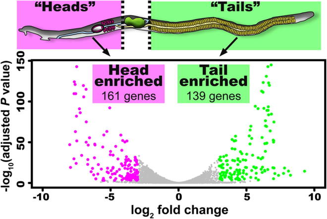
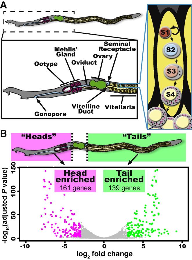
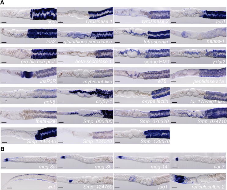
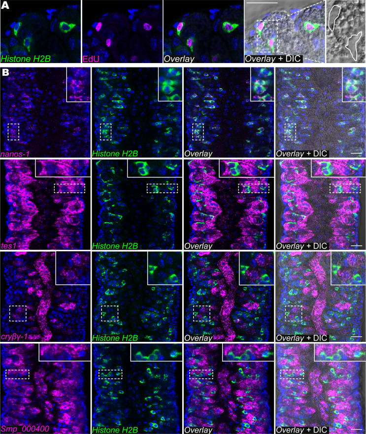
Schistosomes cause significant morbidity and mortality in millions of the world's poorest people. While parasite egg-induced inflammation is the primary driver of host pathology, relatively little is known at the molecular level about the organ systems that participate in schistosome egg production (i.e., testes, ovaries and vitellaria). Here we use transcriptional profiling and in situ hybridization to characterise the vitellarium of Schistosoma mansoni. We uncovered several previously uncharacterised vitellaria-specific factors and defined molecular markers for various stages in the vitellocyte differentiation process. These data provide the framework for future in-depth molecular studies exploring the biology of this important parasite organ.

Keywords: Reproduciton; Schistosoma; Stem cell; Vitellaria.


Figures

None
Graphical abstract
Fig. 1
Fig. 1
Identification of genes associated with the schistosome vitellarium. (A) Cartoon of the schistosome reproductive system and stages of vitellocyte development (inset boxed in cyan). Within the vitellaria, S1 cells proliferate and differentiate to ultimately generate S4 vitellocytes that are rich in lipid (magenta circles) and vitelline (black circles) droplets. These mature vitellocytes pass to the vitelline duct and travel anteriorly to the ootype. (B) Cartoon depicts the strategy to identify vitellaria associated transcripts. To identify genes associated with the vitellaria, RNA from amputated female heads and tails were subjected to RNAseq. The Volcano Plot depicts the 161 head-enriched (magenta dots) and 139 tail-enriched (green dots) transcripts. Genes not reaching our fold-change and significance thresholds are depicted as grey dots.
Fig. 2
Fig. 2
Whole mount in situ hybridization for head- and tail-enriched schistosome transcripts. Expression of (A) tail-enriched or (B) head-enriched transcripts. Gene names are listed below each image. Genes without clear homologues of known function or recognisable domains are listed with their Smp number. Details of genes examined are provided in Supplementary Table S4. Over 75% of tail-enriched transcripts examined by whole mount in situ hybridization were expressed in the vitellaria, whereas the head-enriched transcripts were detected in the oesophageal glands, the ootype, Mehlis’ Gland, and in cells within the parenchyma. Images were captured using a Zeiss AxioZoom.V16 equipped with a transmitted light base and a Zeiss AxioCam 105 Color camera. Anterior is to the left. Scale bars = 100 μm.
Fig. 3
Fig. 3
Fluorescence in situ hybridization identifies markers for unique cell populations within the schistosome vitellaria. (A) 5-ethynyl-2′-deoxyuridine labelling and FISH for Histone H2B. Parasites were treated for 4 h with the thymidine analogue 5-ethynyl-2′-deoxyuridine, fixed and processed for FISH as previously described (Collins et al., 2013). 5-ethynyl-2′-deoxyuridine incorporation following a 4 h pulse appeared to be exclusive for Histone H2B+ cells in the vitellaria, indicating that these cells are the proliferative cells of the vitellaria and are likely to represent S1 cells. Differential Interference Contrast imaging (left) showed that these cells lack the granularity characteristic of differentiated vitelline cells, consistent with the idea that these cells are undifferentiated S1 cells. (B) Double fluorescence in situ hybridization for Histone H2B with nanos-1, tes1, cryβγ-1, or Smp_000400. nanos-1 expression was restricted to Histone H2B+ cells indicating that nanos-1 is likely a marker for S1 cells. tes1, cryβγ-1 and Smp_000400 are not expressed in Histone H2B+ S1 cells; rather, their expression was observed in distinct sets of more differentiated vitelline cells. tes1 expression was highest in cells within the vitellaria and lower in the most differentiated vitelline cells within the vitelline duct. cryβγ-1 was expressed at high levels in cells within the vitelline duct and in cells adjacent to the duct. Smp_000400 appeared to be ubiquitously expressed in all cells of the vitelline lineage with the exception of the S1 cells. Thus, we speculate that cells expressing high levels of tes1 and Smp_000400 are likely to represent an intermediate step in the differentiation process of mature vitellocytes that express cryβγ-1 and Smp_000400. Insets show magnified views of boxed regions. In all images, nuclei are in blue. Images were acquired using an Zeiss LSM700 laser scanning confocal microscope with either a Plan-Apochromat 63×/1.4 Oil DIC or EC Plan-Neofluar 40×/1.30 Oil DIC objective lens. Scale bars = 20 μm.

References

    1. Anders S., Huber W. Differential expression analysis for sequence count data. Genome Biol. 2010;11:R106. - PMC - PubMed
    1. Basch P.F. Oxford University Press; New York: 1991. Schistosomes: Development, Reproduction, and Host Relations.
    1. Berriman M., Haas B.J., LoVerde P.T., Wilson R.A., Dillon G.P., Cerqueira G.C., Mashiyama S.T., Al-Lazikani B., Andrade L.F., Ashton P.D., Aslett M.A., Bartholomeu D.C., Blandin G., Caffrey C.R., Coghlan A., Coulson R., Day T.A., Delcher A., DeMarco R., Djikeng A., Eyre T., Gamble J.A., Ghedin E., Gu Y., Hertz-Fowler C., Hirai H., Hirai Y., Houston R., Ivens A., Johnston D.A., Lacerda D., Macedo C.D., McVeigh P., Ning Z., Oliveira G., Overington J.P., Parkhill J., Pertea M., Pierce R.J., Protasio A.V., Quail M.A., Rajandream M.A., Rogers J., Sajid M., Salzberg S.L., Stanke M., Tivey A.R., White O., Williams D.L., Wortman J., Wu W., Zamanian M., Zerlotini A., Fraser-Liggett C.M., Barrell B.G., El-Sayed N.M. The genome of the blood fluke Schistosoma mansoni. Nature. 2009;460:352–358. - PMC - PubMed
    1. Bobek L.A., Rekosh D.M., LoVerde P.T. Small gene family encoding an eggshell (chorion) protein of the human parasite Schistosoma mansoni. Mol. Cell. Biol. 1988;8:3008–3016. - PMC - PubMed
    1. Chen L.L., Rekosh D.M., LoVerde P.T. Schistosoma mansoni p48 eggshell protein gene: characterization, developmentally regulated expression and comparison to the p14 eggshell protein gene. Mol. Biochem. Parasitol. 1992;52:39–52. - PubMed

Publication types