. 2016 Jun;5(6):764-72.
doi: 10.5966/sctm.2015-0117. Epub 2016 Apr 7.

Reducing Hepatocyte Injury and Necrosis in Response to Paracetamol Using Noncoding RNAs

Affiliations

Reducing Hepatocyte Injury and Necrosis in Response to Paracetamol Using Noncoding RNAs

Dagmara Szkolnicka et al. Stem Cells Transl Med. 2016 Jun.

Abstract

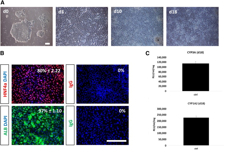
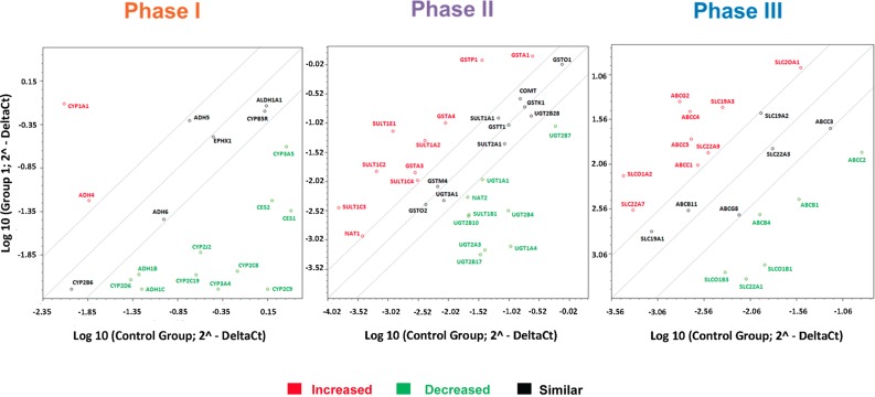
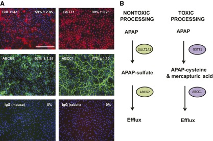
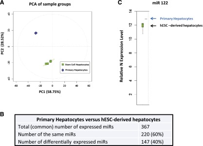
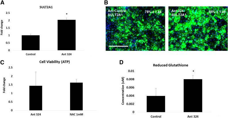
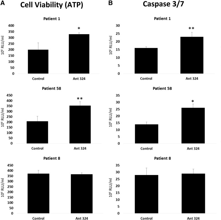
The liver performs multiple functions within the human body. It is composed of numerous cell types, which play important roles in organ physiology. Our study centers on the major metabolic cell type of the liver, the hepatocyte, and its susceptibility to damage during drug overdose. In these studies, hepatocytes were generated from a renewable and genetically defined resource. In vitro-derived hepatocytes were extensively profiled and exposed to varying levels of paracetamol and plasma isolated from liver-failure patients, with a view to identifying noncoding microRNAs that could reduce drug- or serum-induced hepatotoxicity. We identified a novel anti-microRNA, which reduced paracetamol-induced hepatotoxicity and glutathione depletion. Additionally, we identified a prosurvival role for anti-microRNA-324 following exposure to plasma collected from liver failure patients. We believe that these studies represent an important advance for the field, demonstrating the power of stem cell-derived systems to model human biology "in a dish" and identify novel noncoding microRNAs, which could be translated to the clinic in the future.

Significance: The liver performs vital functions within the human body and is composed of numerous cell types. The major metabolic cell type of the liver, the hepatocyte, is susceptible to damage during drug overdose. In these studies, hepatocytes were generated from a renewable resource and exposed to varying levels of paracetamol, with a view to identifying interventions that could reduce or attenuate drug-induced liver toxicity. A novel noncoding RNA that reduced paracetamol-induced hepatocyte toxicity was identified. These findings may represent an important advance for the field.

Keywords: Apoptosis; Drug-induced liver injury; Hepatocyte; MicroRNA; Necrosis; Paracetamol.


Figures

Figure 1.
Figure 1.
Stagewise human embryonic stem cell (hESC) differentiation to the hepatocyte lineage. (A): Phase-contrast imaging demonstrated that cells underwent sequential morphological changes during transit from stem cell (day 0), to definitive endoderm (day 3), to hepatoblast (day 10), to hepatocyte (day 18). (B): Immunocytochemistry demonstrating upregulation of HNF4a and albumin during hepatic specification at day 18. Negative controls performed with corresponding immunoglobulin G. The percentage of positive cells is provided in the top right of each panel. This was calculated from four random fields of view and is shown as ± SD. (C), Cyp3A and Cyp1A2 metabolism were assessed in day 18 hepatocyte-like cells using the Promega pGlo system. Experiments were performed in triplicate and measured on a luminometer. The units of activity quoted are relative light units per milliliter of supernatant per milligram of protein. Scale bars = 100 μm. Abbreviations: ALB, albumin; d, day; DAPI, 4′,6-diamidino-2-phenylindole; HNF4a, hepatocyte nuclear factor 4a; IgG, immunoglobulin G RLU, relative light units.
Figure 2.
Figure 2.
Stem cell-derived (day 18) and primary human hepatocyte gene expression. The scatter plots represent expression of the major metabolic genes involved in phase I, II, and III drug metabolism. Gene expression was performed by using the Human Drug Metabolism RT2 Profiler PCR Array (Qiagen) according to the manufacturer’s instructions. The scatter plot represents the changes in gene expression. The graph plots the log10 of normalized gene expression levels in a control condition, primary human hepatocytes (x axis) versus an experimental condition, human embryonic stem cell (hESC)-derived hepatocytes (y axis). Symbols outside the boundary area indicate fold differences larger than threefold.
Figure 3.
Figure 3.
The expression of enzymes and transporters involved in paracetamol (APAP) metabolism. (A): Representation of protein expression of phase II enzymes (GSTT1 and SULT2A1) and phase III transporters (ABCC1 and ABCG2) in stem cell-derived hepatocytes. The percentage of positive cells is provided in the top right of each panel. This was calculated from four random fields of view and is shown as ± SD. The images were taken at ×20 magnification. Scale bar = 100 μm. (B): Phase II enzymes and phase III transporters play major role in a toxic and nontoxic pathways of paracetamol metabolism. In the nontoxic pathway, paracetamol is metabolized by SULT2A1 enzymes to produce APAP sulfate metabolite that is effluxed from the cell by ABCG2 transporter. In a toxic pathway, paracetamol is metabolized by GSTT1 enzyme to produce APAP cysteine (mercapturic acid) metabolite that is effluxed from the cell by ABCC1 transporter. Abbreviations: ABCC1, adenosine triphosphate-Binding Cassette Transporter Subfamily C member 1; ABCG2, adenosine triphosphate-Binding Cassette Transporter Subfamily G member 2; APAP, acetaminophen; GSTT1, glutathione-S-transferase θ 1; SULT2A1, sulfotransferase 2A1.
Figure 4.
Figure 4.
hESC-derived hepatocytes (hESC-heps) and primary human hepatocytes (PHH) microRNA expression profile. (A): Principal component analysis overview plot demonstrates strong clustering by cell type. (B): Statistical analysis of the microRNA array demonstrates 367 reliably detected microRNAs in both hESC-heps and PHH; 220 microRNAs have a similar expression in both systems and 147 microRNAs are differentially expressed. (C): microRNA 122 is expressed in hESC-heps at the same level as in PHH. The microRNA array was carried out by Sistemic Limited. The RNA samples (four replicates of PHH and four experimental samples of hESC-derived hepatocytes) were run on the Agilent miRNA platform. Abbreviations: hESC, human embryonic stem cell; miR, microRNA; PC1, principal component 1; PC2, principal component 2; PCA, principal component analysis.
Figure 5.
Figure 5.
Antagomir of microRNA 324 upregulated SULT2A1 gene and protein expression and increased cell survival after exposure to paracetamol. At day 17, human embryonic stem cell-derived hepatocytes (hESC-heps) were transfected with the antagomir of the corresponding microRNA at 50 nM for 24 hours. (A, B): Transfection with the Ant-324-5p upregulated SULT2A1 gene expression by twofold (A) and protein expression by 20% (B) in comparison with the scrambled control. At day 17, hESC-hepatocytes were either transfected with antagomirs or treated with 1 mM N-acetylcysteine (NAC) concentration for 24 hours. At day 18, the antagomir-transfected or NAC pre-exposed hESC-hepatocytes were exposed to paracetamol concentration that causes 50% of the cell death (IC50 = 12.85 mM) for another 24 hours. At day 19, the cell viability was measured using CellTiter Assay (Promega) and Glutathione Depletion Assay (Promega). (C, D): The antagomir of microRNA 324 significantly increased ATP to the levels comparable with NAC (fold increase was calculated in comparison with control; the values for Ant ctrl = 2.17 × 10⁸; Ant 324= 3.12 × 10⁸; H2O (vehicle) = 2.38 × 10⁸; NAC = 3.86 × 10⁸ (C) and enhanced reduced glutathione levels (D). Levels of significance were measured by Student’s t test. ∗, p < .05. The percentage of positive cells is provided in the top right of each panel. This was calculated from four random fields of view and is quoted as ± SE. Scale bar = 100 μm. Abbreviations: Ant 324, antagomir 324-5p; Ant ctrl; scrambled antagomir control; ATP, adenosine triphosphate; SULT2A1, sulfotransferase 2A1.
Figure 6.
Figure 6.
Human embryonic stem cell (hESC)-derived hepatocytes transfected with the antagomir 324-5p. At 24 hours post-transfection, transfection, hESC-hepatocytes were exposed to 20% plasma from patients with fulminant hepatic failure for a further 24 hours (patients 1, 8, and 58). ATP levels and caspase 3/7 activation were determined using Promega Glo technology. Units of activity are expressed as RLU per milliliter (n = 4). Levels of significance are quoted and measured by Student’s t test. ∗, p < .05; ∗∗, p < .01. Abbreviations: Ant 324;, antagomir 324-5p; ATP, adenosine triphosphate; RLU, relative light units.

Comment in

References

    1. Olsen AK, Whalen MD. Public perceptions of the pharmaceutical industry and drug safety: Implications for the pharmacovigilance professional and the culture of safety. Drug Saf. 2009;32:805–810. - PubMed
    1. Davies EC, Green CF, Taylor S, et al. Adverse drug reactions in hospital in-patients: A prospective analysis of 3695 patient-episodes. PLoS One. 2009;4:e4439. - PMC - PubMed
    1. Davies EC, Green CF, Mottram DR, et al. Emergency re-admissions to hospital due to adverse drug reactions within 1 year of the index admission. Br J Clin Pharmacol. 2010;70:749–755. - PMC - PubMed
    1. Pirmohamed M, James S, Meakin S, et al. Adverse drug reactions as cause of admission to hospital: Prospective analysis of 18 820 patients. BMJ. 2004;329:15–19. - PMC - PubMed
    1. Szkolnicka D, Farnworth SL, Lucendo-Villarin B, et al. Accurate prediction of drug-induced liver injury using stem cell-derived populations. Stem Cells Translational Medicine. 2014;3:141–148. - PMC - PubMed

Publication types