Review
. 2016 Feb;37(1):27-47.

Fat-Soluble Vitamins: Clinical Indications and Current Challenges for Chromatographic Measurement

Affiliations
Review

Fat-Soluble Vitamins: Clinical Indications and Current Challenges for Chromatographic Measurement

Ali A Albahrani et al. Clin Biochem Rev. 2016 Feb.

Abstract

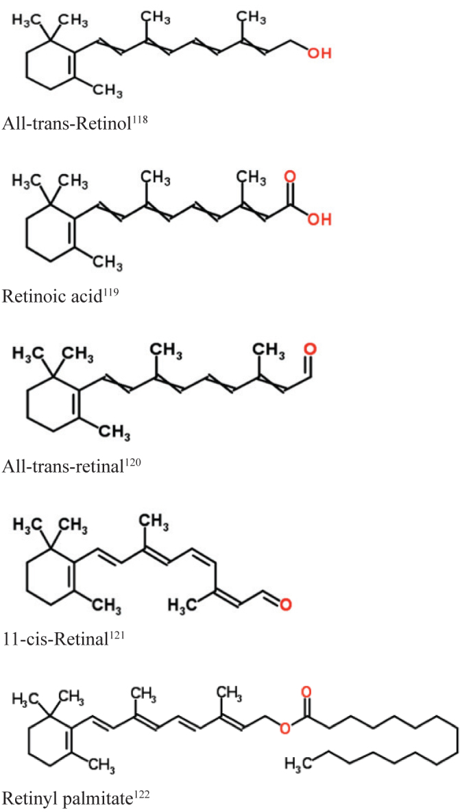
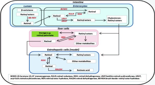
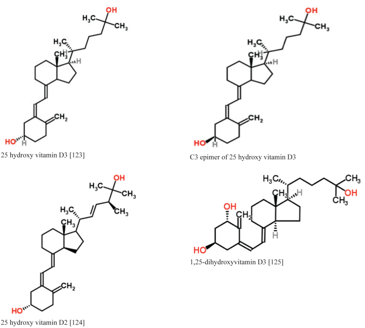
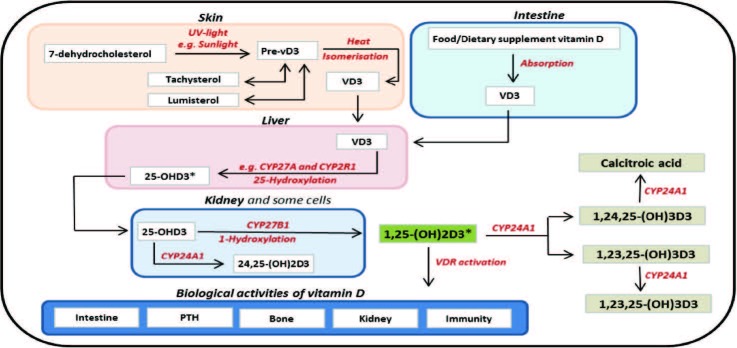
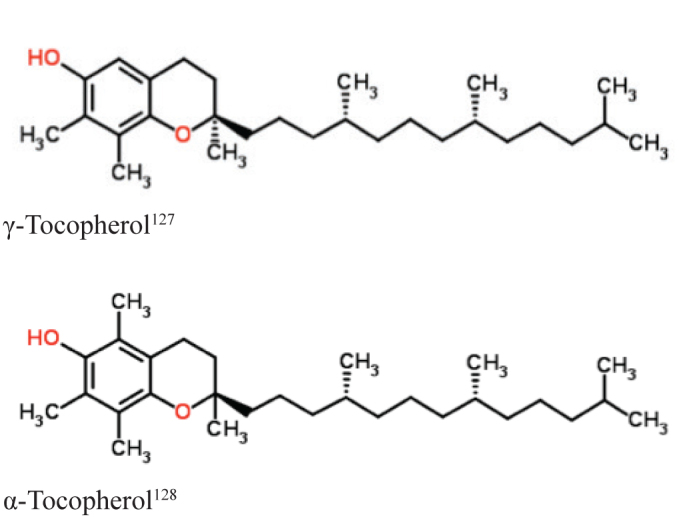
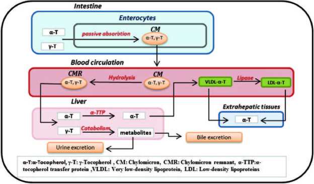
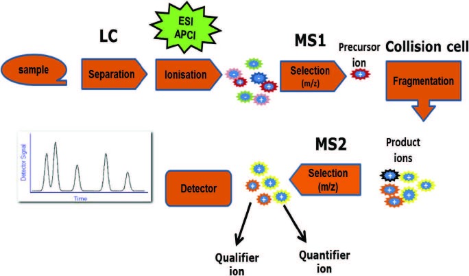
Fat-soluble vitamins, including vitamins A, D and E, are required for a wide variety of physiological functions. Over the past two decades, deficiencies of these vitamins have been associated with increased risk of cancer, type II diabetes mellitus and a number of immune system disorders. In addition, there is increasing evidence of interactions between these vitamins, especially between vitamins A and D. As a result of this enhanced clinical association with disease, translational clinical research and laboratory requests for vitamin measurements have significantly increased. These laboratory requests include measurement of 25-OHD (vitamin D), retinol (vitamin A) and α-tocopherol (vitamin E); the most accepted blood indicators for the assessment of body fat-soluble vitamin (FSV) status. There are significant obstacles to precise FSV measurement in blood. These obstacles include their physical and chemical properties, incomplete standardisation of measurement and limitations in the techniques that are currently used for quantification. The aim of this review is to briefly outline the metabolism and interactions of FSV as a prelude to identifying the current challenges for the quantification of blood vitamins A, D and E.


Figures

Figure 1.
Figure 1.
Chemical structure of vitamin A and its derivatives.
Figure 2.
Figure 2.
General scheme for Vitamin A metabolism. Dietary vitamin A (e.g., retinyl esters and β-carotene) is digested and absorbed through intestinal enterocytes by different mechanisms. In enterocytes, retinol is re-esterified to retinyl esters, which are packed with chylomicrons prior to secretion into the lymphatic system. Through blood circulation, retinyl esters are taken up by liver cells (parenchymal cells), in which retinyl esters are converted to retinol, which can be released to target organs or stored in the liver. Vitamin A is transported through binding with retinol-binding protein (RBP) and thyroxine binding-protein transthyretin (TTR) for extracellular transportation, while intracellular retinol is transported by binding with cellular RBPs (CRBPs).
Figure 3.
Figure 3.
Chemical structure of some vitamin D metabolites.
Figure 4.
Figure 4.
General scheme for Vitamin D metabolism. In the skin, 7-dehydrocholesterol is converted to pre-vitamin D3 under the effects of solar ultraviolet B radiation following isomerisation to vitamin D3 (VD3). Excess amounts of pre-vitamin D3 are converted to lumisterol and tachysterol to circumvent hypervitaminosis D. The VD3 is hydroxylated in the liver by cytochrome P450 enzymes (e.g., CYP27A and CYP2R1) to form 25-hydroxyvitamin D3 (25-OHD3), which is an inactive and storage form of vitamin D3. The 25-OHD3 is further hydroxylated systematically in the kidney (or locally in some cells) to 1,25-dihydroxyvitamin D3 (1,25-(OH)2D3), which is the active form of vitamin D3), by CYP27B1. Most of the biological effects of vitamin D3 are conducted through binding 1,25-(OH)2D3 with a vitamin D receptor (VDR). The 1,25-(OH)2D3 levels might be down-regulated through its conversion to other metabolites such as calcitroic acid and 1,23,25-(OH)3D3., Vitamin D2 is metabolised through similar pathways. *Although C3-epimers of 25-OHD3 and 1,25-(OH)2D3 were reported in human sera, the role of these metabolites is still not clear.
Figure 5.
Figure 5.
Chemical structure of γ-tocopherol and α-tocopherol.
Figure 6.
Figure 6.
General Scheme for Vitamin E metabolism. Dietary vitamin E (mainly γ-tocopherol and α-tocopherol) is absorbed through intestinal enterocytes. In enterocytes, γ-tocopherol and α-tocopherol and other vitamin E forms are packed with chylomicrons prior to secretion into the lymphatic system. Through blood circulation, chylomicrons are hydrolysed and chylomicron remnants are formed. γ-Tocopherol and α-tocopherol are taken up by liver cells, although only α-tocopherol is re-secreted into the bloodstream because of the selective binding of α-tocopherol transfer protein. Blood α-tocopherol is transferred to target tissues by lipoproteins such as very low-density lipoprotein (VLDL) and low-density lipoproteins (LDLs).
Figure 7.
Figure 7.
Schematic of a sample progressing through a liquid chromatography-tandem mass spectrometer. First the LC separates the analytes from its sample matrix, then the analyte will be charged through the ionisation process. The charged molecules (precursor ions) will be selected according to their mass-to-charge ratio (m/z) in the first quadrupole (Q1). The precursor ions are fragmented in the collision cell (Q2); between these ions and high purity gas (e.g., nitrogen gas). Then the fragmented molecules (product ions) can be selected by their m/z in the second quadrupole (Q3).

References

    1. Plum LA, DeLuca HF. Vitamin D, disease and therapeutic opportunities. Nat Rev Drug Discov. 2010;9:941–55. - PubMed
    1. Stephensen CB. Vitamin A, infection, and immune function. Annu Rev Nutr. 2001;21:167–92. - PubMed
    1. Holick MF. Sunlight and vitamin D for bone health and prevention of autoimmune diseases, cancers, and cardiovascular disease. Am J Clin Nutr. 2004;80(Suppl):1678S–88S. - PubMed
    1. Herrmann M. The measurement of 25-hydroxy vitamin D - an analytical challenge. Clin Chem Lab Med. 2012;50:1873–5. - PubMed
    1. Ross C. Vitamin A and carotenoids. In: Shils ME, Shike M, editors. Modern Nutrition in Health and Disease. 10th ed. Philadelphia: Lippincott Williams & Wilkins; 2006. pp. 305–27.