

Reprogramming of Melanoma Tumor-Infiltrating Lymphocytes to Induced Pluripotent Stem Cells
- PMID: 27057178
- PMCID: PMC4707343
- DOI: 10.1155/2016/8394960
Reprogramming of Melanoma Tumor-Infiltrating Lymphocytes to Induced Pluripotent Stem Cells
Abstract
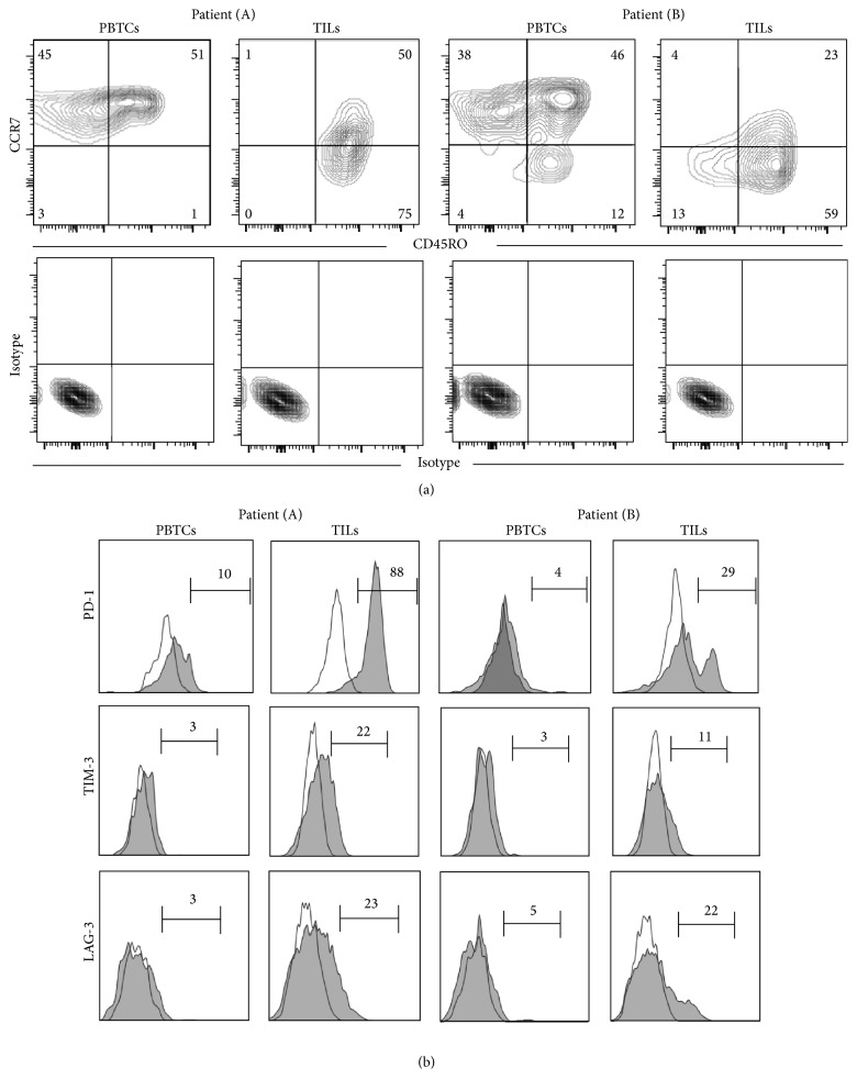
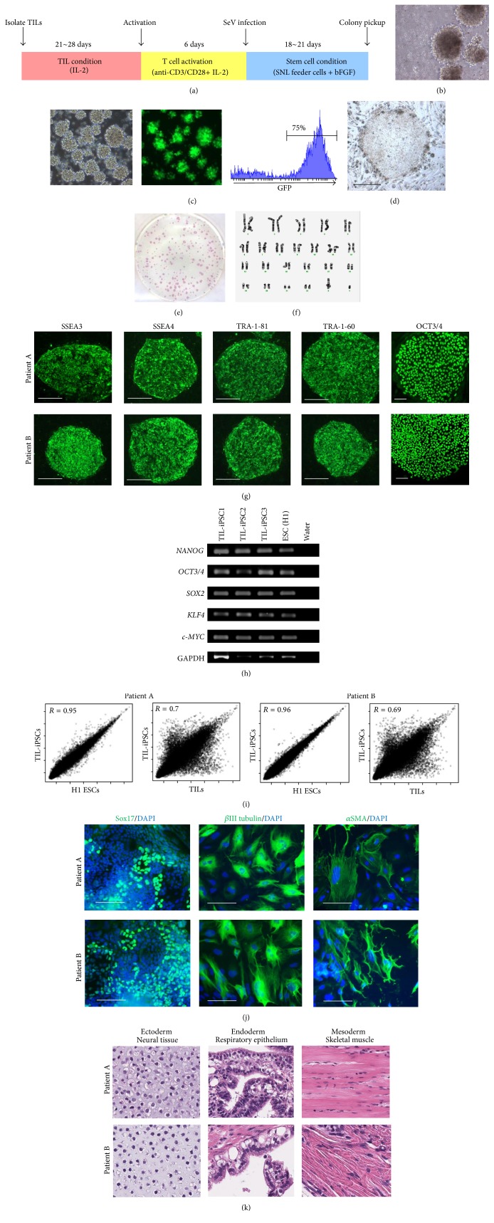
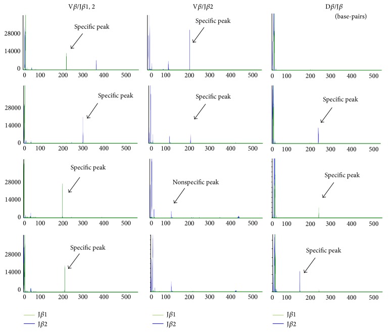
Induced pluripotent stem cells (iPSCs) derived from somatic cells of patients hold great promise for autologous cell therapies. One of the possible applications of iPSCs is to use them as a cell source for producing autologous lymphocytes for cell-based therapy against cancer. Tumor-infiltrating lymphocytes (TILs) that express programmed cell death protein-1 (PD-1) are tumor-reactive T cells, and adoptive cell therapy with autologous TILs has been found to achieve durable complete response in selected patients with metastatic melanoma. Here, we describe the derivation of human iPSCs from melanoma TILs expressing high level of PD-1 by Sendai virus-mediated transduction of the four transcription factors, OCT3/4, SOX2, KLF4, and c-MYC. TIL-derived iPSCs display embryonic stem cell-like morphology, have normal karyotype, express stem cell-specific surface antigens and pluripotency-associated transcription factors, and have the capacity to differentiate in vitro and in vivo. A wide variety of T cell receptor gene rearrangement patterns in TIL-derived iPSCs confirmed the heterogeneity of T cells infiltrating melanomas. The ability to reprogram TILs containing patient-specific tumor-reactive repertoire might allow the generation of patient- and tumor-specific polyclonal T cells for cancer immunotherapy.
Figures
Similar articles
-
Generation of Induced Pluripotent Stem Cells from Human Melanoma Tumor-infiltrating Lymphocytes.J Vis Exp. 2016 Nov 11;(117):54375. doi: 10.3791/54375. J Vis Exp. 2016. PMID: 27911363 Free PMC article.
-
Reprogramming of Tumor-reactive Tumor-infiltrating Lymphocytes to Human-induced Pluripotent Stem Cells.Cancer Res Commun. 2023 May 25;3(5):917-932. doi: 10.1158/2767-9764.CRC-22-0265. eCollection 2023 May. Cancer Res Commun. 2023. PMID: 37377887 Free PMC article.
-
Generation of two induced pluripotent stem cells lines from a Mucopolysaccharydosis IIIB (MPSIIIB) patient.Stem Cell Res. 2018 Dec;33:180-184. doi: 10.1016/j.scr.2018.10.019. Epub 2018 Nov 1. Stem Cell Res. 2018. PMID: 30408744
-
Reprogramming away from the exhausted T cell state.Semin Immunol. 2016 Feb;28(1):35-44. doi: 10.1016/j.smim.2015.10.007. Epub 2015 Nov 14. Semin Immunol. 2016. PMID: 26589493 Review.
-
The Emerging Role of Induced Pluripotent Stem Cells as Adoptive Cellular Immunotherapeutics.Biology (Basel). 2023 Nov 11;12(11):1419. doi: 10.3390/biology12111419. Biology (Basel). 2023. PMID: 37998018 Free PMC article. Review.
Cited by
-
Generation of Induced Pluripotent Stem Cells from Human Melanoma Tumor-infiltrating Lymphocytes.J Vis Exp. 2016 Nov 11;(117):54375. doi: 10.3791/54375. J Vis Exp. 2016. PMID: 27911363 Free PMC article.
-
T Cell Genesis: In Vitro Veritas Est?Trends Immunol. 2016 Dec;37(12):889-901. doi: 10.1016/j.it.2016.09.008. Epub 2016 Oct 24. Trends Immunol. 2016. PMID: 27789110 Free PMC article. Review.
-
Conceptual Development of Immunotherapeutic Approaches to Gastrointestinal Cancer.Int J Mol Sci. 2019 Sep 18;20(18):4624. doi: 10.3390/ijms20184624. Int J Mol Sci. 2019. PMID: 31540435 Free PMC article. Review.
-
The therapeutic potential of multiclonal tumoricidal T cells derived from tumor infiltrating lymphocyte-1derived iPS cells.Commun Biol. 2021 Jun 7;4(1):694. doi: 10.1038/s42003-021-02195-x. Commun Biol. 2021. PMID: 34099861 Free PMC article.
-
Producing proT cells to promote immunotherapies.Int Immunol. 2018 Nov 14;30(12):541-550. doi: 10.1093/intimm/dxy051. Int Immunol. 2018. PMID: 30102361 Free PMC article. Review.
References
Grants and funding
LinkOut - more resources
Full Text Sources
Other Literature Sources
Molecular Biology Databases
Research Materials
