. 2016:2016:5060757.
doi: 10.1155/2016/5060757. Epub 2016 Feb 4.

Chinese Herbal Medicine Fuzheng Kang-Ai Decoction Inhibited Lung Cancer Cell Growth through AMPKα-Mediated Induction and Interplay of IGFBP1 and FOXO3a

Affiliations

Chinese Herbal Medicine Fuzheng Kang-Ai Decoction Inhibited Lung Cancer Cell Growth through AMPKα-Mediated Induction and Interplay of IGFBP1 and FOXO3a

Fang Zheng et al. Evid Based Complement Alternat Med. 2016.

Abstract

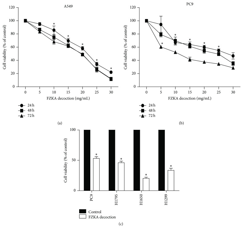
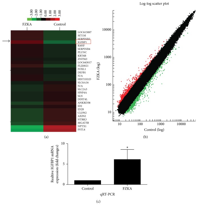
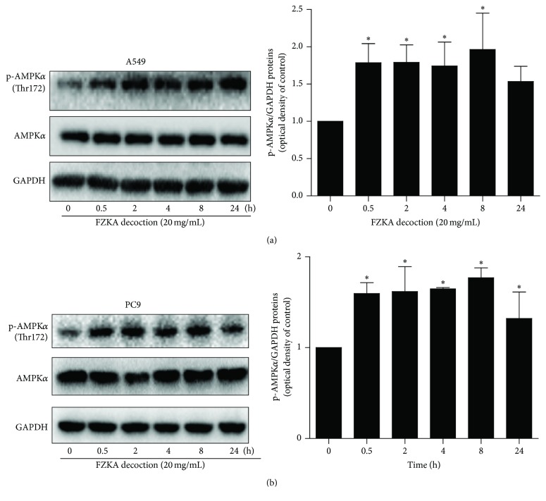
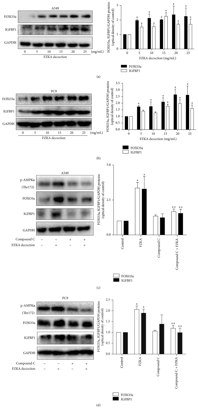
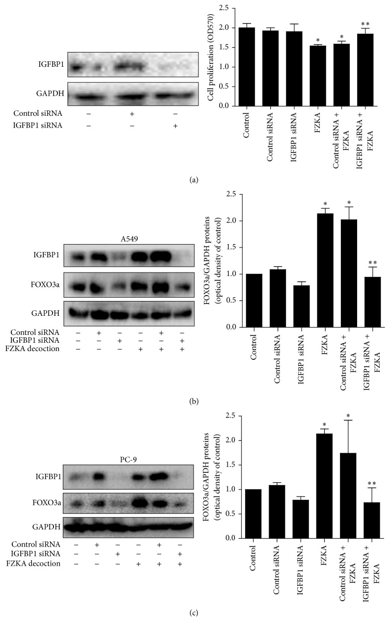
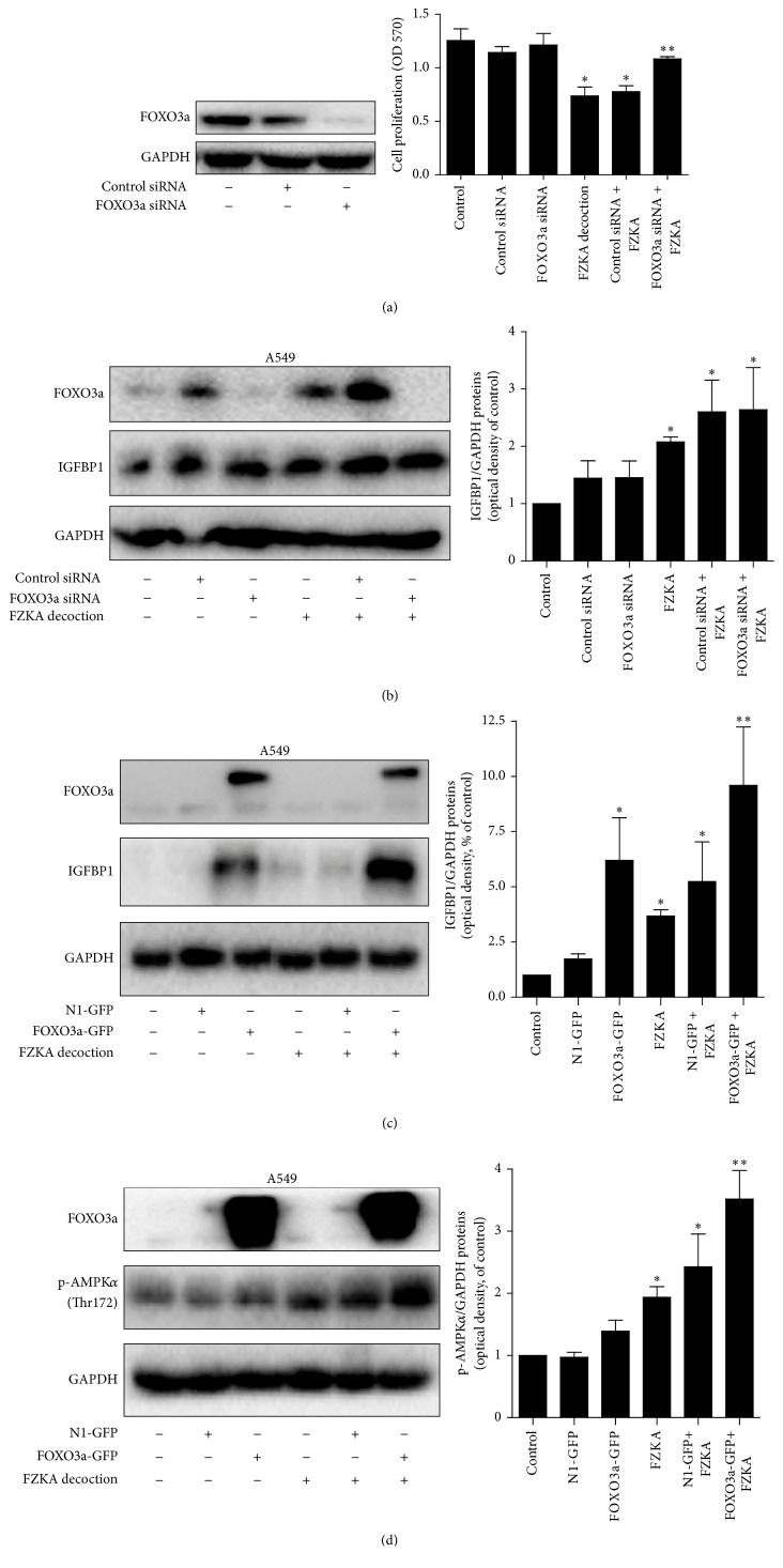
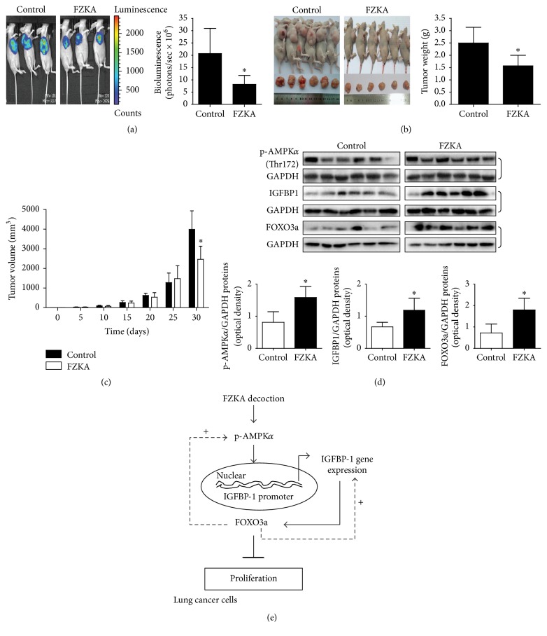
The aim of this study is to investigate the actions of Chinese herbal medicine, called "Fuzheng Kang-Ai" (FZKA for short) decoction, against non-small cell lung cancer (NSCLC) and its mechanisms in vitro and in vivo. We showed that the effect of FZKA decoction significantly inhibited growth of A549 and PC9 cells. Furthermore, FZKA increased phosphorylation of AMP-activated protein kinase alpha (AMPKα) and induced protein expression of insulin-like growth factor (IGF) binding protein 1 (IGFBP1) and forkhead homeobox type O3a (FOXO3a). The specific inhibitor of AMPKα (Compound C) blocked FZKA-induced protein expression of IGFBP1 and FOXO3a. Interestingly, silencing of IGFBP1 and FOXO3a overcame the inhibitory effect of FZKA on cell growth. Moreover, silencing of IGFBP1 attenuated the effect of FZKA decoction on FOXO3a expression, and exogenous expression of FOXO3a enhanced the FZKA-stimulated phosphorylation of AMPKα. Accordingly, FZKA inhibited the tumor growth in xenograft nude mice model. Collectively, our results show that FZKA decoction inhibits proliferation of NSCLC cells through activation of AMPKα, followed by induction of IGFBP1 and FOXO3a proteins. Exogenous expression of FOXO3a feedback enhances FZKA decoction-stimulated IGFBP1 expression and phosphorylation of AMPKα. The reciprocal interplay of IGFBP1 and FOXO3a contribute to the overall responses of FAKA decoction.


Figures

Figure 1
Figure 1
The chromatograms in FZKA decoction by HPLC method. The water extraction of the compound prescriptions of the different groups of the FZKA decoction was qualitatively analyzed by HPLC method as described in Section 2. Conditions: column: C18 column (250 × 4.6 mm, 5 μm); mobile phase: deionized water with 0.1% formic acid (Solvent A) and acetonitrile with 0.1% formic acid (Solvent B). Flow rate: 1.0 mL/min; column temperature: 30°C; injection volume: 10 μL. Four batches of FZKA decoction water extracts including a mixture of three different batches (FZKA1–4) are presented separately.
Figure 2
Figure 2
FZKA decoction inhibited growth of human NSCLC cells in time- and dose-dependent manner. (a-b) A549 (a) and PC9 (b) cells were treated with increased concentrations of FZKA decoction for up to 72 h to examine the cell viability. (c) NSCLC cell lines indicated were treated with FZKA decoction (20 mg/mL) for 48 h. The cell viability was determined using the MTT assay as described in Section 2 and was expressed as percentage of control in the mean ± SD of three separate experiments.  Significant difference as compared to the untreated control group (p < 0.05).
Figure 3
Figure 3
FZKA decoction induced the IGFBP1 mRNA expression in NSCLC cells. (a) Head map compares the fold changes for genes with significantly higher (red) or lower (green) or no expression changes (black) between lung cancer cells in the presence or absence of FZKA decoction for 24 h as determined by NimbleGen Gene chip microarray analysis according to the instruction from the provider. The arrays were scanned with a Roche-NimbleGen MS200 confocal laser scanner, and the obtained images were analyzed using NimbleScan Molecular Annotation System 3.0. (b) Each point on the scatter plot graph represents a gene hybridization signal. The black points represent the ratio that ranged from 0.5 to 2.0, belonging to no different group. The red points represent the ratios that were over 2.0; the green points represent the ratios that were below 0.5. Each row corresponds to a single gene. Color represents different transcript levels, red represents higher gene expression, green represents lower gene expression, and black represents no different gene expression. (c) Total RNA was isolated from A549 cells and a quantitative real-time-PCR assay, as described in Section 2, was used for the detection and quantification of IGFBP1 transcripts. GAPDH was used as internal control.  Significantly different from untreated cells p < 0.05.
Figure 4
Figure 4
FZKA decoction increased phosphorylation of AMPKα in a time-dependent fashion. (a-b) A549 (a) and PC9 (b) cells were exposed to FZKA decoction for up to 24 h, followed by measuring the phosphorylation and protein expression of AMPKα by Western blot. The bar graphs represent the mean ± SD of AMPK/GAPDH of three independent experiments.  Significantly different from untreated cells p < 0.05.
Figure 5
Figure 5
FZKA decoction induced protein levels of IGFBP1 and FOXO3a through the AMPKα pathway. (a-b) A549 (a) and PC9 (b) cells were exposed to increased concentration of FZKA decoction for 24 h. Afterwards, the expressions of IGFBP1 and FOXO3a proteins were detected by Western blot. (c-d) A549 (c) and PC9 (d) cells were treated with Compound C (10 μM) for 2 h before exposure of the cells to FZKA decoction (20 mg/mL) for an additional 24 h. Afterwards, the phosphorylation of AMPKα and expression of IGFBP1 and FOXO3a proteins were detected by Western blot using antibodies against FOXO3a and IGFBP1. The bar graphs represent the mean ± SD of IGFBP1/GAPDH and FOXO3a/GAPDH of three independent experiments.  Significant difference from untreated control cells ∗∗ Significant difference from FZKA decoction treated alone (p < 0.05).
Figure 6
Figure 6
Silencing of IGFBP1 reversed the effect of FZKA decoction on cell growth inhibition and abolished the induction of FOXO3a protein expression. (a) A549 cells were transfected with control or IGFBP1 siRNAs with Lipofectamine RNAiMAX reagent for 24 h, followed by exposure of the cells to FZKA decoction (20 mg/mL) for an additional 24 h. Afterwards, the cells proliferation was detected using MTT assays. The expression of IGFBP1 protein was determined by Western blot. (b-c) A549 (b) and PC9 (c) were transfected with control or IGFBP1 siRNAs with Lipofectamine RNAiMAX reagent for 24 h, followed by exposure of the cells to FZKA decoction (20 mg/mL) for an additional 24 h. Afterwards, the expression of IGFBP1 and FOXO3a proteins was determined by Western blot. The bar graphs represent the mean ± SD of FOXO3a/GAPDH of three independent experiments.  Significant difference from untreated control cells ∗∗ Significant difference from FZKA decoction treated alone (p < 0.05).
Figure 7
Figure 7
Silencing of FOXO3a overcame FZKA decoction-inhibited cell growth; overexpression of FOXO3a strengthened the effect of FZKA decoction on IGFBP1 expression and phosphorylation of AMPKα. (a-b) A549 cells were transfected with control or FOXO3a siRNAs with Lipofectamine RNAiMAX reagent for 24 h, followed by exposure of the cells to FZKA decoction (20 mg/mL) for an additional 24 h. Afterwards, the cells proliferation and expression of FOXO3a and IGFBP1 proteins were detected using MTT assays and Western blot. (c-d) Cells were transfected with control (pEGFP-N1) or FOXO3a (FOXO3a-pEGFP) expression vector for 24 h before exposing the cells to FZKA decoction for an additional 24 h. Afterwards, the expression of FOXO3a and IGFBP1 proteins (c) and phosphorylation of AMPKα (d) were detected by Western blot. Data are expressed as a percentage of total cells. Values in bar graphs were given as the mean ± SD from three independent experiments performed in triplicate.  Significant difference as compared to the untreated control group (p < 0.05). ∗∗ Significant difference from FZKA decoction treated alone (p < 0.05).
Figure 8
Figure 8
The effect of FZKA decoction treatment in orthotopic mice model. (a) The xenografts were assessed by in vivo bioluminescence imaging at the end of the experiments (on day 30). The tumor growth was monitored by injecting luciferin in the mice followed by measuring bioluminescence using IVIS Imaging System. Imaging and quantification of signals were controlled by the acquisition and analysis software living image as described in Section 2. Representative images are shown. ((b) and (c)) The xenografts were harvested on day 30, and the volume and weight of tumors were measured. The bar graphs represented the tumor weight and volume of mice results of mean ± SD.  Significant difference from untreated control (p < 0.05). (d) At the end of the experiments, xenograft tumors were isolated and the corresponding lysates were processed for detecting IGFBP1 and FOXO3a proteins and phosphorylation of AMPKα by Western blot. GAPDH was used as loading control. The bar graphs represented the tumor weight and volume of mice results of mean ± SD.  Significant difference from untreated control group (p < 0.05). (e) The schematic diagram shows that FAKA decoction inhibits growth of NSCLC cells through AMPKα-mediated increase in FOXO3a and IGFBP1 proteins. Moreover, exogenous expression of FOXO3a feedback strengthened FZKA decoction-induced IGFBP1 and phosphorylation of AMPKα. Thus, the reciprocal interplay of IGFBP1 and FOXO3a contributes to the overall response of FAKA decoction.

Similar articles

Cited by

References

    1. Siegel R., Naishadham D., Jemal A. Cancer statistics, 2013. CA Cancer Journal for Clinicians. 2013;63(1):11–30. doi: 10.3322/caac.21166. - DOI - PubMed
    1. Malik P. S., Malik A., Deo S. V., Mohan A., Mohanti B. K., Raina V. Underutilization of curative treatment among patients with non small cell lung cancer: experience from a tertiary care centre in India. Asian Pacific Journal of Cancer Prevention. 2014;15(6):2875–2878. doi: 10.7314/apjcp.2014.15.6.2875. - DOI - PubMed
    1. Li X., Yang G., Li X., et al. Traditional Chinese medicine in cancer care: a review of controlled clinical studies published in Chinese. PLoS ONE. 2013;8(4) doi: 10.1371/journal.pone.0060338.e60338 - DOI - PMC - PubMed
    1. Lichti-Kaiser K., Staudinger J. L. The traditional Chinese herbal remedy tian xian activates pregnane X receptor and induces CYP3A gene expression in hepatocytes. Drug Metabolism and Disposition. 2008;36(8):1538–1545. doi: 10.1124/dmd.108.021774. - DOI - PMC - PubMed
    1. Liu T.-G., Xiong S.-Q., Yan Y., Zhu H., Yi C. Use of Chinese herb medicine in cancer patients: a survey in southwestern China. Evidence-Based Complementary and Alternative Medicine. 2012;2012:7. doi: 10.1155/2012/769042.769042 - DOI - PMC - PubMed

LinkOut - more resources