Review
. 2016:2016:8329296.
doi: 10.1155/2016/8329296. Epub 2016 Feb 11.

A Current Review of the Meniscus Imaging: Proposition of a Useful Tool for Its Radiologic Analysis

Affiliations
Review

A Current Review of the Meniscus Imaging: Proposition of a Useful Tool for Its Radiologic Analysis

Nicolas Lefevre et al. Radiol Res Pract. 2016.

Abstract

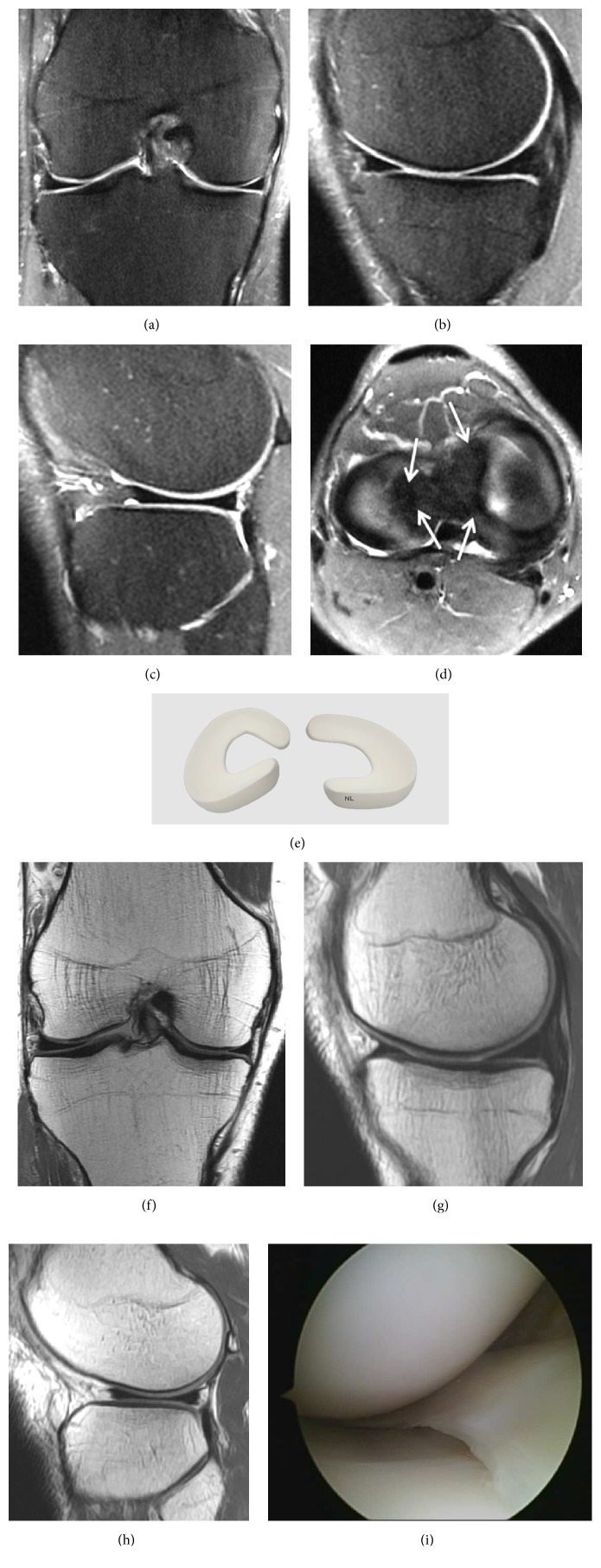
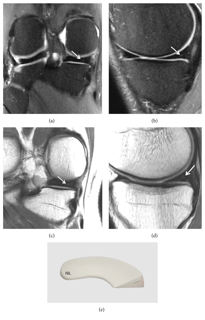
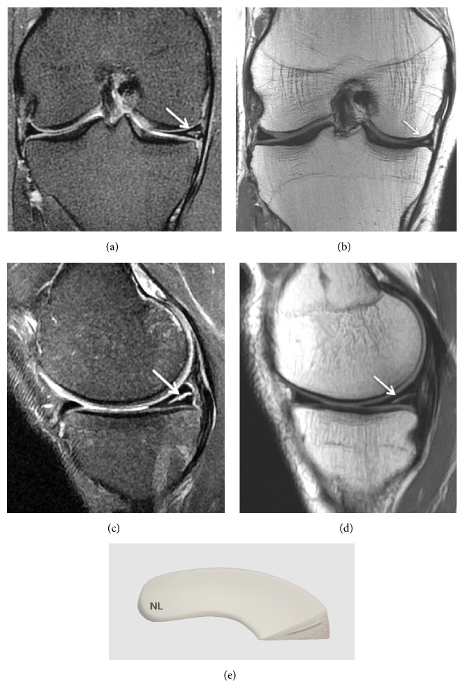
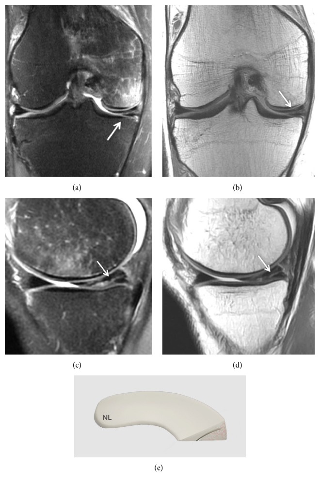
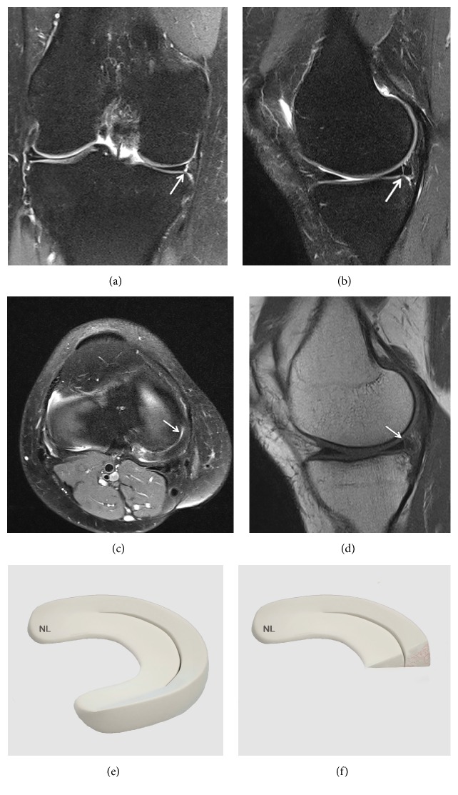
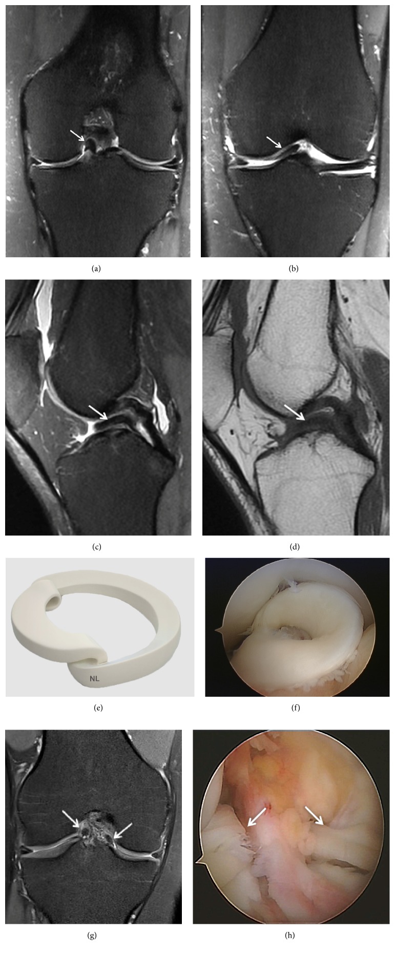
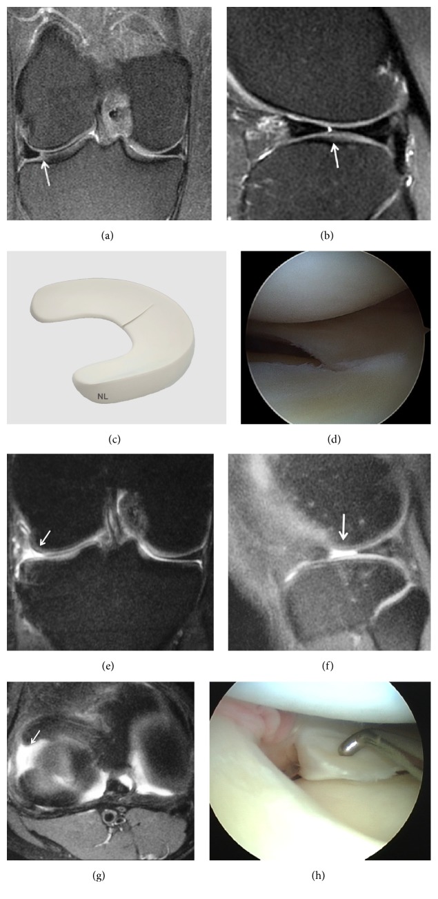
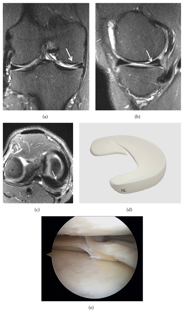
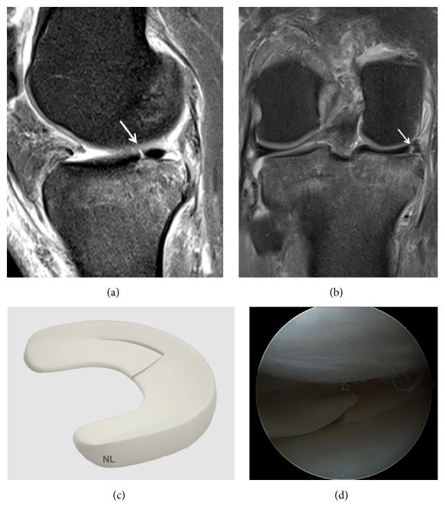
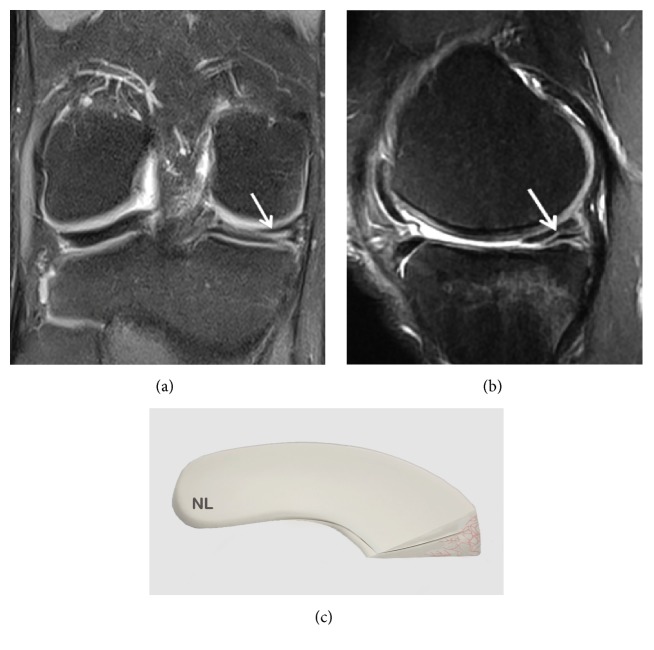
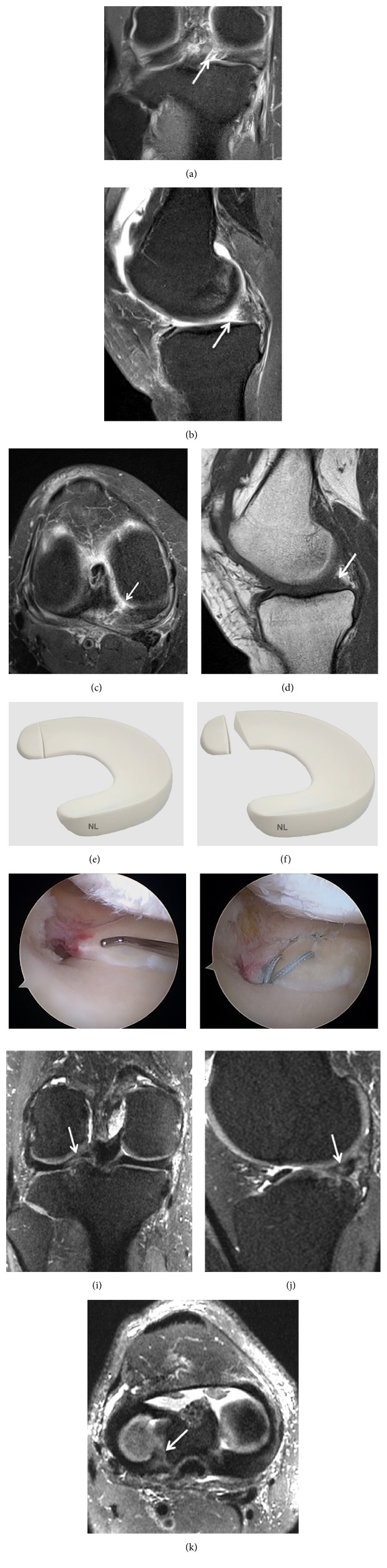
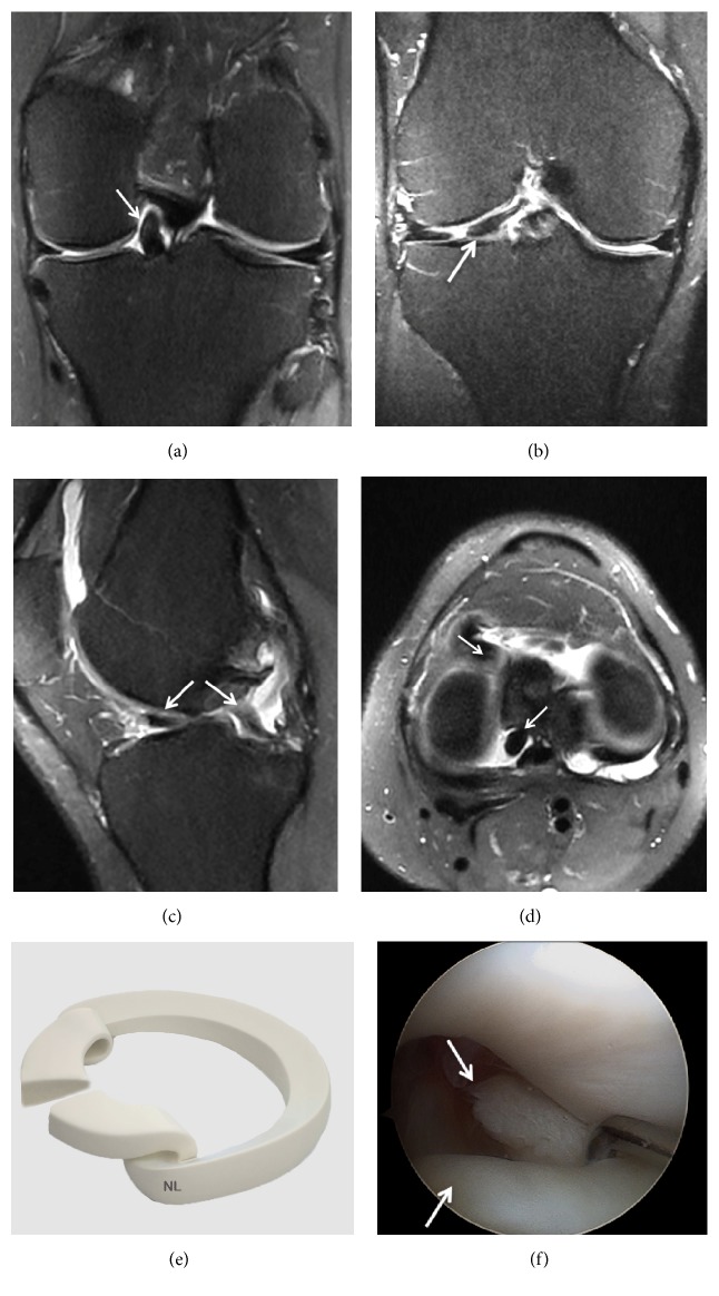
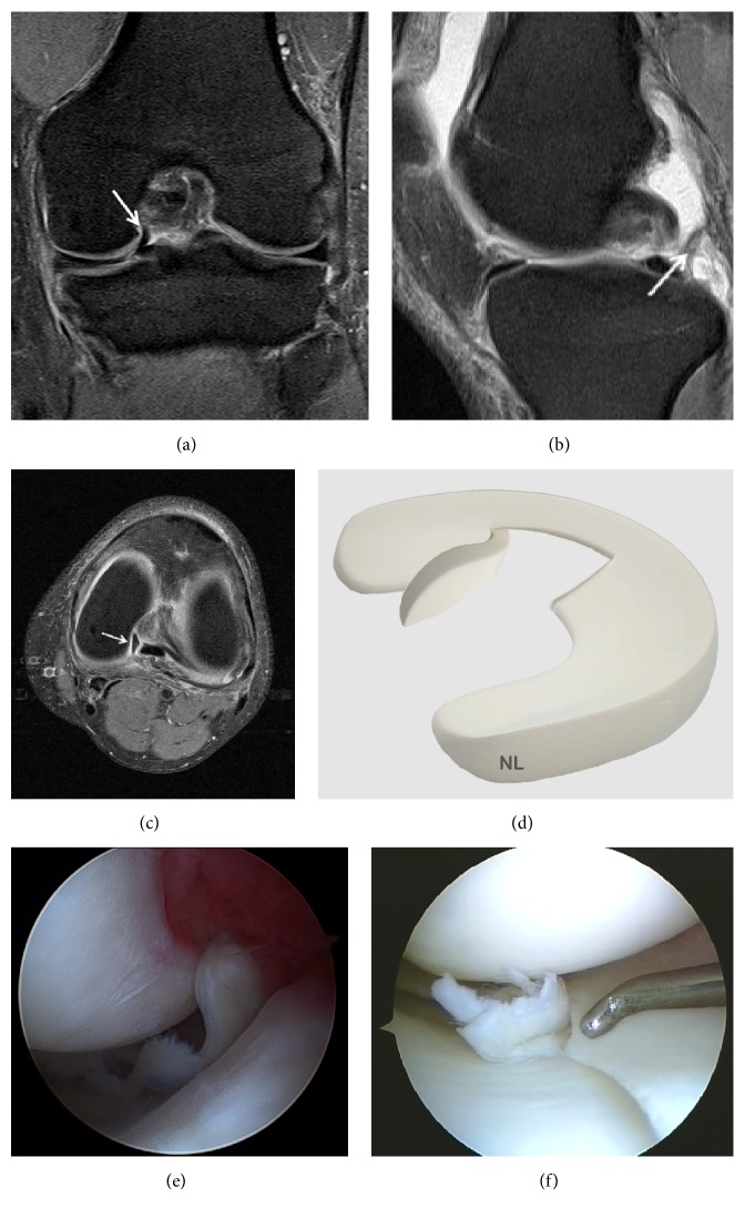
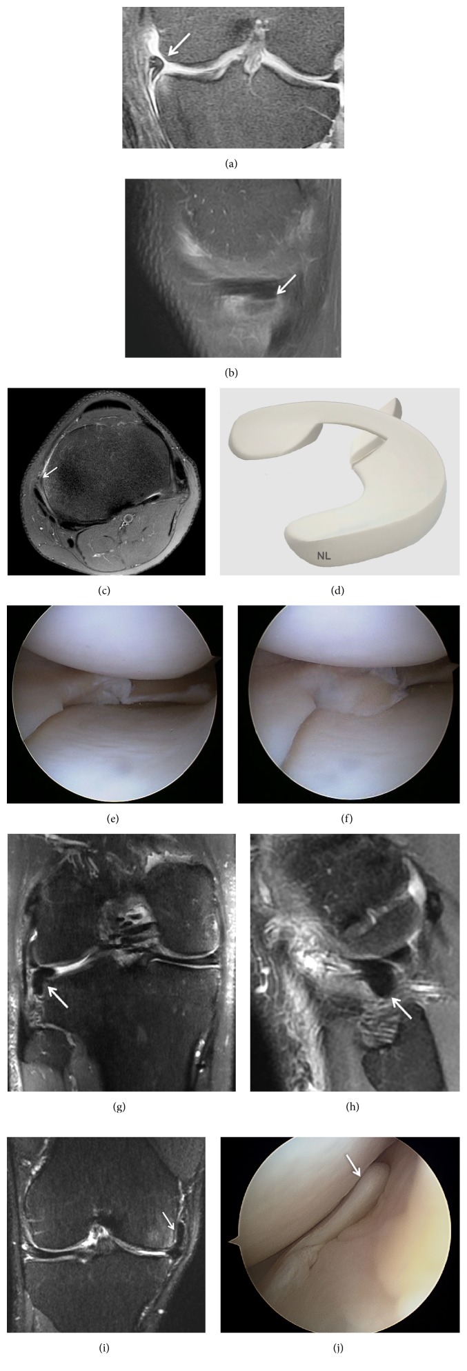
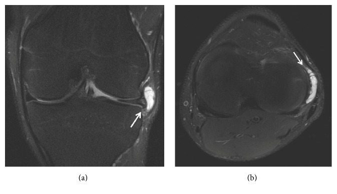
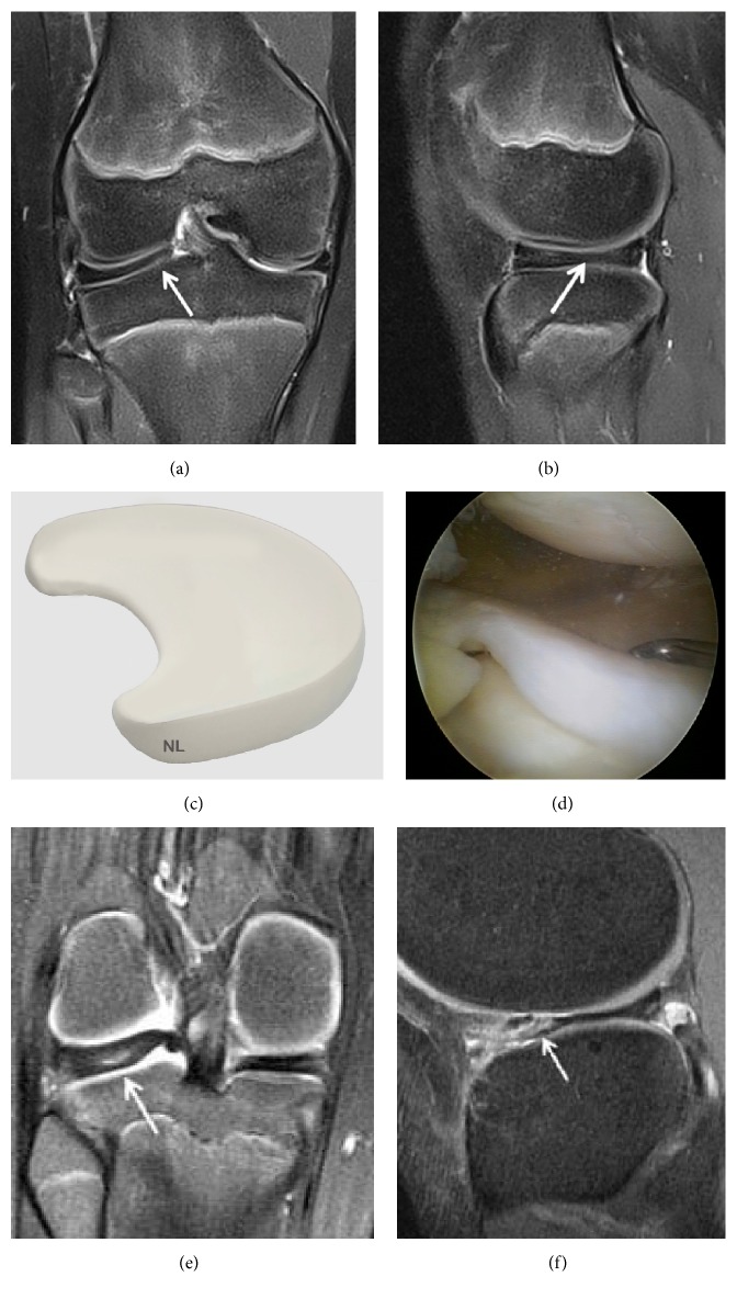
The main objective of this review was to present a synthesis of the current literature in order to provide a useful tool to clinician in radiologic analysis of the meniscus. All anatomical descriptions were clearly illustrated by MRI, arthroscopy, and/or drawings. The value of standard radiography is extremely limited for the assessment of meniscal injuries but may be indicated to obtain a differential diagnosis such as osteoarthritis. Ultrasound is rarely used as a diagnostic tool for meniscal pathologies and its accuracy is operator-dependent. CT arthrography with multiplanar reconstructions can detect meniscus tears that are not visible on MRI. This technique is also useful in case of MRI contraindications, in postoperative assessment of meniscal sutures and the condition of cartilage covering the articular surfaces. MRI is the most accurate and less invasive method for diagnosing meniscal lesions. MRI allows confirming and characterizing the meniscal lesion, the type, the extension, its association with a cyst, the meniscal extrusion, and assessing cartilage and subchondral bone. New 3D-MRI in three dimensions with isotropic resolution allows the creation of multiplanar reformatted images to obtain from an acquisition in one sectional plane reconstructions in other spatial planes. 3D MRI should further improve the diagnosis of meniscal tears.


Figures

Figure 1
Figure 1
Normal meniscus on (a) Coronal T2 FSE Fat Sat MRI; (b) Sagittal T2 FSE Fat Sat MRI (the posterior horn is typically larger than the anterior horn medially); (c) Sagittal T2 FSE Fat Sat MRI (the horns of the lateral meniscus are equal in size and look like opposing triangles); (d) Axial T2 FSE Fat Sat MRI (the shapes of medial and lateral menisci differ, in attachment site (arrow). The medial meniscus is larger and has a more open C-shape configuration, with anterior and posterior tibial attachment sites separated by a greater distance compared to the lateral meniscus which has a more O-shape configuration); (e) three-dimensional diagram of the medial and lateral menisci; (f) coronal proton density weighted sequences; (g) sagittal proton density weighted sequences of the medial meniscus; (h) sagittal proton density weighted sequences of the lateral meniscus; (i) arthroscopic view of meniscus.
Figure 2
Figure 2
Grade 1 meniscal lesion (arrow) (a) Coronal T2 FSE Fat Sat MRI; (b) Sagittal T2 FSE Fat Sat MRI; (c) Coronal proton density weighted sequences; (d) sagittal proton density weighted sequences; (e) three-dimensional diagram.
Figure 3
Figure 3
Grade 2 meniscal lesion on (a) Coronal T2 FSE Fat Sat MRI (arrow); (b) Coronal proton density weighted sequences (arrow); (c) Sagittal T2 FSE Fat Sat MRI (arrow); (d) sagittal proton density weighted sequences (arrow); (e) three-dimensional diagram.
Figure 4
Figure 4
Grade 3 meniscal lesion on (a) Coronal T2 FSE Fat Sat MRI (arrow); (b) Coronal proton density weighted sequences (arrow); (c) Sagittal T2 FSE Fat Sat MRI (arrow); (d) sagittal proton density weighted sequences (arrow); (e) three-dimensional diagram.
Figure 5
Figure 5
A vertical tear in the meniscal tissue communicating with the superior and inferior meniscal articular surfaces completely divides the meniscus into two parts. (a) Coronal T2 FSE Fat Sat MRI (arrow), (b) Sagittal T2 FSE Fat Sat MRI (arrow), (c) Axial T2 FSE Fat Sat MRI (arrow), and (d) sagittal T1-weighted sequence MRI. (e) Three-dimensional diagram shows a vertical and longitudinal tear of the meniscus. (f) Three-dimensional diagram shows a vertical tear.
Figure 6
Figure 6
Displaced bucket-handle tear of the medial meniscus. (a) Coronal T2 FSE Fat Sat MRI shows a displaced bucket-handle fragment of the medial meniscus into the intercondylar notch of the knee (arrow). The remnant of the body of the meniscus is small; (b) Coronal T2 FSE Fat Sat MRI shows a displaced bucket-handle fragment of the anterior medial meniscus (arrow); (c) Sagittal T2 FSE Fat Sat MRI shows the “double PCL sign,” with a displaced fragment of a bucket-handle tear into the intercondylar notch of the knee; (d) Sagittal T1-weighted sequence MRI (arrow); (e) three-dimensional diagram shows a displaced bucket-handle tear of the medial meniscus; (f) arthroscopic view of a bucket-handle tear; (g) Coronal T2 FSE Fat Sat MRI shows a double displaced bucket-handle fragment of the medial and lateral meniscus into the intercondylar notch of the knee (arrow); (h) arthroscopic view of a double bucket-handle tear (arrow).
Figure 7
Figure 7
Radial tear involving the peripheral aspect of the meniscus. (a) Coronal T2 FSE Fat Sat MRI shows the vertical hyperintense signal (arrow) extends to both articular surfaces of the posterior horn of the medial meniscus; (b) Sagittal T2 FSE Fat Sat MRI shows the cleft sign of a radial tear; (c) three-dimensional diagram showing a radial tear involving the peripheral aspect of the meniscus; (d) arthroscopic view of the radial tear; (e) Coronal T2 FSE Fat Sat MRI: a part of the medial meniscus is not identified on the coronal image due to a large radial tear (arrow); (f) Sagittal T2 FSE Fat Sat MRI shows the large radial tear (arrow); (g) axial reconstruction showing the large radial tear (arrow) extending from the free edge into the posterior horn; (h) arthroscopic view of the large radial tear.
Figure 8
Figure 8
Oblique tears are a type of radial tear: (a) Coronal T2 FSE Fat Sat MRI: oblique tear of the body of the medial meniscus (arrow); (b) Coronal T2 FSE Fat Sat MRI: oblique-horizontal tear of the medial meniscus (arrow); (c) Axial T2 FSE Fat Sat MRI reconstruction showing the oblique tear of the posterior part of the medial meniscus (arrow); (d) three-dimensional diagram showing an oblique tear involving the peripheral aspect of the meniscus; (e) arthroscopic view showing a medial meniscus oblique tear.
Figure 9
Figure 9
Radial tear extends towards the periphery to longitudinal meniscal tears; (a) Sagittal T2 FSE Fat Sat MRI (arrow); (b) Coronal T2 FSE Fat Sat MRI shows the longitudinal meniscal tears towards the periphery (arrow); (c) three-dimensional diagram showing the radial tear extends towards the periphery to longitudinal meniscal tears towards the periphery; (d) arthroscopic view showing a medial meniscus tear.
Figure 10
Figure 10
Horizontal tears are also called cleavage or fish-mouth tears. (a) Coronal T2 FSE MRI: horizontal tear (arrow) of the body of the medial meniscus; (b) Sagittal T2 FSE MRI (arrow); (c) three-dimensional diagram.
Figure 11
Figure 11
Meniscus posterior horn avulsion. (a) T2-weighted fat-saturated images showing a complete posterior root tear of the medial meniscus (arrow); (b) Ghost meniscus sign. The posterior horn of the medial meniscus has been replaced with triangular high signal intensity on the T2-weighted fat-saturated sequence (arrow); (c) axial reconstruction showing the large posterior horn avulsion (arrow) with high signal intensity on the T2-weighted fat-saturated sequence; (d) the posterior horn of the medial meniscus is not identified on the sagittal T1 (arrow); (g) arthroscopic view showing a displaced medial meniscus root tear; (h) arthroscopic view showing a suture of the medial meniscus root tear; (i) identification of root tears of the lateral meniscus can be more difficult on the coronal T2-weighted fat-saturated sequence; (j) ghost meniscus sign is less significant on the sagittal T2-weighted fat-saturated sequence (arrow); (k) axial reconstruction showing the posterior horn avulsion (arrow).
Figure 12
Figure 12
Displaced bucket-handle tear of the medial meniscus with tear from the middle part of the meniscus. (a) Coronal T2 FSE Fat Sat MRI: large meniscal fragment (arrow) seen within the intercondylar notch; (b) Coronal T2 FSE Fat Sat MRI: flap tears displaced horizontal under surface tear of the body and anterior horn of the medial meniscus with a flipped fragment (arrow); (c) Sagittal T2 FSE Fat Sat MRI showing a complex tear with a displaced fragment (arrow); (d) Axial T2 FSE Fat Sat MRI reconstruction showing the 2 flap tears of the displaced bucket-handle; (e) three-dimensional diagram; (f) arthroscopic view showing the rupture and the displaced bucket-handle tear.
Figure 13
Figure 13
Meniscal fragments from horizontal meniscal tears can sometimes be displaced in relation to the body of the meniscus, slipping above or below the rest of the meniscal surface. (a) Coronal T2 FSE Fat Sat MRI showing a displaced fragment of the medial meniscus; (b) a meniscal fragment (arrow) is seen posterior to the PCL in Sagittal T2 FSE Fat Sat MRI; (c) axial reconstruction showing meniscal fragment (arrow) on the T2-weighted fat-saturated sequence; (d) three-dimensional diagram showing the meniscal fragments; (e) arthroscopic views of a displaced tear of the medial meniscus in the intercondylar notch; (f) arthroscopic views of a displaced tear of the medial meniscus in the underlying posteromedial tibial plateau.
Figure 14
Figure 14
Meniscal fragments from horizontal meniscal tears displaced under the medial or lateral meniscus. The displaced fragment blocks the peripheral edge of the tibial plateau and the deep part of the MCL or LCL. (a) Coronal T2 FSE Fat Sat MRI showing a displaced horizontal undersurface tear of the body of the medial meniscus with a flipped fragment (arrow) along the undersurface of the native meniscus and extending under MCL; (b) Sagittal T2 FSE Fat Sat MRI showing a displaced fragment of the medial meniscus (arrow); (c) axial reconstruction showing the flipped fragment (arrow) under MCL on the T2-weighted fat-saturated sequence; (d) three-dimensional diagram showing a displaced tear of the medial meniscus; (e) arthroscopic views of a displaced tear of the medial meniscus under the meniscus; (f) arthroscopic view of the medial meniscus tear under the meniscus reduced in intra-articular lesion; (g) Coronal T2 FSE Fat Sat MRI showing a displaced fragment of the lateral meniscus (arrow); (h) Sagittal T2 FSE Fat Sat MRI showing a large fragment of the lateral meniscus under the LCL; (i) complex tear with a displaced fragment (arrow) coursing into the superior recess in Coronal T2 FSE Fat Sat MRI; (j) arthroscopic views of a displaced tear of the medial meniscus into the superior recess.
Figure 15
Figure 15
Lateral meniscal cysts: (a) lateral meniscal cysts are usually located at the anterior meniscal horn (coronal T2 FSE MRI sequences); (b) Axial T2 FSE Fat Sat MRI reconstruction showing the lateral meniscal cysts (arrow).
Figure 16
Figure 16
Discoid lateral meniscus. (a) Coronal T2 FSE Fat Sat MRI showing meniscal enlargement. The lateral meniscal body (arrow) is enlarged and has a more slab-like configuration compared to the normal-appearing triangular medial meniscal body; (b) Sagittal T2 FSE Fat Sat image of the lateral meniscus demonstrating persistence of the bow tie appearance on the more central slices rather than converting into 2 opposing triangles; (c) three-dimensional diagram showing a discoid lateral meniscus; (d) arthroscopic views of a discoid lateral meniscus; (e) posterior cystic degeneration in a discoid lateral meniscus; (f) anterior cystic degeneration in a discoid lateral meniscus.

Similar articles

Cited by

References

    1. De Maeseneer M., Lenchik L., Starok M., Pedowitz R., Trudell D., Resnick D. Normal and abnormal medial meniscocapsular structures: MR imaging and sonography in cadavers. American Journal of Roentgenology. 1998;171(4):969–976. doi: 10.2214/ajr.171.4.9762977. - DOI - PubMed
    1. De Flaviis L., Scaglione P., Nessi R., Albisetti W. Ultrasound in degenerative cystic meniscal disease of the knee. Skeletal Radiology. 1990;19(6):441–445. doi: 10.1007/bf00241801. - DOI - PubMed
    1. Casser H.-R., Sohn C., Kiekenbeck A. Current evaluation of sonography of the meniscus. Results of a comparative study of sonographic and arthroscopic findings. Archives of Orthopaedic and Trauma Surgery. 1990;109(3):150–154. doi: 10.1007/BF00440576. - DOI - PubMed
    1. Gerngross H., Sohn C. Ultrasound scanning for the diagnosis of meniscal lesions of the knee joint. Arthroscopy. 1992;8(1):105–110. doi: 10.1016/0749-8063(92)90143-y. - DOI - PubMed
    1. Rutten M. J. C. M., Collins J. M. P., Van Kampen A., Jager G. J. Meniscal cysts: detection with high-resolution sonography. American Journal of Roentgenology. 1998;171(2):491–496. doi: 10.2214/ajr.171.2.9694482. - DOI - PubMed