. 2016 May 31;7(22):32232-46.
doi: 10.18632/oncotarget.8572.

Avoiding drug resistance through extended drug target interfaces: a case for stapled peptides

Affiliations

Avoiding drug resistance through extended drug target interfaces: a case for stapled peptides

Siau Jia Wei et al. Oncotarget. .

Abstract

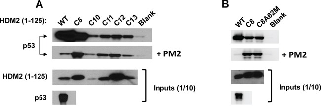
Cancer drugs often fail due to the emergence of clinical resistance. This can manifest through mutations in target proteins that selectively exclude drug binding whilst retaining aberrant function. A priori knowledge of resistance-inducing mutations is therefore important for both drug design and clinical surveillance. Stapled peptides represent a novel class of antagonists capable of inhibiting therapeutically relevant protein-protein interactions. Here, we address the important question of potential resistance to stapled peptide inhibitors. HDM2 is the critical negative regulator of p53, and is often overexpressed in cancers that retain wild-type p53 function. Interrogation of a large collection of randomly mutated HDM2 proteins failed to identify point mutations that could selectively abrogate binding by a stapled peptide inhibitor (PM2). In contrast, the same interrogation methodology has previously uncovered point mutations that selectively inhibit binding by Nutlin, the prototypical small molecule inhibitor of HDM2. Our results demonstrate both the high level of structural p53 mimicry employed by PM2 to engage HDM2, and the potential resilience of stapled peptide antagonists to mutations in target proteins. This inherent feature could reduce clinical resistance should this class of drugs enter the clinic.

Keywords: HDM2; MDM2; cancer resistance; p53; stapled peptide.


Conflict of interest statement

The authors declare no conflicts of interest.

Figures

Figure 1
Figure 1. Selection of PM2-resistant HDM2 by in vitro compartmentalisation
1. HDM2 expression constructs (blue and purple bars) appended with 2CONA p53 response element (“RE”, green) and HA-tag coding sequence (black) and p53 expression construct (red bar) are segregated into aqueous emulsion compartments along with PM2 (cyan helix). Protein expression occurs within compartments. PM2 inhibition of HDM2 results in no HDM2-p53-DNA complex formation (left bubble), whereas resistant HDM2 can form the complex (right bubble). 2–3. The emulsion is broken and complexes captured with anti-HA antibody. DNA encoding resistant HDM2 variants is amplified by PCR. 4. Selectants further evaluated by secondary pull-down assay or subjected to further rounds of selection.
Figure 2
Figure 2. Selected HDM2 variants display in vitro PM2-resistance phenotype
A. In vitro pull-down assay showing reduced inhibition by PM2 (10 μM) to binding of p53 for indicated parental HDM2 variants and WT HDM2 (residues 1-125). Note: exposure time for HDM2 inputs is 8 hours and 1 second for all other panels. B. in vitro pull-down assay showing little impact upon reversion of the M62A mutation to PM2 binding in HDM2-C8 (residues1-125). Blank indicates background p53 binding in absence of HDM2. Note: exposure time for HDM2 inputs (developed using film) is 8 hours and 10 second for all other panels (digitally acquired).
Figure 3
Figure 3. Sequence alignment of selectant HDM2 clones showing PM2 resistance
Mutated residues are highlighted in red, with those present in more than one selectant boxed. The M62A mutation (green) was incorporated into the selection library.
Figure 4
Figure 4. PM2 resistance comes at cost of reduced interaction with p53
In vitro pull-down assay showing reduced inhibition by PM2 (10 μM) to indicated point mutants (asterisk) derived from HDM2-C8 (residues 1-125). The point mutants L34P, F55L, Y60C and C77R also show reduced inhibition by Nutlin (10μM). Blank indicates background p53 binding in absence of HDM2. Note: exposure time for HDM2 inputs is 8 hours (developed using film) and 3 minutes for all other panels (digitally acquired).
Figure 5
Figure 5. PM2 resistance is also seen when point mutants are introduced into full-length HDM2
A. In vitro pull-down assay showing reduced inhibition by PM2 (10 μM) to indicated C8-derived point mutants (asterisk) present in full-length HDM2 The point mutant F55L also shows reduced inhibition by Nutlin (10μM). Blank indicates background p53 binding in absence of HDM2. B. As in A, additionally showing levels of wild type and indicated HDM2 variants co-eluted off beads after pull-down following mock (panel 3) and PM2 treatment (panel 4). Note exposure time for p53 pull-down in absence of treatment (panel 1) is 5s and 10 minutes for pull-down after PM2 treatment (panel 2, developed using film). Exposure time for HDM2 input and HDM2 (+ indicated variants) eluted off beads after pull-down is 30 seconds (digitally acquired).
Figure 6
Figure 6. PM2 shows reduced inhibition of selected HDM2 variants in p53/MDM2-null DKO cells
A. Wild-type and HDM2-C8 (full-length) were co-transfected with p53 and p53-reporter gene, and reporter gene activity measured in the presence of PM2 (20 μM) or Nutlin (10 μM). p53 activity is denoted as percentage of that observed when p53-alone co-transfected with reporter gene. Shown below are Western blots indicating expression levels of HDM2 variants and p53 cotransfected into DKO cells. B and C. As in ‘A’, with wild-type HDM2 and indicated HDM2-C8 derived point mutants (full length) co-transfected into DKO cells. p53 activity is denoted as percentage of that observed when p53-alone co-transfected with reporter gene. Shown below are Western blots indicating expression levels of HDM2 variants and p53 cotransfected into DKO cells.
Figure 7
Figure 7. Stapled peptide recapitulates key p53 signature residues that interact with HDM2 N-terminal domain
Overlay of p53 peptide (green) and MO6 stapled peptide (cyan, with staple moiety in grey) when bound to HDM2 N-terminal domains. The relative configurations of the key F19 and W23 residues are conserved, with some deviation in the orientation of L26. Adapted from 1YCR and 4UMN. Shown below is alignment of p53 peptide, PM2 and MO6, with signature residues shaded and residue differing between PM2 and MO6 highlighted in red.
Figure 8
Figure 8. Projection of HDM2-C8 mutants onto HDM2 N-terminal domain structure
Shown is structure of M06 stapled peptide (cyan, grey) bound to HDM2-M62A N-terminal domain (pink). The residues contributing to the resistance phenotype are coloured yellow, and the rest are purple. Adapted from 4UMN.
Figure 9
Figure 9. The staple moiety makes favourable contacts with F55 in the N-terminal domain of HDM2
Left: Overlay of p53 peptide (green) and MO6 stapled peptide (cyan, staple in gray) bound to HDM2 N-terminal domain (magenta, surface representation). The positions of the F55 and I99 residues are indicated in yellow. Right: Same as left, highlighting the relative orientation of the p53 peptide and MO6 stapled peptide L26 side chains in respect to I99 in HDM2.

References

    1. Phillips C, Roberts LR, Schade M, Bazin R, Bent A, Davies NL, Moore R, Pannifer AD, Pickford AR, Prior SH, Read CM, Scott A, Brown DG, Xu B, Irving SL. Design and structure of stapled peptides binding to estrogen receptors. J Am Chem Soc. 2011;133:9696–9699. - PubMed
    1. Walensky LD, Kung AL, Escher I, Malia TJ, Barbuto S, Wright RD, Wagner G, Verdine GL, Korsmeyer SJ. Activation of apoptosis in vivo by a hydrocarbon-stapled BH3 helix. Science. 2004;305:1466–1470. - PMC - PubMed
    1. Brown CJ, Quah ST, Jong J, Goh AM, Chiam PC, Khoo KH, Choong ML, Lee MA, Yurlova L, Zolghadr K, Joseph TL, Verma CS, Lane DP. Stapled peptides with improved potency and specificity that activate p53. ACS Chem Biol. 2013;8:506–512. - PubMed
    1. LaRochelle JR, Cobb GB, Steinauer A, Rhoades E, Schepartz A. Fluorescence correlation spectroscopy reveals highly efficient cytosolic delivery of certain penta-arg proteins and stapled peptides. J Am Chem Soc. 2015;137:2536–2541. - PMC - PubMed
    1. Kim YW, Grossmann TN, Verdine GL. Synthesis of all-hydrocarbon stapled alpha-helical peptides by ring-closing olefin metathesis. Nat Protoc. 2011;6:761–771. - PubMed

MeSH terms